Feb
17
2024

Ashwagandha’s Benefits

Medical news today had an article in October 2023 that reviewed ashwagandha’s benefits. I thought it would be useful to discuss this topic in an abridged version. Often things get distorted on the Internet and a reality check helps to separate facts from fiction. Ashwagandha (botanical name: Withania somnifera) is a small shrub that grows in Southeast Asia, Africa and some areas of India. An alternative name is Indian ginseng.  Ashwagandha has been part of Ayurvedic medicine for more than 3000 years.

About ashwagandha’s effectiveness

Ashwagandha is part of natural medicines that go under the name of adaptogens. The effective substances of ashwagandha were isolated. Researchers named them withanolides. They are alkaloids and consist of polyoxygenated steroidal lactones with 28-carbon ergostane skeletons.

People are taking ashwagandha supplements to counter stress and fatigue. But it also increases testosterone, which increases muscle mass. In addition, there is an increase of libido in men as well as in women.

People with insomnia benefit from the relaxing and sleep-inducing effect of ashwagandha supplements. Ashwagandha stimulates the immune system. It also significantly counters the stress response, as it lowers cortisol levels.

The active chemical components of ashwagandha

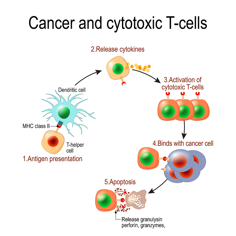
Various researchers pinpointed the diverse actions of ashwagandha extracts to withanolides, which are the pharmacologically active ingredients. There are at least 25 different chemical structures of withanolides, which are depicted in this reference: This explains the diverse effects of ashwagandha extracts on the immune system, the stress hormone axis, the libido brain centers and testosterone production. One human study showed that within 96 hours of ashwagandha extract administration the immune system was activated; T cells and natural killer cells were measured and were significantly elevated in number in comparison to controls:

Specific clinical trials showing effects of ashwagandha

Effects on the brain

In a small clinical study oral intake of ashwagandha improved performance on cognitive tasks, executive function, attention, and reaction time.

Dr. Amala Soumyanath is a professor of neurology in the School of Medicine at Oregon Health & Science University. She stated: “These laboratory studies show that ashwagandha extracts can act on neurotransmitter pathways including those involving serotonin and Gamma-aminobutyric acid (GABA), and also affect systems like the hypothalamic-pituitary-adrenal (HPA) axis and the sympathetic-adrenal-medullary (SAM) axis.”

What does this mean? It means that ashwagandha contains a mix of withanolides that calm down the brain, particularly when it is stressed. At the same time, it increases serotonin and GABA, which has antidepressant and calming effects. Clinical trials have measured cortisol levels in volunteers and found that cortisol is lowered within a few weeks. This is the strongest evidence that ashwagandha in fact counteracts stress.

Libido and muscle strength

A small human study found that muscle strength was higher after exposure to oral ashwagandha in comparison to controls.

Ashwagandha showed a significant increase of maximum oxygen consumption (VO2max) in healthy adults and athletes, which is a direct measurement of exercise fitness. A clinical trial with 50 overweight males aged 40-70 years, showed that 16 weeks of oral ashwagandha increased their testosterone level by 14.7%. In addition, there were improvements in fatigue, vigor, and sexual and psychological well-being. There was also an increase of 18% of DHEA-S a precursor of testosterone. The authors felt that a larger study with a longer observation time would be beneficial to confirm this study. Overall, the objective effects of ashwagandha are in the mild to moderate category. The mild increase in testosterone affects libido only marginally. Some articles in social media are vastly exaggerating the objective findings.

Health risk of ashwagandha

There are downsides as well. Ashwagandha stimulates the immune system and in doing so may exacerbate autoimmune diseases. It also can increase thyroid hormones, which is important to know for people with hyperthyroidism or hypothyroidism. Men who have hormone sensitive prostate cancer should avoid ashwagandha, because it elevates testosterone.

Researchers pointed out that gastrointestinal upsets, such as diarrhea, nausea, and vomiting due to ashwagandha are common side effects. The higher the dosage and the longer the exposure is, the more likely it is that the patient experiences side effects. Researchers reported a few cases of reversible liver toxicity cases due to ashwagandha. At this point the effects of concomitant medications taken at the same time as ashwagandha is not known.

Ashwagandha’s Benefits

Ashwagandha’s Benefits

Conclusion

Lately social media is full of ashwagandha’s benefits. Rosy reports of increased libido, extra energy and boosts of confidence circulate on the Internet. Men are told that in order to build up muscle bulk with exercise they must take ashwagandha. This review checked the facts. There is a 14.7% rise in testosterone from taking ashwagandha and this may contribute to an increase of muscle mass with exercise, but this is not a huge effect. Also, the mild increase of libido due to the increase of testosterone is not as significant as reported in the social media. The active ingredients of ashwagandha are the withanolides, which researchers isolated. They do help to deal with stress-related problems like lack of energy and insomnia. In addition, they stimulate the immune system to give a stronger response.

Side effects of ashwagandha

But ashwagandha also has some side effects. It can make auto immune reactions worse. It can interfere with both hyperthyroidism and hypothyroidism. Men who have hormone sensitive prostate cancer must avoid ashwagandha because of the testosterone increase. Gastrointestinal upsets, such as diarrhea, nausea, and vomiting from ashwagandha supplementation are common side effects. So far scientific evidence is based on relatively small clinical trials. Scientists must conduct much bigger trials that are going on for several years to provide more in-depth information on the effects of ashwagandha.

Feb
04
2024

Beef and Dairy May Cause Cancer and MS

New cancer research suggests that chronic virus particles in beef and dairy may cause cancer and MS (multiple sclerosis). The Medical journal Medscape.com had a review article that summarized this line of research.

Papillomaviruses and cervical cancer

Harald zur Hausen, M.D., D.Sc., a German virologist, detected that papillomavirus causes cervical cancer. He was given the Nobel prize in Medicine in 2008 for “his discovery of human papillomaviruses causing cervical cancer”. In the meantime, we know that there are two strains, namely HPV-16 and HPV-18 that are carcinogenic. We also learnt that papillomavirus causes oropharyngeal cancer, anal cancer, penile cancer and vulvar cancer. Since then, HPV vaccines are commonly in use for cancer prevention.

Evidence for causation of colorectal cancer

Professor Harald zur Hausen and his wife, Professor Ethel-Michele de Villiers, continued work on looking for viral particles in many other cancers. This led them to state that colorectal cancer was due to a latent viral infection. He determined that, when women were breastfeeding their infants for prolonged periods of time (about 1 year), they transmitted oligosaccharides with the breast milk to their offspring, which gave lifelong prevention against colorectal cancer to their children. He also showed that colon cancer patients had round particles in their intestinal mucous membranes, which consisted of single-stranded DNA rings.

Persistent viruses

The researchers said that they came from viruses and they named them bovine meat and milk factors (BMMF). In the same areas in the intestine, they detected acid radicals from oxidative stress typical for chronic inflammation. They postulated that infants who are weaned from breast milk prematurely, and started on cow milk formulas ingest BMMF. This infects the lining of the gut where a chronic subclinical BMMF infection gets established. Decades later the patient comes down with colorectal cancer. They established that children who are breast fed for 1 year do not get BMMF particles in the lining of their guts or colorectal cancer. They also don’t get MS in adult life.

Criticism of two regulatory agencies in Germany

The above results of professor zur Hausen and his wife were published in February of 2019. This led to a lot of press releases questioning these results. In Germany the consumption of beef and milk products is popular. The DKFZ (Deutsches Krebsforschungszentrum) felt that an “all-clear signal” was necessary. The Bundesinstitut für Risikobewertung (BfR) stands for the German Federal Institute for Risk Assessment, situated in Berlin. It is a substructure of the German government responsible for food safety. The second agency is the Max Rubner Institute (MRI) in Karlsruhe, also known as Federal Research Institute of Nutrition and Food. Both agencies started to investigate the facts of Prof. zur Hausen’s research. At the end of November 2022 the BfR and MRI made a joint statement. They stated the following:

  • BMMF were not new viral agents, but variations of already known DNA sequences.
  • BMMF commonly occurred in a variety of animal- and plant-based foods.
  • BMMF were not capable of infecting human cells.
  • It was true that consumption of red and processed meat correlates with the incidence of intestinal tumors.
  • However, consumption of dairy products are linked to a reduced risk of intestinal tumors.
  • There is no evidence that breast cancer would be associated with the consumption of beef or dairy.
  • They stated that milk products and beef are valuable supplementary diet components for infants due to their micronutrients. They further stated that dairy and beef products are safe for people of all ages.

Evidence for causation of multiple sclerosis

Professor zur Hausen and his wife, Professor Ethel-Michele de Villiers both worked at the German Cancer Research Center in Heidelberg. They pursued research about MS. MS is usually attributed to IgG1 and IgG3 autoantibodies that destroy Schwann cells. The Hausen research team found the following perplexing facts:

  • MS was associated with the consumption of dairy products and beef products.
  • They isolated ring-shaped DNA molecules (BMMF) from dairy and cattle blood.
  • Epstein Barr virus (EBV) also plays a role in initiating MS. MS patients have higher antibody titers against EBV.
  • One research paper noticed that MS patients in Antarctica secreted EBV in their saliva in the winter month. However, vitamin D3 stopped the viral excretion. This is very interesting as vitamin D is an important immune response stimulator.

More evidence:

  • They isolated BMMF particles from lesions of MS patients.
  • They noticed the old fact that MS is more common further away from the equator. Vitamin D production from sun exposure reduced cases of MS.
  • Prolonged breast feeding up to one year prevents life threatening rotaviruses and noroviruses in the newborn. The reason is exposure to oligosaccharides in breast milk. The mother starts producing breast milk in the middle of her pregnancy. It protects mother from tumors (including breast cancer), from MS later in life as well as type 2 diabetes.
  • The researchers formulated the hypothesis that both EBV and BMMF are responsible in patients to form MS lesions in the brain when vitamin D levels are low. A good dose of vitamin D3 every day may be helping to keep EBV and BMMF in a dormant phase.

Discussion

At this point there is no consensus why an increased consumption of beef and processed meat causes more colorectal cancer. The question is whether BMMF particles cause colorectal cancer or whether meat consumption experiences metabolization into carcinogenic substances? Either way it would be desirable to cut down on your red meat consumption.

With respect to MS, we know that autoantibodies are destroying the Schwann cells. But the question is why the immune system produces autoantibodies. Could it be that persistent EBV viruses switch the immune system from a normal to an autoantibody mode? Would BMMF be an additional factor?

Beef and Dairy May Cause Cancer and MS

Beef and Dairy May Cause Cancer and MS

Conclusion

Professor zur Hausen, a virologist from Germany and his wife Professor Ethel-Michele de Villiers researched persistent viruses. Professor zur Hausen detected the connection of papilloma viruses to cervical cancer. He received the Nobel prize in medicine 2008. They proposed the theory that newborns in their first year of life have an immature immune system. If they are fed cow’s milk and/or beef during the first year they accumulate bovine meat and milk factors (BMMF), which can subsequently lead to colon cancer or to MS as an adult. Two top German institutes banded together to criticize Prof. zur Hausen’s research.

Criticism of Professor zur Hausen’s research

The Bundesinstitut für Risikobewertung (BfR) stands for the German Federal Institute for Risk Assessment, situated in Berlin. It is a substructure of the German government responsible for food safety. The second agency is the Max Rubner Institute (MRI) in Karlsruhe, also known as Federal Research Institute of Nutrition and Food. These research institutes concluded that not all of the findings of Professor zur Hausen were valid. I listed 7 of their concerns. On the other hand, they agreed that it was true that consumption of red and processed meat correlated with the incidence of intestinal tumors. Time will tell which parts of the research ultimately will be valid and which are not.

Incoming search terms:

Sep
09
2023

How the Immune System affects Parkinson’s Disease

This article explains how the immune system affects Parkinson’s disease (PD). Notably, in the past physicians thought that Parkinson’s disease was due to a degenerative change of the substantia nigra. This explained why balancing was a problem, why shaking of the hands occurred and why falls happened often. It it important to realize that nobody thought about the immune system.  And no-one knew that an autoimmune process could be behind Parkinson’s disease.

T cells that react to a damaged protein called alpha-synuclein

There are specific changes in the immune system approximately 10 years before Parkinson’s disease symptoms occur in patients who come down with the disease. Researchers from the La Jolla Institute for Immunology showed that T cells play a key role in causing PD. They react to a damaged protein called alpha-synuclein build up in the dopamine-producing brain cells. Laboratory physicians can assay this through a simple blood test, which becomes a screening tool for early Parkinson’s disease. The reactive T cells stay around for about 10 years, then fade away. There seem to be other immune factors that weaken the initial aggressive phase of the T cells.

The role of inflammation in Parkinson’s

When the immune system malfunctions chronic inflammation can develop. In farmers exposed to pesticides the later development of Parkinson’s disease was observed. The researchers thought that the pesticides caused an irritation of the immune system leading to chronic inflammation. There is evidence that the gut bacteria are different in Parkinson’s disease patients when compared to normal controls. The gut absorbs the metabolites of the abnormal gut bacteria and causes chronic inflammation. In an attempt to stop the inflammatory process, the immune system can develop autoimmune antibodies, which can cross react with cells of the substantia nigra. This in turn can cause Parkinson’s disease.

Lifestyle factors that people can change to prevent PD

Dr. Rebecca Gilbert, vice-president and chief scientific officer for the American Parkinson Disease Association (APDA) commented on the importance of lifestyle changes. She said: “It makes intuitive sense that instituting lifestyle modifications that potentially decrease inflammation may decrease the risk of Parkinson’s disease. Exercise, for example, has been shown to reduce inflammation and is probably one of the many reasons that exercise reduces the risk of Parkinson’s disease and also improves established Parkinson’s disease.” She commented further: “Also, we should avoid things like excessive alcohol and nicotine that we know have negative effects on the immune system,” she added. “And managing our stress as best as possible can slow and help maximize outcomes of many diseases.”

Changing diet can help postpone Parkinson’s disease

With regard to the best diet that will help Parkinson’s disease patients she said: “The MIND diet emphasizes whole grains, vegetables, nuts, legumes, and berries. Fish is the preferred protein and olive oil is the preferred fat. Recently a study showed that adherence to the MIND diet and the Mediterranean diet had an association with later onset of Parkinson’s disease.”

The gut connection to Parkinson’s disease

According to the WHO the global prevalence of Parkinson’s disease has doubled in the last 25 years. At this point we do not know why this is so. But many investigations have shown that there is a significant difference in the gut bacteria composition of healthy controls and Parkinson’s disease patients. There is a 30% difference between the bacterial composition of healthy controls and patients with Parkinson’s disease. This has led to Braak’s Hypothesis of Parkinson’s Disease. This hypothesis says that an unknown pathogen enters through the nose, the person swallows it and it ends up in the gut. Absorption gets it into the gut wall and it migrates through the vagus nerve into the central nervous system where it leads to accumulation off alpha-synuclein in the substantia nigra. This destroys the dopamine producing cells in that region causing the symptoms of PD.

Can any diet fight gut dysbiosis?

  • In 2022 study they found that flavonoids, the pigments of fruit were associated with a lower mortality of patients with Parkinson’s disease.
  • In an earlier study of 2018 researchers determined that a protein from fish with the name parvalbumin helped Parkinson’s patients to stop producing alpha-synuclein. PD patients suffer from clumping of alpha-synuclein, which causes their symptoms.
  • Restriction of refined carbohydrates “especially diets with a low glycemic index, rich of vitamins and polyphenols, a Mediterranean diet for example, can be recommended”.

Regular exercise to prevent Parkinson’s disease

Regular physical exercise maintains body function and muscle strength. Dr. Emer MacSweeney said: “Being physically active is one of the best things you can do for your body. Exercise helps protect against many diseases and keeps the heart, muscles, bones, and brain in optimum condition. Exercise promotes the oxygenation of the brain and stimulation of multiple neurochemicals.”

Several studies showed that patients with PD deteriorate slower, if they exercise regularly. Part of that response is due to the release of endorphins and serotonin, but we do not know all of the positive mechanisms of exercise at this time.

How the Immune System affects Parkinson’s Disease

How the Immune System affects Parkinson’s Disease

Conclusion

Recent research changed what we know about Parkinson’s disease (PD). Braak’s Hypothesis of Parkinson’s Disease states that an unknown pathogen enters through the nose, gets swallowed and ends up in the gut. From there it gets taken up into the gut wall and migrates through the vagus nerve into the central nervous system. There it leads to accumulation of alpha-synuclein in the substantia nigra. This destroys the dopamine producing cells in that region causing the symptoms of PD. But we also know that chronic inflammation can aggravate the symptoms of PD patients. When the composition of the gut bacteria deteriorates, this too will make PD patients worse.

Lifestyle changes help to postpone Parkinson’s disease

A healthy diet, like the MIND diet, DASH diet or the Mediterranean diet have beneficial effects on PD patients. Many studies also found that regular physical exercise is a stabilizing factor in PD patients. There are still many gaps in what we know about the causation of PD. But the above summarized factors are a good start.

Incoming search terms:

Jan
14
2023

Cortisol is the Number 1 Longevity Hormone

At the 30th American Anti-Aging Academy Medicine Conference in Las Vegas one topic was that cortisol is the number 1 longevity hormone. Dr. Thierry Hertoghe, an endocrinologist from Brussels, Belgium gave a comprehensive lecture on Saturday, Dec.10, 2022. He explained that cortisol from our adrenal glands is vital to our health. Certainly, it is important for our quality of life and for health in general.  Indeed, cortisol keeps us alive when various circumstances challenge our health. Specifically, cortisol is on the bottom of the hormone cascade of corticotropin releasing hormone (CRH) from the hypothalamus/ Adrenocorticotropic hormone (ACTH) from the pituitary gland/and cortisol from the adrenal glands. It is important to realize that this is the hormone axis of the stress reaction that Dr. Selye first described.

Production of cortisol

Sedentary men produce 30 mg of cortisol per day, women 20 mg/day. Under stressful conditions ACTH production increases and causes an increase of cortisol production in the adrenal glands. To clarify, stressed men produce 40 to 60 mg per day and stressed women produce 30 to 40 mg/day. In addition, the older a person is, mononuclear leukocytes have less type 1 and type 2 glucocorticosteroid receptors.

Action of cortisol

Dr. Hertoghe pointed out that cortisol increases energy and stress resistance. It causes blood sugars to increase and also elevates blood pressure. The immune system gets stronger, allergies and inflammation decrease. Cortisol shapes our bodies: when there is cortisol deficiency the body is thin; with normal cortisol production the body has a normal configuration. Your body develops swelling and obesity with cortisol excess, but when cortisol is missing the body gets frail and thin.

Cortisol deficiency

Low cortisol production leads to low blood sugar and low blood pressure. This diminishes glycogen stores in the liver and muscle mass. A lack of circulating cortisol reduces gluconeogenesis in the liver, which is an alternative pathway of glucose production. One of the hallmark symptoms is mental fatigue, particularly in times of stress. With cortisol deficiency the person feels fatigue in the second part of the day.

Chronic fatigue syndrome

The peak age for chronic fatigue syndrome patients is 31 years. The mean duration of the illness lasts 7 years. 58% of patients have free serum cortisol levels at 20:00 hours, which is less than normal. In 40% of patients the total cortisol level is less than normal.

Burnout

With burnout the urinary free cortisol is diminished. Patients with cortisol deficiency have a hyper sensitivity to stress.

A lack of cortisol leads to feeling no energy and having no stress resistance. The person feels tense and has negative thinking. A person who has sufficient cortisol is full of energy, expresses emotions and handles stress well. With enough cortisol a person is relaxed, has positive feelings and finds solutions everywhere.

Adrenal crisis

Dr. Hertoghe explained that an adrenal crisis happens when there is not enough cortisol production in the adrenal glands to support the metabolism and the cardiovascular system. Gastrointestinal diseases, infectious diseases or stressful events like surgery, exposure to heat, strenuous physical activity, major pain or pregnancy can all trigger an adrenal crisis. When glucocorticoid therapy is withdrawn, this can also cause an adrenal crisis.

Addison’s disease

With Addison’s disease there is a lack of energy, muscle weakness, loss of appetite and unintentional weight loss. The reason for the symptoms is that the adrenal glands are not producing enough cortisol and aldosterone. There was a more than 2-fold increased risk of dying from Addison’s disease compared to a normal population in a Swedish study.

In another study the serum cortisol levels were determined in patients with heart attacks. The ones who had extremely low levels of cortisol were at almost a 9-fold risk of dying within 30 days compared to patients with a normal serum cortisol level. One of the famous patients with Addison’s disease was J.F. Kennedy. He collapsed in London, England and was finally diagnosed correctly in a British hospital after having suffered for several years without the correct diagnosis. This disease is not always easily diagnosed.

Cortisol therapy

With an adrenal crisis or Addison’s disease cortisol therapy can be life-saving. The physician uses physiological doses of glucocorticosteroid hormones (cortisol and others) and in emergency situations higher than normal doses. Dr. Hertoghe said that survival data were gathered with various severe disease conditions as follows.

Coronary heart disease survival

Dr. Hertoghe reported about a study that observed 1090 adults over 11 years. There was a 40% reduction of mortality when patients were treated with cortisol following a heart attack compared to controls who did not get cortisol treatments. The researchers also found that there was a 39% lower stroke risk when treatment with cortisol was instituted.

In a clinical trial with 39 patients who had heart attacks those patients treated with pharmacological doses of methylprednisolone (3 grams daily) experienced a significant reduction in infarction size.

In another study with 1118 patients Dr. Hertoghe reported a 28% reduction in mortality over 28 days compared with a placebo group. In this study medical researchers gave 2-3 grams of methylprednisolone early on into the heart attack. In the same study, after 6 months there was a 33% mortality reduction.

Stroke survival

Following a stroke, low cortisol levels (270 nmol/L) and high cortisol levels (550 nmol/L) both had an association with a poor long-term prognosis 1 year after follow-up.  However, the study also showed that patients with normal cortisol levels (between 270 nmol/L and 550 nmol/L) following a stroke had a very good long-term prognosis.

AIDS survival

AIDS patients belong into the group of chronic diseases that are associated with low cortisol levels in the blood. There are also clinical signs of low cortisol, namely dark circles around the eyes, hollow cheeks, brownish pale skin with pigment spots, hand creases that are pigmented and weight loss. Severely ill AIDS patients often have severe lung infections (Pneumocystis carinii pneumonia) due to their immune deficiency. A study involving 23 AIDS patients used methylprednisolone in the treatment group and no methylprednisolone in the placebo group. The clinicians administered 40 mg of methylprednisolone every 6 hours for 7 days in addition to antibiotic treatment for 3 weeks. They compared this to the placebo group without methylprednisolone. 75% of the treatment group survived until the hospital discharge compared to only 18% of the placebo group.

Sepsis and septic shock

Treatment of patients with septic shock with ACTH (the stress hormone) infusions showed that 88% had a cortisol response among survivors. The ultimate non-survivors only had a 27% cortisol response of their adrenal glands when stimulated by intravenous ACTH. Dr. Hertoghe pointed out that this suggests that some patients with septic shock may have relative adrenocortical insufficiency. This would explain why the adrenal glands of septic shock patients can be non-responsive to ACTH. In these cases, it is important that the physician treat with either intravenous cortisol or with synthetic glucocorticoids.

Treating septic shock with intravenous hydro-cortisol

One study showed that treating septic shock with intravenous hydro-cortisol gave the following results compared to placebo:

  • 7- fold higher septic shock resolution when compared to the placebo group
  • 8 – fold survival in the intensive care unit
  • 2 – fold higher hospital survival
  • 2 – fold better septic shock reversal in 7 days
  • 8 – fold better survival in the next 28 days.

Overall, this study clearly revealed that septic shock recovery is much better with intravenous hydro-cortisol treatment.

Cancer survival

A study found that breast cancer patients with metastases had flat saliva cortisol curves when compared to patients without breast cancer. If left alone breast cancer patients with a flat cortisol curve die earlier than those who used cortisol supplementation.  The researchers found out that the natural killer cells (NK cells) in these patients were missing. These are special lymphocytes that can kill cancer cells with their enzyme vesicles.

Glucocorticoid treatment

Dr. Hertoghe talked about short-term high-dosed glucocorticoid therapy and the difference between synthetic cortisol equivalents (Dexamethasone etc.) and the natural cortisol. He also talked about adding small amounts of these hormones: DHEA, fludrocortisone (0.1 mg daily orally), and bioidentical vasopressin to optimize cortisol replacement.

Cortisol is the Number 1 Longevity Hormone

Cortisol is the Number 1 Longevity Hormone

Conclusion

One of the vital hormones in our bodies is cortisol, the stress hormone. There is a hormone cascade starting with CRH in the hypothalamus, which stimulates ACTH release from the pituitary gland. This in turn releases cortisol from the adrenal glands. The end product, cortisol is the number 1 longevity hormone. It helps us to survive any stressful event. It gives us energy, stimulates the immune system and helps us to overcome dangerous illnesses. Dr. Hertoghe, an endocrinologist from Belgium gave a detailed lecture at the 30th American Anti-Aging Academy Medicine Conference in Las Vegas on Dec. 10, 2022. Dr. Hertoghe discussed how to survive a heart attack or stroke, sepsis, AIDS or Addison’s disease (=adrenal gland insufficiency) by adding appropriate cortisol doses or equivalent synthetic glucocorticosteroids. One thing is clear: we all need cortisol until the end of our lives.

Dec
17
2022

Mast Cell Activation Syndrome

Mast cell activation syndrome (MCAS) also goes by the name of systemic macrocytosis. That is to say it is a syndrome where mast cells are multiplying abundantly and secreting the inflammatory substances histamine, leukotrienes and cytokines. Certainly, people who suffer from MCAS can get severe anaphylactic reactions, but an epinephrine injection can often stop this.  Indeed, the physician must look for potential triggering factors like alcohol, spicy foods, exercise, insect stings, possible heavy metal accumulation or certain medications. In some cases heavy metal accumulation could also be a factor that triggers mast cells to release histamine.  In these cases a series of 20 chelation therapy sessions would be stabilizing.

Symptoms of mast cell activation syndrome

  • When there is skin involvement in patients with mast cell activation syndrome, they get flushing, itching and skin rashes.
  • With gastrointestinal involvement patients experience nausea and vomiting, bloating, abdominal pain, diarrhea and reflux (GERD).
  • Patients with neurological symptoms develop brain fog, headaches, cognitive problems, tremors and anxiety/depression.
  • When MCAS affects your endocrine glands, you may develop bone pain, bone lesions or weak bones.
  • Patients where the heart is affected may be fainting, their blood pressure may fluctuate between with high or low readings and they may experience heart palpitations.
  • When your respiratory system is affected, your lungs may be wheezing and you may develop nasal congestion.
  • More symptoms
  • The most dangerous symptom is anaphylaxis. This is a life-threatening allergic reaction where your air way entry could close off.
  • Any of these symptoms can get triggered by heat, cold or temperature changes. Stress, friction, insect bites or stings can also trigger a reaction. Additional factors can be environmental odors or perfumes, certain foods or medicine, alcohol and contrast dyes.

Diagnosis of mast cell activation syndrome

The most appropriate specialist to see is an allergist or immunologist. Other specialists could be a dermatologist, gastroenterologist, hematologist or endocrinologist. You will need blood tests like a serum tryptase level, which is a marker for mast cell burden. It is best to get a baseline tryptase level and also get a tryptase level after a mast cell reaction. In addition, you need a 24-hour urine collection for a number of mast cell activators. Depending on where your mast cell activation syndrome is located you need a skin or bone marrow biopsy.

More possible tests

The physician may decide to do an endoscopy or colonoscopy of the gastrointestinal tract. The pathologist can do several staining procedures with biopsy material to specifically look at mast cells. If there is a strong family history of mast cell activation syndrome the physician may decide to do genetic tests. In order to assess mast cell damage, your doctor may order a bone density test and bone scans including CT scans of the abdomen and chest.

Treatment of mast cell activation syndrome

The treatment of mast cell activation syndrome consists of a combination of multiple steps. First, if there is a life-threatening anaphylactic reaction, the patient applies an epinephrine injection. The physician taught the patient earlier how to do an epinephrine injection. This stops the sudden, rapid release of mediators from mast cells. After the epinephrine injection the patient needs transport to the nearest ER of a hospital for follow-up care. It is important that any patient with this syndrome should carry injectable epinephrine( an Epi-Pen) at all times.The purpose of treatment against mast cell activation syndrome is to block reactivity of mast cells or to stop the effects of mast cell mediators.

A number of medications are available to do this.

You can lower your risk of getting mast cell activation syndrome by watching your diet. Here is a list of the foods that will protect you:

Mast Cell Activation Syndrome

Mast Cell Activation Syndrome

Conclusion

Mast cell activation syndrome is a complex disease entity. Often there are several factors that contribute to this. Conventional medicine still cannot offer a treatment modality that will cure this condition, the only possibility is to control it. The physician must therefore use a combination of treatment modalities in order to help the patient with this condition. In cases of heavy metal accumulation several treatments with chelation therapy are beneficial. With an acute anaphylactic reaction, the

Mast Cell Activation Syndrome applies an epinephrine injection, which will stabilize the condition. But the patient should now follow this up with a series of blood tests in the emergency department of a hospital.

Treating mast cell hyperactivity

The purpose of treating mast cell activation syndrome is to block reactivity of mast cells or to stop the effects of mast cell mediators. H1 and H2 antihistamines help for gastrointestinal hyperactivity. Cromolyn sodium and ketotifen are mast cell stabilizers. Leukotriene inhibitors such as montelukast help to stabilize the mast cells to not secrete cytokines, which cause inflammation. Aggressive mast cell disease may require chemotherapy treatment, similar to what is needed to treat cancer.

The purpose of treatment is to help the patient control the mast cell hyperactivity. At this time medical science does not have all the answers. Unfortunately, at this point conventional medicine has no cure for this syndrome, but it can be managed with a lot of attention to the symptoms.

Aug
13
2022

New Immunotherapy Approach against Cancer

A recent publication reported about a new immunotherapy approach against cancer. The model it dealt with was a very vicious brain cancer with the name glioblastoma. The results of this research were subsequently transferred to another vicious cancer, osteosarcoma, which is a form of bone cancer with a very poor prognosis. Researchers have to do further clinical experiments to establish this new immunotherapy in osteosarcoma patients. Physicians completed the following experiments and clinical studies.

Oncolytic virus Delta-24-RGD can lead to remission in glioblastoma patients

Researchers at the University of Navarra, Pamplona, Spain together with The University of Texas MD Anderson Cancer Center in the US investigated glioblastoma patients. They found that treatment of glioblastoma patients with oncolytic viruses Delta-24-RGD led to a greater than 3-year remission in 20% of cases. Normally, patients with a glioblastoma survive only 9 months on average. 12% had a greater than 95% reduction in the size of the tumor. This was a phase 1 clinical study with 37 patients who had recurrent malignant glioblastoma. The authors said: “Oncolytic adenoviruses are attractive therapeutic agents because they can kill tumor stem cells and induce cell death by several mechanisms, including direct lysis, expression of toxic proteins, induction of cytokines, and T-cell–mediated immunity.” The particular oncogenic virus that the researchers used was an adenovirus Delta-24-RGD.

Transferring glioblastoma results to a cure for osteosarcoma

The same researchers wanted to see whether the cure rates of treating patients with glioblastoma was transferable to other cancer patients. In particular they were interested in patients with osteosarcoma, which is a similarly vicious cancer. Advanced osteosarcoma has a survival rate of 27% after 5 years. The researchers first did experiments with a human osteosarcoma cell line in tissue culture and at the same time a murine osteosarcoma cell line. Later they tested the action of oncolytic viruses Delta-24-RGD in a mouse model.

Experiments with osteosarcoma cells in tissue culture

The advantage of such experiments is that you can control all the parameters easily in a Petri dish. But critics say that this is far removed from osteosarcoma behavior in humans. Researchers found that the oncolytic virus Delta-24-RGD killed many osteosarcoma cells in vitro. They also were able to insert a new gene into the oncolytic virus, which was equally effective in killing osteosarcoma cells. They called this virus Delta-24-ACT.

Curing osteosarcoma in a mouse model

Next the researchers tested effectiveness of the oncolytic viruses, Delta-24-ACT and Delta-24-RDG in mice. They injected osteosarcoma cells from tissue culture into the tibia of mice. Tumor growth was subsequently measured. The experimental groups were given two oncolytic virus infections, the control group did not. On day 10 and 18 the researchers could see that controls had faster growing tumors compared to the experimental groups. The experimental groups had less tumor side effects. And the experimental mice survived longer than the controls. Further research showed that the oncolytic viruses produced a 4-1BBL protein, which stimulated the animals’ immune system to fight the osteosarcoma.

New immunotherapy approach against cancer: Effector T cells

Researchers could prove that in mice treated with oncolytic viruses it was the special protein (4-1BBL) that stimulated T lymphocytes to become killer T cells. They in turn attacked the osteosarcoma cells.

New immunotherapy approach against cancer: The need for human research

Doing research in humans is more complicated than in a mouse model. But in order to improve survival rates in patients with osteosarcoma human research is absolutely essential. However, research is complex and the effects of oncolytic viruses is only in the 20% range with regard to increasing survival. This requires more research. It may be that instead of oncolytic viruses a stimulatory protein would arm T cells to become killer T cells that fight the cancer.

New Immunotherapy Approach against Cancer

New Immunotherapy Approach against Cancer

Conclusion

Glioblastoma patients had a better survival after treatment with oncolytic viruses Delta-24-RGD. Researchers translated this type of research to another cancer, osteosarcoma. This also has a poor prognosis, Researchers did experiments in tissue culture and in a mouse model. They were able to show that oncolytic viruses produced a 4-1BBL protein, which stimulated the animals’ immune system to fight the osteosarcoma. Specifically, the protein armed T lymphocytes and turned them into killer T lymphocytes. These destroyed osteosarcoma cells in tissue culture or in the mouse model. It is encouraging to see positive results in a laboratory setting of a tissue culture. The step further in an animal experiment is also a positive achievement. More research will improve the cure rates of osteosarcoma. The effective treatment of osteosarcoma in humans is still far away! The next step is human research that shows improvements in patients’ survival rates.

Jun
18
2022

Tick Bites Can Render You Allergic to Red Meat

Tick bites can render you allergic to red meat. This comes from the alpha-gal syndrome, which is a type of food allergy. It is a tick with the name of Lone Star tick that transmits this syndrome in the southeastern United States.

Deer carries the Lone Star tick into other parts of the US. The bite of the tick transfers a sugar molecule called alpha-gal into the person’s body. Subsequently the person develops a sensitivity to red meat, like beef, pork and lamb. Red meat membranes are rich in the sugar alpha-gal. The allergy can also be directed against other mammal-related products like milk protein. Often the person is unaware of this type of allergy, alpha-gal syndrome. In this case people continue to get exposed to red meat and mammal products, and the immune reactions become more severe over time. Anaphylactic reactions that are not due to food allergies have a high probability to be due to alpha-gal syndrome.

More details about the alpha-gal syndrome

Alpha-gal is the abbreviation for Galactose-alpha-1,3-galactose, which is a carbohydrate. It is part of most mammalian cell membranes, except for primates. The immune system in humans recognizes it as a foreign body and produces anti-alpha-gal antibodies. It is the bite of the lone star tick in North America or the castor bean tick in Sweden that can start the allergy to alpha-gal. If a person has frequent anaphylactic reactions, the physician should think of alpha-gal syndrome, which could be the underlying cause.

Sensitization of the human host

When the lone star tick bites mice, rabbits or deer it takes up alpha-gal sugar. Subsequently, when the tick bites a human, the alpha-gal sugar is injected into the human host together with its saliva. This alarms the immune system and antibodies are produced. When the human host later consumes meals with red meat, the body reacts to the previous sensitization to alpha-gal sugar by the tick bite. The antibody response to alpha-gal sugar from further red meat meals becomes even stronger than before. The only relief for human host from immune reactions is to switch to a diet that is free of red meat.

Allergic symptoms

The alpha-gal allergy can manifest itself by skin rashes, welts, skin itchiness, swelling, shortness of breath, headaches, belly aches, diarrhea and vomiting. In serious cases an anaphylactic reaction can occur, which in some cases can be lethal.

Protein allergies versus carbohydrate allergies

Until 2009 medical science believed that allergies would only be due to proteins. One such example are allergic reactions to peanuts. It is the peanut protein that can cause allergies. Subsequently, the alpha-gal allergy became known, which involves the sugar galactose-alpha-1,3-galactose. This was the first sugar molecule that researchers could demonstrate to mount an allergic reaction, from which the human host could turn sick.

Tick Bites Can Render You Allergic to Red Meat

Tick Bites Can Render You Allergic to Red Meat

Conclusion

The Lone Star tick in the southeastern US carries the sugar galactose-alpha-1,3-galactose (for short alpha-gal) which originates from bites of mammals that are not primates (cattle, pigs and lambs). When the tick bites a human, the immune system produces antibodies against alpha-gal. This can produce skin rashes, welts, skin itchiness, swelling, shortness of breath, headaches, belly aches, diarrhea and vomiting. But when the person recovers from the tick bite, a lifelong sensitivity against reed meats remains. Every time a sensitized person consumes a red meat meal the same symptoms, as originally experienced from the tick bite, return.

Abstinence from red meat

The only remedy for the alpha-gal syndrome is to abstain from red meat. The cell membranes of the muscle of red meat contain the sugar alpha-gal. Seafood, chicken, eggs and turkey meats are OK for consumption. But the patient has to be diligent about not making any dietary mistakes. If intermittent red meat exposure continues, a more severe allergy can develop. These have the name of “anaphylactic reactions”, where the patient is in danger of suffocating or even die from it.

Apr
24
2022

Immune System Responses against Covid-19 or anti-Covid Vaccination

This article is about immune responses against Covid-19 or anti-Covid vaccination.

Investigations of the immune system showed that people who had Covid-19 and subsequently were vaccinated had an immune response that lasted for at least one year. In the following a discussion follows about the details why this is so.

The WHO confirmed more than 430 million cases of Covid-19 since the pandemic started. 4.9 billion people or 63.9% of the world population received at least one vaccination as of late February 2022.

How vaccination against Covid-19 works

The vaccine attacks the spike protein of the Covid-19 virus (SARS-CoV-2). The body produces antibodies through B cell activation. These are lymphocytes that originated from the bone marrow (hence B cells). They are capable of producing large amounts of antibodies that target the spike protein of the virus. It takes at least two vaccinations with the Pfizer vaccine to get a good antibody response. A booster vaccine brings the antibody titer even higher.

Cell-mediated response by killer T cells

When the virus enters a cell, the antibodies are no longer effective. Here the T killer cell comes into play. T killer cells are lymphocytes that were thymus processed (hence the name T cells) and activated by the vaccine. T killer cells function as a second line of defence. These immune cells specialize in detecting virus-infected cells. They immediately destroy these cells before the virus has a chance to replicate and shed more virus copies into the blood stream. It is the T cell response that prevents hospitalizations and deaths. Media coverage misled the public to believe that the bulk of the immune response would be from antibody production. However, the truth is that the T cell response is what is responsible for recognizing Covid-19 variants and why people survive Covid-19 with very little complications.

Long term immunity

When people had a Covid-19 infection the activated B cells that produced antibodies and the T killer cells get converted into memory cells. With a re-infection the memory cells can reactivate themselves to turn again into active B cells producing antibodies and T killer cells. The key for immunity with multiple vaccinations is also the memory cell pool. Those with two Pfizer vaccinations and a booster vaccine are much more immune to Covid-19 than those who only received one or two vaccinations.

Waning immunity

Following an infection with Covid-19 the immunity comes to a peak at 3 months after the start of the infection. The CDC said that people who are 90 days post-infection do not require a quarantine when they meet someone with an active Covid infection. By about 6 months immunity is declining. This is also true for people after double vaccination and a booster vaccine.

Hybrid immunity

The immune response after either a natural infection with Covid or following vaccinations with anti-Covid vaccines lasts only about 6 months. On the other hand, scientists observed that people recovering from Covid infections who subsequently received an anti-Covid vaccine just once had a very strong immune response that lasts for over a year. This is called hybrid immunity. This type of immune response triggers a very strong antibody response that is lasting longer. Immunologists are now researching whether the hybrid immunity is achievable with an improvement to vaccines. Results of this will not be available for several years.

Immune System Responses against Covid-19 or anti-Covid Vaccination

Immune System Responses against Covid-19 or anti-Covid Vaccination

Conclusion

The immune response to Covid lasts for about 6 months; the same is true for the immune response after a vaccination. But when a person comes down with Covid and he or she receives a vaccination one month later, the immune response lasts for over one year. Scientists call this hybrid immunity. At this point experts recommend to have a booster vaccine every 6 months. But immunologists are researching for ways to incorporate the mechanisms of hybrid immunity into vaccines, which likely will not be available for several years.

Avoid deliberate exposure to Covid-19

Some persons who read about the response after a Covid infection and a vaccination misinterpreted this. They took this as an “invitation” to expose themselves to the virus in the hope to boost their immunity. However, a Covid infection remains an illness with potentially serious consequences. There is no way to predict whether the course of an infection will be mild or extremely severe. In addition, a significant number of patients come down with “long covid” and struggle with breathing problems, lack of energy and more. Covid is still here, and our best protection remains the full vaccination including boosters and hygienic measures. This involves masks, frequent hand-washing and disinfection of surfaces in daily life.

Mar
07
2022

T-Cell Immunotherapy Cures Chronic Lymphocytic Leukemia

T-cell immunotherapy cures chronic lymphocytic leukemia after 10 years. Researchers were able to use the blood of leukemia patients and modify immune cells to attack their cancer cells. Specifically, they introduced a chimeric antigen receptor into immune cells from patients in the lab. Subsequently they transfused the modified immune cells back into the leukemia patients. After 10 years researchers detected the same active cytotoxic T lymphocytes in both chronic lymphocytic leukemia patients, which contained the chimeric antigen receptor marker. This means that active cytotoxic lymphocytes, that also have the name of killer T cells, continued to eliminate any pathological cells from the lymphocytic leukemia patients.

The immune system explained

The immune system can respond with two major responses. The B lymphocytes originate from the bone marrow and turn into antibody producing plasma cells. With viruses this system works very well as it inactivates viruses that the immune system recognizes don’t belong into the body. The other branch of the immune system are the thymus-processed T cells. These are important to eradicate cancer cells. They are also called CD4 cells or cytotoxic T lymphocytes. Often tumor cells produce specific proteins that suppress the immune cells. But the researchers of these two chronic lymphocytic leukemia patients managed to introduce a chimeric antigen receptor into the CD4 cells that specifically targeted the leukemia cells. The immune system in these patients was working optimally and remained active for 10 years.

Some statistics regarding chronic lymphocytic leukemia

Here are some statistics of chronic lymphocytic leukemia (CLL). There were about 61,090 new cases of leukemia and 23,660 deaths from leukemia in 2021 in the US. Among these were 21,250 new cases of chronic lymphocytic leukemia (CLL). There were about 4,320 deaths from CLL. The average lifetime risk of getting CLL is 1 in 175 people or 0.5% of the population. The risk of getting CLL is slightly higher for men than women. CLL is a leukemia of older people, the average age at the time of diagnosis is 70 years. CLL is rare under the age of 40 and extremely rare in children.

Potential serious side effects of T-cell immunotherapy

Dr. David Porter, one of the authors of the study published in Nature said that this type of immunotherapy can have serious side effects. He added that therapies have become safer over the years. Oncologists are giving immunotherapies like the one which I described to hundreds and thousands of patients.

Here are the more common side effects.

  • The tumor lysis syndrome: when the tumor cells are all attacked at the same time, there is a lot of tumor cell destruction and the contents of the cells end up in the blood. This makes the patient rather sick for a few days. There can be serious electrolyte abnormalities that have to be countered with intravenous fluids. The toxins can also cause kidney damage, which physicians monitor closely.
  • Cytokine release syndrome: With this syndrome people develop a high fever, nausea, vomiting, much like a severe flu. They also develop muscle aches and joint pains. Patients can develop extremely low blood pressure. This occurs because fluid leaks into the lungs, which also causes problems breathing.
  • Neurologic toxicity: There can be a loss of speech and thought disturbances. Seizures can develop and the patients may turn comatose. Nevertheless most patients recover from this spontaneously.

Details of one case of CLL with successful treatment

Doug Olson was one of the patients who was studied in the publication in Nature. His original diagnosis was chronic lymphocytic leukemia when he was 49 years old. For 6 years he did not need much treatment. But then his leukemia flared up and chemotherapy got his CLL into remission for 5 years. Generally, leukemia behaves this way that treatment gets it into remission (meaning the leukemia is controlled). But on another occasion, it gets into a relapse, which means the leukemia flares up again. 11 years after the original diagnosis of the CLL there was a rapid decline due to another relapse. In a bone marrow biopsy 50% of the white blood cells were CLL and 50% were normal.

Infusion of CAR-T cells

He received his first infusion of CAR-T cells in September of 2010. Following this he became very sick and the oncologist hospitalized him for three days. One week later the oncologist could not find any more cancer cells in his body. But the cancer specialists were very reluctant to call it a cure at that time. Fast forward 10 years. And now there are still no cancer cells in Doug’s body. The blood analysis showed that active CAR-T cells are in Doug’s blood monitoring for him that no CLL cells reoccur. Now, 21 years after the initial diagnosis of his CLL the oncologists are convinced that the T-Cell Immunotherapy was what cured Doug.

Discussion

CLL is a special form of blood cancer. Chemotherapy has been successful in increasing survivor rates over the years. But the end of the patient with CLL comes from a final relapse of this leukemia form, which eventually no longer responds to chemotherapy. The researchers in this publication used a novel immunotherapy approach, where they introduced a chimeric antigen receptor into immune cells of patients in the lab. Subsequently they transfused this back into the leukemia patients.

T-cell Immunotherapy used surveillance T cells successfully

These modified immune cells became the “surveillance team” that eradicated new CLL cells and destroyed them on an ongoing basis. This immune therapy is getting rid of the last CLL tumor cell. The two cases described in this paper and investigated thoroughly after 10 years of immunotherapy intervention were completely free of CLL cells in their bone marrow biopsies. Two cases are not enough data, but it is a powerful result for a pilot study. Oncologists have to produce much larger clinical trials with more patients. This establishes that this new immunotherapy is superior to conventional chemotherapy and indeed prolongs survival compared to chemotherapy alone.

T-Cell Immunotherapy Cures Chronic Lymphocytic Leukemia

T-Cell Immunotherapy Cures Chronic Lymphocytic Leukemia

Conclusion

This pilot study showed that the immune system can be stimulated to suppress and eradicate leukemia (CLL) cancer cells. The authors introduced a chimeric antigen receptor into immune cells that were taken from patients. The researchers obtained blood samples. Then they introduced a chimeric antigen receptor into immune cells in the lab. Subsequently they injected these CAR-T cells back into the CLL patients. In these patients the CAR-T cells behaved like surveillance cells, which eradicated leukemia cancer cells on an ongoing basis. After 10 years of follow-up in two patients in this pilot study the clinicians could not find any CLL cancer cells in their bone marrows, but the CAR-T cells were still present. This type of study is encouraging as it is a model for immunotherapy of other cancers. It is a promising start, but obviously researchers need to do more studies to fine-tune cancer immunotherapy.

Feb
20
2022

Stimulating the Immune System Leads to Better Cancer Survival

Notably, conventional medicine has nothing to offer against advanced cancer, but stimulating the immune system leads to better cancer survival.

Dr. Hoffer’s survival experiment with incurable cancer patients

The following is a description of a 9-year follow-up of incurable cancer patients. They were given supplements known to stimulate the immune system and their survival rates were recorded. Ref. 1 describes the experiment by Dr. Hoffer, the father of orthomolecular medicine. It is important to realize that this is a branch of medicine that uses large doses of vitamins and minerals. This can rectify metabolic changes in various diseases. Dr. Hoffer treated 131 advanced cancer patients between 1976 and 1988 with a mixture of mega vitamins and minerals. There was a control group (not taking any supplements) and the experimental group.

Results regarding incurable cancer patients over 9 years

In fact, the results of this 9-year follow up study are depicted in the image below. The Y-axis represents the % of survival (at the zero point of time 100 % of each group were alive), the X-axis shows the time of survival in years. To clarify, the group of cancer patients taking meta vitamins is depicted with orange columns, the control group with blue columns. At 7 years of follow-up none of the controls survived. Explicitly, there was an 8-year survival advantage of the mega vitamin group versus the control group (control group 28% survival at year 1 of follow-up, mega vitamin group 34% survival at year 9 of follow-up).

List of supplements patients in the experimental group took daily

With this in mind, here is the detailed list of the supplements that Dr. Hoffer instructed his experimental group cancer patients to take daily.

Vitamin C, 10,000 to 40,000 mg orally daily; B3 vitamin (niacin or niacinamide) 300 to 3,000 mg; vitamin B6 (pyridoxine) 200 to 300 mg; folic acid 1 to 30 mg; vitamin E succinate 400 to 1,200 IU; Coenzyme Q10 300 to 600 mg; selenium 200 to 1,000 micrograms daily; zinc 25 to 100 mg; calcium and magnesium supplement (2:1 ratio); mixed carotenoids as carrot juice; multivitamins and minerals.

Ref. 1 (page 347) explains that in this case the Mayo Clinic did a study where they “duplicated” Dr. Hoffer’s study by using only high doses of vitamin C. It is important to realize that they failed to show any cancer fighting effect. However, they neglected to include all of the other cancer fighting supplements listed above. Vitamin C is an antioxidant that stimulates the immune system partially, but does not fight cancer by itself.

Strengthen your immune system by taking 14 supplements

In the following I like to share what I found when I investigated what supplements are necessary for optimal immune responses. The Linus Pauling Institute wrote a detailed review of the literature on the topic regarding “Immunity in depth”. It is published by the Oregon State University.

Essentially, there were 14 supplements that are listed below that were critical for the immune system to fully respond.

In the following I listed the 14 supplements, but, if they were present in Dr. Hoffer’s clinical cancer trial, I inserted them right after each item. 8 out of 14 supplements overlapped between Dr. Hoffer’s supplements and the supplements necessary to stimulate the immune system. There is a total overlap of 57%.

  • Vitamin A: mixed carotenoids as carrot juice
  • Vitamin B6: vitamin B6 (pyridoxine) 200 to 300 mg
  • B12 vitamin
  • Folic acid: folic acid 1 to 30 mg
  • Vitamin C: Vitamin C, 10,000 to 40,000 mg
  • D3 vitamin: Ray Schilling’s answer to Can vitamin D lower your risk of CoVID-19?
  • E vitamin: vitamin E succinate 400 to 1,200 IU
  • Iron
  • Copper
  • Selenium: selenium 200 to 1,000 micrograms daily
  • Magnesium: calcium and magnesium supplement (2:1 ratio)
  • Zinc: zinc 25 to 100 mg
  • Omega-3 fatty acids
  • Probiotics

Dr. Hoffer’s additional vitamins and minerals were: multivitamins and minerals; Coenzyme Q10 300 to 600 mg; and vitamin B3 (niacin or niacinamide) 300 to 3,000 mg. The 5 items that were missing in Dr. Hoffer’s clinical trial were vitamin B12, vitamin D3, iron, copper and probiotics.

Discussion

During the Covid epidemic the importance of the immune system for survival became very clear. One of the current mysteries regarding the immune system is why some people develop only very mild symptoms with Covid, while others get deadly sick. The other question has been around much longer: when it comes to cancer survival, why are there long-term survivors with some advanced cancers, but others perish. I believe that the key is how well the immune system is functioning. Dr. Hoffer’s end stage cancer survival trial achieved a 34% survival of cancer patients at year 9 of the clinical trial. At that time 100% of the control group were dead. Indeed, this is a remarkable finding.

Supplementation with vitamins and minerals prolonged cancer survival

The only difference was the supplementation with 57% of the Oregon University list of supplements necessary to stimulate the immune system. One of the more important supplements, namely vitamin D3 was not even included and yet there was a 34% survival in the experimental group after 9 years. Conventional medicine concentrates on surgery, radiotherapy and chemotherapy as the major therapeutic tools to fight cancer, but there is rarely if at all the mention of supplements. Ordinarily end stage cancer patients live on average 3 to 6 months.

Mayo Clinic’s attempt to jeopardize Dr. Hoffer’s cancer survival findings

When the Mayo Clinic got wind of Dr. Hoffer’s clinical trial they quickly attempted to “duplicate” the findings, but they left everything out except mega doses of vitamin C. Then they proclaimed that Dr. Hoffer’s data were flawed. In reality they failed to duplicate the findings, because they were poor copycats. Vitamin C is a powerful antioxidant, but it won’t be of help to cancer patients on its own. As the Oregon State University publication showed, there are 14 supplement that are necessary to work in symbiosis to stimulate to immune system to fight cancer.

It is significant that there was a 57% congruence between Dr. Hoffer’s list and the Oregon State University list of supplements to stimulate the immune system. Future cancer clinicians should revisit Dr. Hoffer’s clinical findings and finetune them to increase the long-term cancer survival times. For one, the supplement list should include vitamin D3, probiotics and omega-3 fatty acids.

Stimulating the Immune System Leads to Better Cancer Survival

Stimulating the Immune System Leads to Better Cancer Survival. (Image source). 

Conclusion

Dr. Hoffer did a clinical trial that lasted 9 years between 1976 and 1988. Some patients were recruited earlier than others, but all were observed for a total of 9 years. He treated end-stage cancer patients with vitamin and mineral supplements. A control group that did not take any supplements was included in the trial. After 9 years the experimental mega vitamin group had a survival of 34%. None of the controls that did not take any supplements were still alive after 7 years. The literature by the Oregon University showed that 14 supplements are necessary to support the immune system. Dr. Hoffer’s clinical trial used 57% of these supplements. I am postulating that the good results of the mega vitamin group with respect to cancer survival likely comes from a strengthening of the immune system with the supplements.

The future of cancer treatments

Cancer treatments are entering a new phase where with the help of multiple treatment modalities combined (photodynamic therapy or PDT, immunostimulation, oxygen therapy and low-dose laser activated chemotherapy) it is now possible to cure many cancers that were untreatable in the past. The tunnel vision approach of conventional oncology with only a combination of surgery, chemotherapy and radiotherapy is obsolete for cases where cancer has metastasized. At this point the methods described here are promising, but have to be still considered experimental until larger clinical trials confirm Dr. Hoffer’s findings.

Reference

Ref. 1: Andrew W. Saul, PhD: “The Orthomolecular Treatment of Chronic disease”, Basic Health Publications Inc., Laguna Beach, CA 92651, 2014.

Dr. Hoffer’s cancer survivor experiment (part of the above) was previously published here.