Aug
25
2018

The Downside Of Living To 100

A review article has examined longevity and reviewed the downside of living to 100. In their 80’s about 10% of the population live in nursing homes, but among centenarians 55% are residing in nursing homes. They are often very lonely, as their social circles have shrunk as they aged.

Common diseases of older people

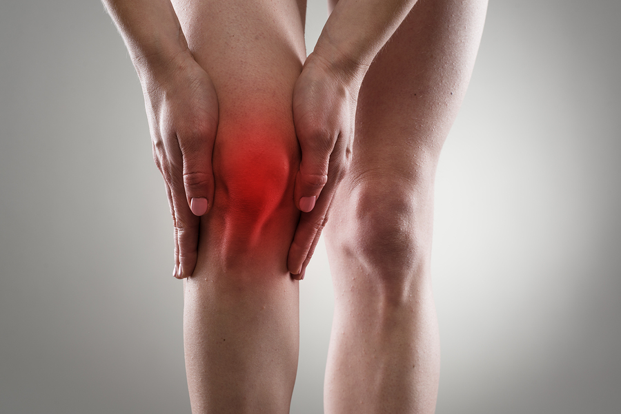
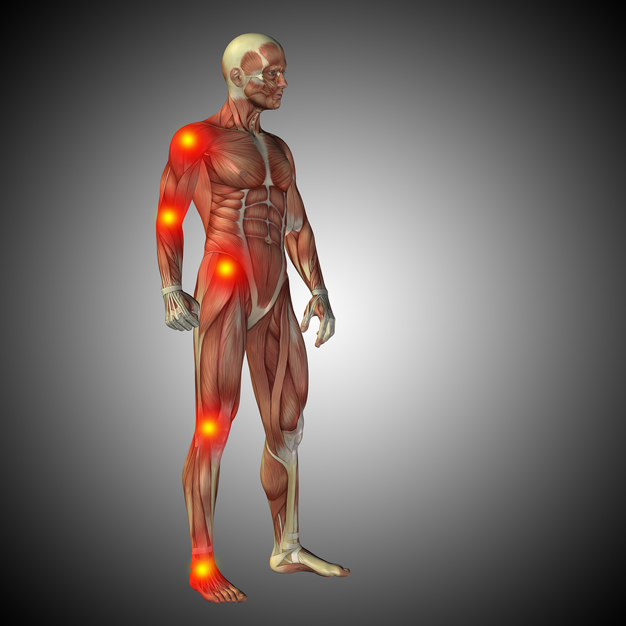
Osteoarthritis makes it difficult for people to get around, it causes chronic pain and it can also be the reason for falls. In 1990 there were 213.4 cases of osteoarthritis per 100,000. 26 years later, in 2016 there were 232.1 cases of osteoarthritis per 100,000 people.

Chronic obstructive pulmonary disease (COPD) has been falling, because less people smoke cigarettes now. Statistics show 1667 cases of COPD per 100,000 in 1990, but only 945 cases of COPD per 100,000 in 2016.

Diarrhea and common infections have dropped sharply from 8951 per 100,000 in 1990 to 3276 per 100,000 in 2016.

What other common diseases do older people get?

There are a number of common diseases that affect the elderly.

Osteoarthritis

Osteoarthritis of the hips and the knees are common, but it can affect every joint in the body. In the end stage knee replacements or hip replacements may be necessary. But before a total knee replacement or total hip replacement can even come into consideration, the person’s heart needs a thorough checkup to ensure that it is safe for the patient to undergo surgery under a general anesthetic.

Heart disease

Older people often have heart disease.

When coronary arteries are narrowed, heart attacks occur. Cardiologists can place stents, so that previously narrowed coronary arteries receive normal blood flow. Following such a procedure the patient may live for another 10 to 15 years.

There are also heart valve calcifications. The aortic valve is particularly endangered. A heart surgeon may be able to replace a diseased aortic valve by a porcine valve.

The nervous system of the heart transmits electrical signals from the sinus node to the muscle fibers, which can get diseased. Heart rhythm problems may necessitate the insertion of a pacemaker.

Finally, the heart may enlarge, but pump less blood than before. This condition is congestive heart failure. The 5-year survival for this condition is only 50.4%. Unfortunately there is very little the doctor can do for patients like this.

Cancer

The older we get, the more DNA mutations we accumulate. At one point cancer develops. If the diagnosis happens at an early stage there is a good chance that surgery can remove a cancerous growth, and the patient survives. But there are cancers that are notoriously difficult to recognize in the early stages. These are: cancer of the pancreas, kidney cancer, stomach cancer and certain types of leukemias.

Respiratory diseases

Those who smoked earlier in life may develop chronic obstructive pulmonary disease (COPD). It is a chronically disabling lung disorder. Often these individuals have to carry an oxygen tank with them wherever they go. The 5-year survival rate for people with COPD is 40 to 70%.

Osteoporosis

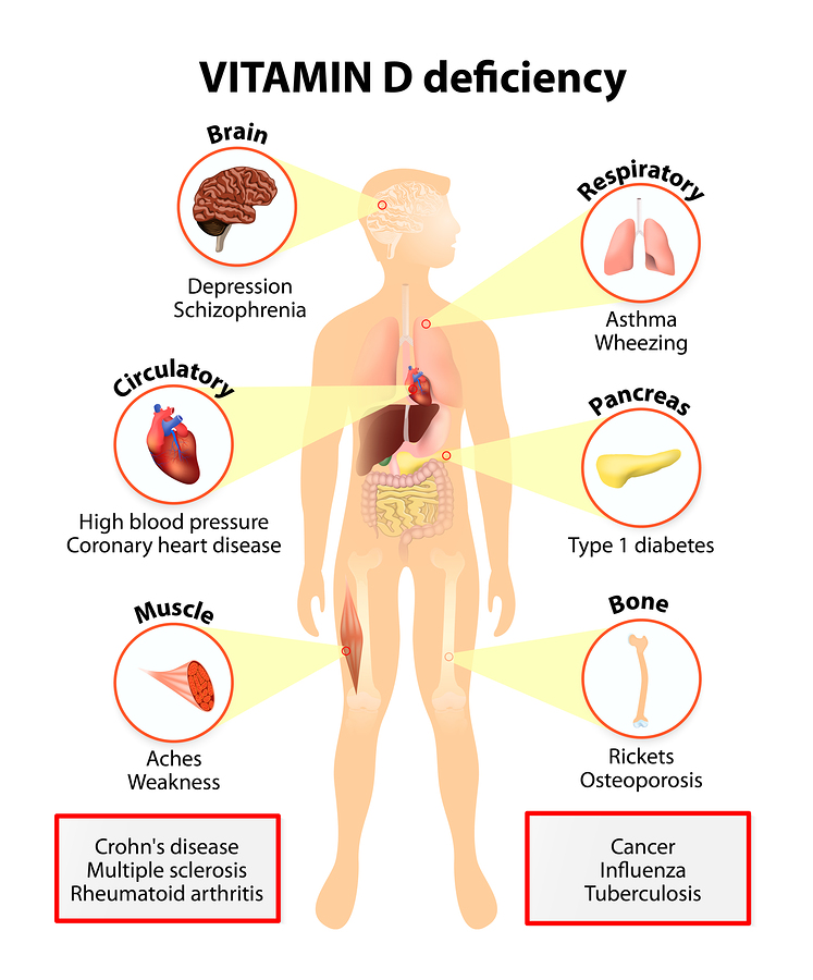
Osteoporosis is a disease where the bone is brittle. Spontaneous bone fractures can occur at the wrists, the upper thigh bone (femoral fractures) or in the vertebral bones. Women in menopause are hormone deficient and this contributes to calcium depletion of the bones. Lately research has shown that vitamin K2 and vitamin D3 are necessary for a normal calcium metabolism. Briefly, 200 micrograms of vitamin K2 and 5000 IU of vitamin D3 every day are the necessary dosage that the body can absorb calcium from the gut, eliminate it from the blood vessels and deposit it into the bone. Calcium is present in milk products and milk. If a person does not consume enough milk products a supplement of 1000 mg of calcium daily does make sense.

Alzheimer’s

The older we get, the more likely it is an onset of Alzheimer’s or dementia. Between the ages of 90 to 94 there is a yearly increase of Alzheimer’s of 12.7% per year. The group from age 95 to 99 years has a yearly increase of Alzheimer’s of 21.2% per year. Persons aged 100 years and older have an increase of Alzheimer’s by 40.7% per year. What this means is that essentially there is a doubling of Alzheimer’s every 5.5 years. We do not have all of the answers why this is happening and why Alzheimer’s develops. But we do know that diabetics are more likely to develop Alzheimer’s. High blood sugar levels and high insulin levels seem to lead to the precipitation of the tau protein in the brain, which causes Alzheimer’s.

Diabetes

When diabetes is not well controlled, there is accelerated hardening of the arteries. This can cause heart attacks and strokes. Longstanding diabetes can affect the kidneys (diabetic nephropathy, kidney damage) and can lead to hardening of the leg arteries. Often the only treatment left is a below knee amputation. Blindness from uncontrolled diabetes is common and pain from diabetic neuropathy as well.

Diabetics have an average life expectancy of 77 to 81 years. However, if they pay attention to their blood sugars and manage their diabetes closely they can live past the age of 85.

Falls and balance problems

As people age, their balance organ is not functioning as well. Also, people with high blood pressure medication may have postural hypotensive episodes that can lead to falls.

There may be a lack of cognitive functioning and misjudging of steps, ledges and irregularities in the floor. When a person has brittle bones from osteoporosis and they fall, a hip fracture is very common. At a higher age surgery for a hip fracture is dangerous. It can have a mortality of 50%.

Obesity

A person with obesity has a life expectancy that is 10 years less than a person without obesity. The reason for this is that with obesity This is so, because the risk of heart attacks, strokes, cancer, arthritis and diabetes is increased.

Depression

Older people often get depressed. It even has its own name: involutional depression. People can get into a state of mind, where they think negatively. Depressed people feel that they have nothing to live for. They lost friends; they are shut in because they can’t drive a car any more. This type of depression needs treatment by a psychologist or psychiatrist. The danger of leaving depression untreated is that the person may get suicidal. In older people depression is often precipitated by physical health problems.

Oral health

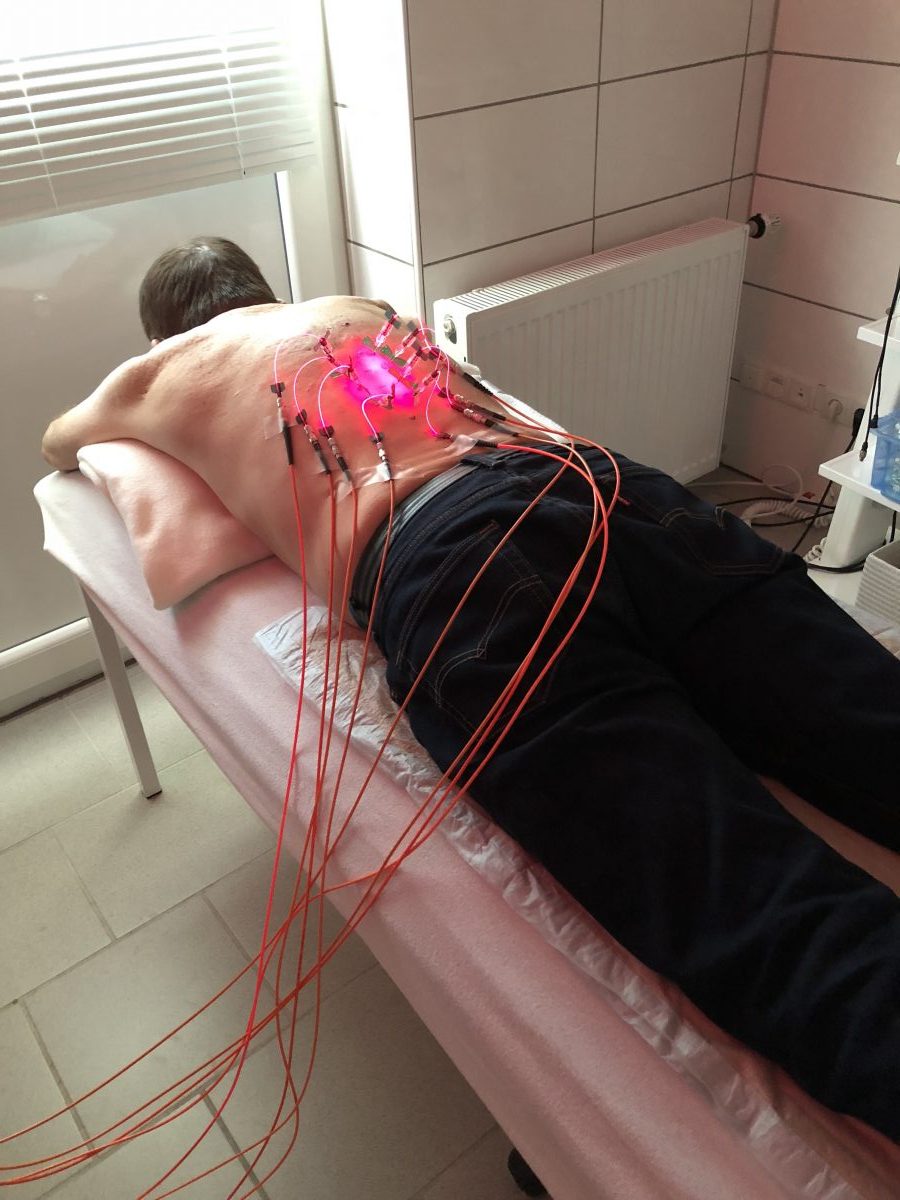
When teeth are not looked after, gingivitis and periodontitis can develop. Infected gums can shed bacteria into the blood and this can affect the heart valves. Endocarditis, the infection of heart valves, is a cardiological emergency. Prolonged antibiotic therapy is necessary to overcome this condition.

Poverty

Poverty has real consequences. The aging person may not have access to the optimal medical care facility because of a lack of funds. But even at a younger age there is evidence that people are healthier when they are wealthier.

Shingles

Older people often get shingles, even if they had chickenpox or shingles as a child. This is evidence that the immune system is getting weaker. Shingles in an older person should alarm the treating physician that there could be an underlying cancer. Due to that knowledge a cancer-screening tests should be part of the medical exam. In addition, a varicella vaccine should be offered to the patient to build up immunity.

The Downside Of Living To 100

The Downside Of Living To 100

Conclusion

Living to 100 is often glorified in the press. Maybe you have seen a 90-year old jogger completing a marathon, or you saw an 85-year old couple ballroom dancing. But what they don’t show you is what I summarized here, the less glamorous things about living to 100. You may get a heart attack or a stroke. Osteoarthritis may affect you how you walk. Congestive heart failure may make you get short of breath when you walk upstairs. Then there are various cancer types that are difficult to diagnose early.

If you have smoked in the past, you may suffer from chronic obstructive pulmonary disease (COPD), which leaves you breathless.

Other illnesses

Osteoporosis can lead to spontaneous fractures. Because the bone has a lack of calcium, this is difficult to treat and takes a long time to heal.

Alzheimer’s is ever so much more common when you approach the year 100. There are other medical conditions you can get: obesity, diabetes and depression. When you get shingles for the second time, it may mean that your immune system is getting weak and a cancer-screening test should be done.

There are some downsides when you approach the age of 100.

Know your risks and be vigilant

You may keep your physician busy checking out various age-related illnesses, but more importantly, get regular check-ups and tests. Any condition is easier to treat with an earlier diagnosis! The message for anybody reading this is very simple. Prevention through healthy living is something you can actively pursue. Keep your body and your mind busy. Enjoy time with friends and family instead of living a solitary existence. See the glass that is half full instead of viewing it as half empty. Stick to a healthy diet. Knowing all the risks is not a scare but a call to being vigilant. Knowledge is powerful and will help you to enjoy your golden years feeling well and happy.

Jun
23
2018

Low-Dose Laser Activated Stem Cell Therapy

Low-dose laser activated stem cell therapy is a treatment solution for those with degenerative joint disease. Degenerative joint disease or osteoarthritis typically affects the major joints like the knees or the hips. Low-dose laser can activate stem cells. In doing so this therapy can also offer a solution for those with chronic back pain due to degenerative changes in the discs or facet joints.

Osteoarthritis in the spine

Last year my family doctor diagnosed osteoarthritic changes in the discs and facet joints of my lower back. I visited Dr. Weber in Germany and he treated me with low-dose laser activated stem cell therapy in November 2017. A prior blog explained the details of this treatment.

New symptoms of back pain in the thoracic spine

During the spring and summer of 2018 I noted that there was some residual back pain in my dorsal spine (also known as thoracic spine). This was just above the previously treated back pain, which had been fine since November 2017. My family doctor arranged for an MRI scan of the thoracic spine confirming moderately severe degenerative changes in the discs and facet joints of the lower thoracic spine. This was not really a surprise because of a family history of these kinds of degenerative problems on my mother’s side. I thought that I should go back to Dr. Weber in Germany. He had given me relief from my back pain in the lumbar spine with low-dose laser activated stem cell therapy.

Additional left knee pain

I also had developed pain in my left knee, which got worse from kneeling or walking on uneven ground. There was definitive grinding in my left knee when my physician palpated the knee joint while moving the lower leg. My right knee did not have any pain, and there was no grinding in it.

First day of my treatment in May 2018

I will not explain in detail the process of the treatments. You can read about it in my prior blog.

However, I will describe the overall treatment schedule.

1. General assessment by Dr. Weber

Before any treatment Dr. Weber went over the history of my thoracic spine pain and the pain in my left knee. He projected the result of the MRI scan of my thoracic spine onto a large TV screen. I could see the degenerative changes of many discs and facet joints on both sides in the lower 6 levels of my thoracic spine.

2. Liposuction to remove fat as a mesenchymal cell source

The treatment started with anesthetizing the area where the physician intended to harvest fat from my right lower buttock area. Next the fat went into a cell separator to separate stem cells and fat. The fat is not necessary for the procedure, only the stem cells.

3. Venipuncture to harvest blood for PRP

Blood was drawn from one of my arm veins for preparing PRP (platelet rich plasma). This fraction of the blood is necessary to activate the stem cells from either fat cells or bone marrow.

4. Left knee and lower thoracic spine injections

Dr. Weber used an intraarticular needle to inject a mixture of the fat derived (mesenchymal) stem cells and PRP. After the injection into my left knee, the physician removed the syringe but left the needle in place.

Through the needle the doctor inserted very fine sterile glass fibers for intraarticular laser treatment. This consisted of five laser colors using low-dose laser beams. The colors were yellow, blue, green, infrared and red. The significance of the various colors and how deep they penetrate into tissues was discussed under this blog.

Stem cell therapy of lower thoracic spine

Stem cell therapy of lower thoracic spine

Thoracic spine injection

Next Dr. Weber determined first the depth of the lower thoracic spine. An ultrasound machine showed him that he could not exceed 18 mm in length when injecting needles into my back. This would ensure that he did not puncture my lungs. Dr. Weber explained to me that some people had a 2- or 4-inch subcutaneous fat layer. Dr. Weber used 12 interstitial injection needles to inject 6 levels of my lower thoracic spine (6 on each side). This step is depicted in the image on the left, where my wife took a photo of the low-dose laser treatment after the insertion of the 12 interstitial needles . This was stimulating the injected mesenchymal stem cells.

At the end of the first day I received an infrared light treatment over my thoracic spine for 20 minutes, followed by a treatment in a light therapy bed for 20 minutes. These latter treatments were necessary in addition to the laser treatments to stimulate the stem cell activity further.

Second day of my treatment in May 2018

The second day was only a half-day treatment. Dr. Weber had kept half of the fat-derived stem cells and of the PRP preparation in the fridge overnight.

Another injection of a mixture of fat-derived mesenchymal stem cells and PRP into my left knee followed, as well as an injection along the lower thoracic spine. Essentially, this was a repetition of the treatments of the previous day for both my left knee and the lower thoracic spine. Dr. Weber explained that there is merit in doing it this way. He said it would increase the success rate of the low-dose laser activated stem cell therapy.

The reasons behind low-dose laser activated stem cell therapy

A group of dentists have shown that mesenchymal stem cells from bone marrow, dental pulp, periodontal ligament or adipose tissue showed stimulation by low-dose laser therapy.

Detailed research from Japan has shown that low-dose laser therapy releases various growth factors from mesenchymal stem cells, from osteoblast cells and other cells including skin cells. This can promote wound healing and helps stem cells to build up cartilage in joints.

Bone marrow stem cell stimulation

The stimulation of bone marrow by low dose laser therapy also releases bone marrow derived stem cells into the blood. This way these stem cells can contribute to the healing process in joints. Dr. Weber used this method to stimulate release of bone marrow-derived stem cells into my system. He punctured my pelvic bone with an interstitial needle. Subsequently he introduced glass fibers through the interstitial needle into the bone marrow space. Five colors of laser, namely yellow, blue, green, infrared and red were used to stimulate the stem cells of my bone marrow. Dr. Weber explained that low-dose laser activated bone marrow stem cells can easily leave the bone marrow and travel via the circulatory system. This is how they reach the area where they are needed.

History of stem cell therapy

Dr. Michael Weber published a book entitled “Medical low-level-laser therapy, foundations and clinical applications”, 2nd edition, June 2015. On page 541 he explains the beginning of stem cell research by Dr. James Till and Dr. Ernest McCulloch in Toronto/Ont in 1961. He explained further how activation of mesenchymal stem cells by low-dose laser light improved cell viability and cell growth. Barboza et al. also researched these topics.

How do I feel about low-dose laser activated stem cell therapy?

Within only 1 ½ weeks I noticed that my thoracic spine pain disappeared. My left knee pain disappeared within 2 weeks. I am aware that there is a consolidation phase of possibly 3 to 6 months which is necessary to build up the full amount of cartilage. But it is the relief of pain that I was hoping for. Rather than treating my osteoarthritis with pain pills and wait until a total knee replacement, I have now a second chance to regain full mobility without pain. Now I feel more confident about aging without the “usual aches and pains” and staying free from disease.

Low-Dose Laser Activated Stem Cell Therapy

Low-Dose Laser Activated Stem Cell Therapy

Conclusion

I described how Dr. Weber treated my mid back and left knee with mesenchymal stem cells. These were activated by platelet rich plasma (PRP) and low-dose laser therapy. Stem cells from fatty tissue are called mesenchymal stem cells. They are useful for building up lost hyaline cartilage, the coating of bone in joints. With degenerative arthritis, also called osteoarthritis, this layer is getting thinner, sometimes to the point where bone rubs on bone. But stem cell treatment with mesenchymal stem cells can rebuild hyaline cartilage. This is part of regenerative medicine where the body’s own stem cells can help to cure disease.

My family has a strong history of osteoarthritis. But fortunately I seem to respond to regenerative medicine using low-dose laser activated stem cell therapy.

Feb
18
2018

Causes Of Back Pain And Their Treatment

There are many possible causes of back pain and their treatment is described in this blog. I have listed the 10 most common causes of back pain below. I also added brief therapeutic suggestions.

Facet joint disease (lower back strain)

When there is a misalignment of the facet joints, the joint capsules of these small joints are pulled, which can cause severe back pain. Chiropractic or physiotherapy treatments initially followed by active exercises will help (facet joint pain).

Degenerative disc and facet joint disease

Degenerative changes of the disc material between vertebral bodies and osteoarthritis of the facet joints can cause back pain. This is due to irritation of the nerve roots. Anti-inflammatory medication and physiotherapy treatments often help; swimming will be beneficial as well. End stage intractable disease, if confined to one or two levels, may respond to fusion surgery by a spinal surgeon. While you research the surgical option, I suggest you investigate about the possibility of stem cell therapy with PRP (platelet rich plasma). This is less invasive and will likely heal better than conventional surgery.

Spondyloarthropathies

This is a family of chronic inflammatory joint diseases of the spine . The physician treats the underlying disease and uses anti-inflammatories to control inflammation. Most of all, a person with this condition needs a referral to a rheumatologist for more specific therapy.

Osteoporosis

Osteoporosis is a bone condition that can lead to compression fractures of the spine. These in turn can irritate one of the nerve roots to lea to radiating pain into one or both legs. The physician will treat the underlying hormone disbalance, if present. Regular walking and swimming are used to remobilize. Avoid alcohol, caffeine and stop smoking. Supplements like calcium, bisphosphonates, vitamin D in higher doses and vitamin K2 can help re-calcify the bone.

Scoliosis

Scoliosis is often congenital. This spinal curvature leads to back pain and hurts more the more the spine is curved. Treatment: bracing during growth spurts; good posture; strengthening exercises; in severe cases corrective surgery by a spinal surgeon through the use of Harrington rods.

Spinal stenosis

Spinal stenosis is often the end condition of severe degenerative arthritis of the spine. It is more common in the older generation. As this link explains, there is a narrowing of the channel through which the spinal cord travels. In the past decompression surgery for severe cases was the only means to free spinal cord and nerve roots. In the last few years stem cell therapy is a new addition as an alternative to orthopedic surgery. The advantage of stem cell therapy is that there is no scarring following the procedure and the stem cells function like a biological knife from inside.

 Posttraumatic fibromyalgia

Some people develop lower back pain following traumatic injuries. Instead of resolving their muscle sprain, they end up with chronic pain. Often low dose antidepressants and cognitive therapy will help. Mild physical exercises may help to rehabilitate the patient and return to normal muscle mobility. Unconventional therapy like prolotherapy, dry-needling and low-dose laser therapy (Dr. Weber) may be useful.

Disc herniation with or without sciatica

With disc herniation the back pain is intense, which often prompts the orthopedic surgeon to do an early discectomy (back surgery). But long term studies have shown that only in 3% of all acute back pain cases surgery is necessary; many cases heal on their own. In this case the new regenerative medicine approach of stem cell therapy with PRP will give superb clinical results. Stem cells will rebuild the disc and also take care of any arthritis in the small joint along the spine.

Spondylolisthesis and other congenital malformations

There are 4 grades of slipped vertebral body diseases (spondylolisthesis), where grade IV is the most severe case. Grade I to IIIA do not need surgery, stage IIIB and IV need fusion with instrumentation by a spinal surgeon.

Bone metastases

In stage 4 of many cancers metastases travel through the whole body including the skeleton. The underlying cancer needs treatment, often chemotherapy is required. Unfortunately cancers with bone metastases have a very poor prognosis. Here is a link how to manage symptoms of bone metastases.

Causes Of Back Pain And Their Treatment

Causes Of Back Pain And Their Treatment

Conclusion

I have listed the ten most common ones of back pain. For muscle spasms, simple physiotherapy or chiropractic treatment are often very helpful. Early degenerative changes in joints and discs will often respond to anti-inflammatory medication. But anti inflammatory drugs can be hard on your liver and your kidneys. If your back pain is getting more chronic, your doctor may want to arrange for an MRI scan of the area that causes pain. If this shows degenerative change in the discs and facet joints it is time to contact a regenerative medicine clinic. They specialize in stem cell and platelet rich plasma (PRP) treatments. This may be what you need to restore your back to normal. Conventional surgical methods are often the choice, but they are not always successful. Surgery can leave scarring behind, which by itself can give you chronic back pain.

Feb
03
2018

Drugs Are Not An Escape

We have a drug epidemic, but drugs are not an escape. Along with this epidemic there have been thousands of lethal overdoses. I like to review why so many people use street drugs or prescription pain pills. It started years ago with heroin injections had become more popular. But then heroin was getting a bad name, it became illegal, and prescription pain pills replaced heroin. Percocet, OxyContin and morphine prescriptions took off. Together with it the number of people with addiction increased drastically. The latest fashion craze is Fentanyl.  Fentanyl works faster than morphine, but also wears off faster. One of the deadly side effects is respiratory depression. This  means that a person cannot breathe and as a result will die. This is what is happening right now and what creates the news headlines.

Pain receptors

There are pain receptors all over the body, in muscles, organs and the skin. They are called nociceptors. Many medical conditions lead to pain in various parts of the body. For instance, with end stage degenerative arthritis bone rubs on bone. This causes a lot of pain in joints like the knees or the hips. Physicians often prescribe narcotics like OxyContin to control the pain. It may help for some time. But after a few weeks patients complain that the strength of OxyContin has worn off. The same dosage that gave them relief from the pain initially just does not give them relief any more. This observation is crucial. It is exactly what people who use OxyContin as illicit street drug found as well. The drug wears off, because the opioid receptors are getting weaker.

Pain receptor resistance

Nociceptors or opioid receptors are pain receptors. These receptors report pain to the body, and they have connections through the spinal cord to the brain. When an opioid is interacting with the pain receptor it inhibits acute pain to a large extent. But when the pain becomes chronic, the pain receptor is not working as well. More and more of the pain medication is necessary to achieve pain relief.  Eventually the opioid medication is not working to relieve pain any more. The name for this phenomenon is “pain receptor resistance”. Scientific work behind why pain receptors get lazy in responding to opioids is still ongoing. In the meantime it simply is an observation. This is the reason why drug addicts are tending to increase the dosage of OxyContin or of Fentanyl. The toxic threshold of Fentanyl that is exceeded in the process is the reason for the deaths.

Insulin resistance

You may have heard of insulin resistance in diabetics. This is a very similar problem. With insulin resistance the diabetic no longer has a 100% response to insulin. This means that blood sugar is not going through cell membranes effectively as it should when the insulin receptor is working. The blood sugar values stay high and make the diabetes worse.

With pain receptor resistance the opioid is not giving the relief from pain it used to give. No wonder that the patient is feeling frustrated that the pain medication is no longer working properly.

Therapeutic index

The layperson does not normally know that there is a dosage for any drug where it becomes toxic. There is also a lower dosage where the drug is becoming effective. The difference between the two dosages is the “therapeutic window” or the “therapeutic index”.

Morphine has a therapeutic index of 70:1. Compare that to alcohol with a therapeutic index of only 10:1. Fentanyl has a therapeutic index of 300:1.

The danger of Fentanyl

One would think that Fentanyl should be safer than morphine, because the therapeutic index is larger. But the onset of Fentanyl is much faster than that of morphine and by the time drug users take Fentanyl they often are desperate for the drug to take their pain away and they overdose, which causes respiratory depression. If they are not intubated by an anesthetist and connected to artificial respiration, they die very quickly. Janssen Pharmaceutica first developed Fentanyl in 1959. Fentanyl was strictly part of the pre-anesthetic medicine for patients before going into surgery requiring a general anesthetic. The anesthetist was right there and would intubate the patient. That’s why we never heard of deaths from Fentanyl in the past.

If a person gets no pain relief from any opioid because of receptor resistance, the tendency is to increase the dosage. But this is what pushes the person into toxic ranges and this is what causes death from inadvertently suppressing the ability to breathe. People can get into trouble with alcohol overdoses, but the ordinary person usually knows how to handle alcohol. Fentanyl is a lot different due to the fact that respiratory depression (not being able to breathe) is one of the early symptoms that hits you suddenly.

What drugs do to your brain

Dr. Daniel Amen, a psychiatrist and brain researcher has used SPECT brain scans to produce images of different clinical conditions. This link shows the effect of a stroke, of Alzheimer’s disease and of drug abuse on the brain. Dr. Amen says that a brain can be rehabilitated. He has other SPECT images showing the images of an addicted brain and that of a rehabilitated brain. Spect scans are expensive.  But they may be worth it, to impress a drug addict to stop the drugs and get rehabilitated.

Big Pharma and opioid drugs

In 1995 the FDA approved the use of OxyContin for chronic pain. Perdue Pharma, the drug company that had developed this opioid convinced the FDA that OxyContin stayed in the blood for 12 hours and they claimed that by being active longer than other opioids this would lead to better pain control and less addiction. It turns out that this was a slick sales pitch, however it was not true. Pain receptor resistance is as much a problem with OxyContin as with any other opioid. And the drug is as addictive as all the others. But the problem is that the FDA had approved OxyContin. What’s more, Purdue Pharma sent aggressive drug representatives to all the practicing physicians misleading them as well as the FDA that OxyContin would not be as addictive as other opioids.

Penalty for misrepresentation of OxyContin

In 2007 Purdue Pharma had to pay a fine of 634 million $ for misrepresenting the addiction potential of OxyContin. But the drug company was allowed to continue to market OxyContin. Since 2015 Purdue Pharma has been cashing in 2.4 billion $ annually from the sales of opioids including OxyContin. In the meantime Fentanyl, despite its danger of suppressing the ability to breathe, has taken off with regard to sales. Fentanyl also has taken over in terms of causing deaths across the US and other countries.

Alternative treatment of pain

According to Ref. 1 chances are that 70% of patients with back pain will get better with a few visits to an acupuncturist. In 1972 Dr. Ulett’s laboratory at the University of Missouri succeeded in getting the first NIH research grant for the study of acupuncture in the US. During these studies they found remarkable facts, the most important perhaps that electrical stimulation of acupuncture needles resulted in a doubling of the effectiveness of traditional Chinese acupuncture. After extensive research Dr. Ulett stated: ”The ancient practice of traditional Chinese acupuncture is now obsolete” (Ref.1). in the meantime the more effective electro-acupuncture has replaced acupuncture needles with the use of electrical skin pads.”

Other applications for electroacupuncture

Beside pain relief many other applications exist for electro-acupuncture. Addiction medicine makes use of electro-acupuncture in weaning people from morphine or heroine etc. It is effective to treat psychiatric illness, particularly depression. It is useful in relieving nausea and vomiting due to chemotherapy with cancer treatments or associated with pregnancy without affecting the pregnancy.

Prolotherapy

Prolotherapy has been successful for the treatment of back and neck pain, for arthritic pain and pain from chronic muscle spasm. When prolotherapy is done by itself 12.5% Dextrose is injected into the area of injury. Dr. Fields said that the reason it works is that there is activation of local stem cells in the injured area in the area of the Dextrose injection site. These stem cells will do the healing (details explained in an interview with Dr. Reeves). Further improvement of this technique and better results are possible by injecting a small amount of platelet rich plasma (PRP) very focally to an area of ligament rupture. PRP is obtained by centrifuging blood from the patient’s vein. The red blood cells are not necessary, but the platelet fraction and some of the plasma are part of the the PRP preparation.

It is often striking how much pain control occurs after only one or two prolotherapy treatments.

Stem cell therapy with PRP and low-dose laser therapy

This is a more direct way to get stem cells where the doctor wants them to do their work. Stem cell therapy with PRP and low-dose laser activation is the latest in terms of controlling pain. At the 22nd Annual Anti-Aging Conference in Las Vegas (Dec.10 to 14, 2014) Dr. Purita gave an overview of this method of pain control. He discussed the importance of the proper harvesting of PRP. He explained that apart from white blood cells (WBC) and platelets an important component of PRP are very small embryonic like stem cells (VSELs). They are visible through the microscope. The missing link has been the observation that white blood cells produce inflammatory substances, which have been detrimental in stem cell injections with PRP in the past. There was a poor survival rate of stem cells.

Low dose laser activation of stem cells and PRP

Photo-activation of the PRP before injection leads to anti-inflammatory behavior of the WBC in PRP. Dr. Purita calls this “light activated PRP”, which leads to the best results with stem cell/PRP injections.

Soft laser stimulation with red, green and blue soft lasers have also been effective to improve tissue healing significantly when stem cells and PRP are used together with light activation. The main sources for good stem cells are the fat tissue (from the “love handles”) and the bone marrow (obtained from pelvic bone). The stem cells mesenchymal stem cells (from fatty tissue) and bone marrow stem cells. The mix of all of this can mend osteoarthritic joints, degenerative disc of the spine, in short all musculoskeletal injuries that may be painful. The hallmark of healing with stem cells is that chronic pain disappears.

Curative treatment versus symptomatic treatment

It is one thing to treat a patient for pain symptomatically by prescribing OxyContin or other opioids. It is a completely other thing when the physician cures the pain by one of these alternative methods. Let us assume that a patient has a fall of a few step from a ladder and sustains a contusion of the lower back. This can be painful and may require a few pain pills. But if it does not improve within one week and diagnostic tests show that there is no fracture, electroacupuncture may be the solution to treat the pain rather than the use of an opioid. If this does not help, consider the combination stem cell/PRP/low-dose laser therapy. Chances are better than 80% that this will help. The patient will no longer suffer of pain. With healing life can go on: no pain pills. No addiction. No death.

Drugs Are Not An Escape

Drugs Are Not An Escape

Conclusion

Conventional medicine treats pain symptomatically instead of treating the cause. It gets the physician to prescribe opioids, which can be the beginning of a series of tragic events. It can lead to drug addiction, dose acceleration, toxic effects of opioids including possible death through asphyxiation from no longer being able to breathe.

Stem cell therapy

The physician who concentrates on treating causes will use alternative methods to treat pain. The most effective of these methods is stem cell therapy. It is a mixture of adipose mesenchymal stem cells with bone marrow stem cells that are part of the therapy. The addition of platelet rich plasma is another part of the mix, and the final step is activation of stem cells with low-dose laser therapy. The cell mix is injected in the critical area. The end result is that the patient’s pain disappears, and it leads to a cure. The only situation where short-term opioids are justifiable is following surgical procedures. But even in these cases the narcotics should only be necessary for a few days to avoid serious side effects.

Reference 1: George A. Ulett, M.D., Ph.D. and SongPing Han, B.M., Ph.D.: “The Biology of Acupuncture”, copyright 2002, Warren H. Green Inc., Saint Louis, Missouri, 63132 USA

More info about electroacupuncture: https://www.askdrray.com/electro-acupuncture-twice-as-effective-as-conventional-acupuncture/

More about prolotherapy stem cell therapy: https://www.askdrray.com/prolotherapy-and-stem-cell-therapy/

Jan
06
2018

Lyme Disease The Great Imitator

Dr. Pamela Smith talked about Lyme disease the great imitator when she gave a presentation. This was at the 25th Congress of the American Academy of Anti-Aging Medicine, Dec. 14-17, 2017, which I attended. Dr. Smith gave a talk about how to approach a complex patient when multiple systems are affected. Part of that talk dealt with Lyme disease, which I will review below in some detail.

Transmission of Lyme disease

Lyme disease is one of the fastest growing infectious diseases in the US. As a result about 200,000 new cases of Lyme disease occur in the US every year.

The transmission occurs through ticks that that carry a spirochete, called Borrelia burgdorferi.

This bacterium, much as syphilis, which also is caused by a spirochete, produces imitator disease patterns. Clinically it can be a challenge to diagnose Lyme disease.

The common way of transmission to humans is by infected ticks that bite the skin. But Dr. Smith said that transmission of the spirochete can also occur by breastfeeding, blood transfusions, in vitro fertilization and finally by sex. Although originally Lyme disease infected ticks were found on deer, other species can also be carriers. Ticks from mice, foxes, raccoons, songbirds, chipmunks, and squirrels can also transmit Lyme disease.

Clinical presentation of Lyme disease

Only 30 – 40% of adults with Lyme disease have the characteristic rash of the “bull’s-eye lesion” (erythema migrans). With children this presentation is even less common (only 10% have erythema migrans). If there is a bull’s-eye lesion, this will last from one week to several months. A laboratory test using an enzyme-linked immunosorbent assay (ELISA test) can confirm the diagnosis of the disease.

Disseminated early or late Lyme disease

Fatigue, headaches and weakness can be non-specific symptoms of Lyme disease. Furthermore, other non-specific symptoms like back pain, muscle and joint pains as well as chills can detract the physician from diagnosing Lyme disease. In addition irregular heartbeats, nausea, vomiting, swollen lymph glands, memory loss, gait problems, bladder and kidney problems are other symptoms. Finally, liver problems, sore throat, fever, seizures, depression, dementia, hallucinations, mood swings and arthritis can be other symptoms.Even eating disorders, verbal aggression, schizophrenia and suicide can be symptoms of Lyme disease.

Common symptoms that have a link to Lyme disease

Common symptoms of Lyme disease include headaches, fatigue, joint pain and swelling of joints, stiffness of the neck or back. There can be difficulties with concentration, speech or writing. Further symptoms are sleep disturbances, numbness or tingling of arms or feet and forgetfulness. 

Lyme disease development

Borrelia burgdorferi can be found inside body cells and outside of cells as biofilms. This form makes them resistant as it allows Borrelia burgdorferi to exchange DNA and makes them resistant to antibiotics. There are also two major forms of Borrelia burgdorferi, namely cell-wall forms and cystic forms. Once the patient has been bitten by the infected tick Borrelia can quickly change shape into the more difficult to treat cystic form. Within 24 hours Lyme disease can spread to other parts of the body. Common such areas are the eyes, brain tissue and glial cells, heart, collagen, synovial fluid of joints and skeletal muscle fibers.

Lyme disease can also complicate many other diseases. These are ALS, Alzheimer’s disease, fibromyalgia, MS, bipolar disorder, neurological disease, heart disease (Lyme carditis) and autism.

Treatment of Lyme disease

  1. Dr. Smith said that Lyme disease is often complicated by dysfunctional gut flora. She prefers to start patients on a sugar-free and gluten-free diet. The patient also has to take probiotics.
  2. 75% of Lyme disease patients show a cure after three weeks of Doxycycline 100 mg twice per day. Alternatively cefuroxime 500 mg twice per day is a medication of choice.
  3. Cefuroxime only treats the cell‐wall forms. Doxycycline treats the intracellular forms. Metronidazole or tinidazole will help to eradicate the cystic forms of Lyme disease.
  4. Grapefruit seed extract is another treatment modality if the patient is allergic to Metronidazole. It eradicates the cystic form of Lyme disease.
  5. Serrapeptase from whole leaf stevia extract will also help to eradicate Borrelia biofilms and persisters.
  6. Monolaurin, a coconut oil extract is effective in treating all three morphological forms of Borrelia burgdorferi.

Patients with neurological symptoms

Patients with neck stiffness, headaches or neuropathy need treatment for a longer period of time. These patients also need monitoring for recurrent Lyme disease at the end of the treatment.

Case presentation of a patient with Lyme disease

Dr. Smith presented one of her patients with Lyme disease in detail. She was a 45-year old executive. She suffered from extreme fatigue. It took quite a few tests to find out that her antibody titers against Lyme disease were very high.

Here is her long list of symptoms: hair loss, four urinary tract infections in quick succession, brain fog, extreme fatigue, systemic pain, musculoskeletal pain, anxiety and depression, eczema, psoriasis, itching, stomach ache, trouble eating, weight loss of 12 pounds, flu, strep presented like meningitis.

Comprehensive treatment of patient with Lyme disease

Dr. Pamela Smith instituted a comprehensive treatment protocol. It turned out that she had developed gastritis, which was the reason for her weight loss. This needed conventional treatment. After the treatment with antibiotics, her energy picked up, and her appetite came back. She also engaged in yoga and other self-awareness programs. She deliberately slowed down her lifestyle activities. Her symptoms were mostly gone or significantly diminished. She was able to function. She experienced energy, joy, and could focus again. The only symptoms left were some mild pain, some bladder problems, some limitations with her diet and mild brain fog.

Husband had Lyme disease

Part of the work-up was to test her husband for Lyme disease. He tested positive. He was also treated although he was entirely asymptomatic. When his treatment was finished, the doctor tested him for a specific antibody and this came back as negative. This meant that he now was free of Borrelia burgdorferi and would no longer be able to infect her. The doctor thought that it was most likely through sex that she had contracted Lyme disease. The problem is that some people are completely asymptomatic, but nevertheless they can be carriers of Lyme disease.

Lyme Disease The Great Imitator

Lyme Disease The Great Imitator

Conclusion

Lyme disease, the great imitator, has become a more common disease in the US and around the world. Years back Lyme disease was often overlooked. But lately physicians have diagnosed Lyme disease earlier as diagnostic tests have improved. With earlier treatment a lot of suffering of the patient can be prevented. But in many cases symptoms are confusing as Lyme disease involves several organ systems. This makes the diagnosis more difficult. By diagnosing Lyme disease earlier, treatment can start at an earlier stage, and the patient will soon return to a state of wellness.

Dec
09
2017

Stem Cells Cure Back Pain

A person with chronic back pain has several treatment options, but only stem cells cure back pain. Stem cell treatment has been available in the US and Canada and many other countries for approximately 10 years.

I come from a family with a strong history of back pain (mother, maternal grandmother and maternal grandfather). They all got their back pain in their mid to late 40’s. From my growing up years I remember that they complained about chronic back pain on and off. Sometimes they had to cancel events they wanted to attend because they could not tolerate sitting. In those times there were no CAT scans or MRI scans. If you had back pain, you just had to put up with it.

My personal experience

Given my family history of back pain I was surprised that my back pain was only a more persistent problem in the last 1.5 years, but not earlier. Normally a monthly chiropractic adjustment would keep my back symptoms under control. But in the last 1.5 years I needed to see a chiropractor more often than that. I took omega-3 fatty acid supplements for the past several years (two capsules twice per day) thinking that this should halt the development of degenerative arthritis in the lower back joints. When I turned 71, it was clear to me that I was now at the point where my immediate relatives were when they were in their late 40’s. Therefore, diet, exercise, weight loss, good nutrition and supplements can only do that much for you. If there is a familiar disposition, it will eventually catch up with you.

Conventional medicine’s approach to lower back pain

I have practiced as a general practitioner for 16 years in the past. In addition I joined Workers’ Compensation for another 16 years as a medical advisor. From this clinical activity I knew of hundreds of cases first hand what the steps were in the treatment of chronic back pain. First of all, physiotherapy treatments or chiropractic treatments were the treatment protocol. In minor back pain cases this would often help the pain symptoms. Furthermore, if residual pain persisted, the patients received anti-inflammatory medication (non-steroidal anti-inflammatory drugs or NSAID’s). Finally, if symptoms continued to persist, a CT scan or MRI scan was necessary for assessment. If it showed moderate changes like my findings, the patient received intermittent physical therapy, chiropractic therapy or acupuncture therapy. 

Surgical procedures for chronic lower back pain

If there were more severe degenerative changes or spinal stenosis with severe degenerative changes, a referral to an orthopedic surgeon or neurosurgeon would be necessary. But this was often the point of no return. If the surgeon felt that the condition was severe enough to do back surgery, various procedures could follow. For disc herniations irritating one of the nerve roots, laparoscopic discectomy was the treatment of choice. For severe spinal stenosis or intractable pain from end stage facet joint disease instrumentation was an option.

Fusion surgery

Under a general anesthetic the surgeon makes an incision in the patient’s back over the lumbar spine. The surgeon identifies the diseased disc level and places stabilizing stainless steel plates over the affected facet joints or the narrowed disc space. Many people think that fusion surgery would be the end of their trouble. In many cases this can actually be the beginning of chronic back trouble. The problem is that the body is designed to move. If the surgeon takes movement away in one area of the spine, the levels above and below have to work harder. It often takes only a few months or a couple of years, and the patient is back with excruciating pain from degenerative changes in the levels above and below the previous surgery. What does the surgeon usually do? He does more fusion surgery above and/or below the previous area of surgery.

Alternatives to back surgeries

New treatment options have opened up new possibilities. On the one hand there is prolotherapy treatment that I have described under this link. On the other hand stem cell therapy is another popular regenerative technique. Prolotherapy strengthens tissues, relieves pain and increases the range of motion in joints. There is 80 to 85% full pain relief and more than 80% improvement in range of motion. Prolotherapy promotes the healing of torn ligaments and tendons. There are many suitable conditions that lend themselves to the treatment with prolotherapy like the hip, knee, shoulder, ankle, neck, lower back and elbow. With prolotherapy the physician uses hyperosmolar dextrose injections into the affected area. Current thinking is that this irritates the tissues, which mobilizes local stem cells to heal the area.

In my case I had two prolotherapy treatments of my lower back, but it did not change my lower back pain.

MRI scans of my lumbar spine

We needed to find out what was happening in my lower back. My general practitioner ordered MRI scans of my lower back in summer of 2017. There are 5 levels of the lumbar spine from L1 to S1. In my case one level of 5 was normal. The other levels showed bulging of the discs. The scans also showed signs of arthritis in the small joints adjacent to the spine. Lucky for me, there was no sign of spinal stenosis. It was not good news: overall 4 levels of my lumbar spine showed signs of  degenerative disc changes. At the same levels I also had arthritic changes in the facet joints. This was enough to consider some intervention, or I would be headed for trouble in the future.

Stem cell treatment for chronic back pain

Following the failed prolotherapy for my lower back pain I needed to figure out what to do next. The MRI scans had shown degenerative changes in the discs of the lower 4 levels of the lumbar spine. There also was arthritis in eight facet joints (two on each side of each of the four L2 to S1 levels). Conventional medicine would have offered corticosteroid injections into the facet joint areas. My experience with many patients who had this procedure was that the effect of the corticosteroid injections wore off after 3 to 6 months. If a patient had more than 3  injections, there usually was a point of no return, and fusion surgery would be next.

Best therapy for my own chronic lower back condition

For me there was no question that stem cell therapy would be the best fit for treating my back condition. In addition platelet -rich plasma and low-level laser therapy could activate the stem cells. This would be the ideal non-invasive treatment option to treat my chronic lower back pain. I had met Dr. H. Michael Weber before. He is a well-known laser expert from Germany who has a double certification as an engineer and as an internist treating various clinical conditions with laser and stem cell therapy. In addition he is an expert of regenerative medicine methods. Also, he invented and designed the laser machines himself. I set up an appointment in the fall of 2017 at his clinic in Lauenförde, Germany.

First day of stem cell treatment

On the first day fat tissue was removed under a local anesthetic from my lower left buttock area. Next a cell separator divides the tissue into connective tissue, fat cells and mesenchymal stem cells. Two blood samples were also taken from me for processing platelet rich plasma (PRP). PRP is a natural stem cell activator. Growth factors and anti-inflammatory cytokines were also part of the mix together with the stem cells.

The very same afternoon I received the stem cell mix by injection. Eight needles, four on each side, were necessary to administer the stem cell combination. I also had a treatment on a light therapy bed with red light to activate stem cells in general. The stem cell injection was a pain free procedure, as I received a shot of a  local anesthetic in the area before. After that the physician inserted laser applicators through the interstitial needles.

Laser activation of injected stem cells

The next step was to use laser treatments with 5 different colors (infrared, blue, red, yellow and green) for 10 minutes for each of the 8 interstitial needles. The laser activation and the PRP mixed with the mesenchymal stem cells were the two main stem cell activators. They are crucial for activating the stem cells. But growth factors and anti-inflammatory cytokines also aided stem cell activation.

Second day of stem cell treatment

On the second day I received an infrared light treatment over my back for 20 minutes. Following that I received light therapy bed treatment for 20 minutes. The physician told me  that all of this was to activate the stem cells further. The next step was a bone marrow low-dose laser therapy.

Bone marrow stem cell activation by low-dose laser therapy

Often stem cell therapists mix mesenchymal stem cells from fat tissue with bone marrow stem cells which they harvest before from pelvic bone marrow. Dr. Weber told me that he would do a direct bone marrow laser activation of the pelvic bone marrow instead. He anesthetized the tissue above the pelvic bone. Following this he made a small hole into the pelvic bone through which he inserted a laser applicator into the bone marrow cavity. 5 different colored lasers were again applied for 10 minutes each to activate the bone marrow stem cells. Studies have shown, as Dr. Weber stated, that low-dose laser activates bone marrow stem cells. They can be found in the blood circulation within 1 hour. This is similar to mixing stem cells in a Petri dish and then injecting it as a mix, except it is a less invasive approach.

Further activation of stem cells

Following these procedures Dr. Weber felt that another light bed therapy was necessary for 20 minutes. He also gave me a Weber medical laser watch called “Regenerate+”. This device fits on the wrist. It is programmed to generate a number of different lasers to shine against the underside of the wrist. This is the area where the ulnar and radial arteries run close to the surface. This device will shine the laser lights for 30 minutes, and the laser light reaches the arterial blood. The circulating stem cells from the stem cell therapy are receiving a further boost this way. Dr. Weber told me to use this device twice a day on an ongoing basis. The Weber medical laser watch stimulates the immune system.  Jet lag also responds to, and it can stimulate stem cells as they circulate in the blood.

Stem Cells Cure Back Pain

Stem Cells Cure Back Pain

Conclusion

Medical tourism is flourishing. I have become a medical tourist myself because I did not want to get crippled by conventional medicine regarding my lower back pain. Two days after my stem cell treatment my back pain was significantly improved. There was mild pain in the area of the fat liposuction site. Four days after the treatment the lumbar spine pain was gone. Innumerable chiropractic treatments and two prolotherapy treatments had not given me relief. Now stem cell therapy in Germany has taken my chronic back pain away in only a few days. I realize that the healing process will take 3 to 6 months to complete, but as a patient what counts most is pain relief.

What, if someone criticizes me for choosing stem cell treatment?

It is difficult to argue with success. Whether somebody criticizes me for having followed a non-conventional treatment protocol does not matter to me. My question back would be: what do you do when conventional methods fail? Are you willing to suffer chronic pain and swallow pain pills that could either get you addicted or have serious side effects? I would try stem cell therapy again, if I had a problem that does not respond to conventional therapy.

Oct
28
2017

Take Enough Vitamin D3

Many people supplement with 300 to 400 IU of vitamin D3, but do they take enough vitamin D3? There is a simple way of finding out: ask your doctor to order a 25-hydroxyvitamin D blood test.   This will show whether the gut absorbed enough of the essential vitamin. It will also show whether or not your vitamin D3 capsules or tablets were strong enough. It is now generally accepted that a good range of the vitamin D blood level is between 50 and 80 ng/ml. Unfortunately many Americans who come down with various diseases have blood levels of less than 30 ng/ml. Here are some facts about what a lack of vitamin D3 can cause.

Increased risk of mortality with lower vitamin D levels in ICU patients

  1. A New England Journal study from 2009 reported about 1100 patients in Intensive Care Units (ICU). Their average vitamin D blood level was only 16 ng/ml. They tracked the mortality rates depending on the vitamin D blood level. Insufficient vitamin D levels showed an association with a mortality rate of 45%. An intermediate level had a mortality rate of 35%. And a satisfactory level of vitamin D had a mortality of only16%. Between the low level of vitamin D and the normal level there was a 3-fold difference in mortality!
  2. Another study from 2015 repeated the mortality study with 135 ICU patients. Researchers correlated Vitamin D blood levels with mortality rates of patients. When vitamin D levels were below 12 ng/ml, there was a mortality rate of 32.2%. Patients with higher levels of vitamin D had a mortality rate of 13.2%. The authors concluded that vitamin D blood levels were an independent risk factor for mortality. Patients less than 12 ng/ml had a 2.4-fold higher risk of dying than patients with normal vitamin D levels.

Do patients with multiple sclerosis take enough vitamin D3?

Perhaps one of the earliest results of vitamin D3 research was the following observation. More than 90% of patients with multiple sclerosis were deficient in vitamin D blood levels. Their levels were below 20 ng/ml. Other researchers showed that vitamin D could directly tone down the aggressiveness of the immune cells of MS patients. These were the ones that attacked the myelin sheath. As a result of this knowledge it is important for MS patients to take high enough vitamin D3 supplements. When they reach good vitamin D blood levels their MS is better controlled.

Canada as a northern country has 291 MS patients per 100,000 people. Contrast this to 110-140 MS patients per 100,000 people in the northern US (between the 37th parallel and the US/Canadian border). In addition south of the 37th parallel there are only 57-78 cases of MS per 100,000 people. Researchers have concluded that the less sun light people get, the higher the rate of MS in the population will be. However, instead of sun exposure you can supplement with vitamin D3 capsules to get the blood vitamin D levels up to the range of between 50 and 80 ng/ml.

Do stroke patients take enough vitamin D3?

Strokes are very common. About 6.8 million Americans survive a stroke and live with various disabilities. 15% die shortly after their stroke. 40% are left with moderate to severe disabilities. Many require special care.

  1. Studies have shown that patients with the lowest level of vitamin D have the poorest functional outcomes. Moreover, for every 10 ng/ml decrease in vitamin D levels the odds of a healthy recovery 3 months after the stroke fell by about half. This was independent of age and the initial stroke severity.
  2. In another 2015 study from South Korea 818 stroke patients took tests to evaluate whether they had adequate vitamin D blood levels. There was a clear division between those whose levels were higher than 10 ng/ml or lower. When the vitamin D level was higher, there was a 90% better recovery from their stroke after 3 months. In comparison those whose vitamin D levels were below 10 ng/ml had poor recovery rates. Experts say that vitamin D levels should stay in the range between 50 and 80 ng/ml. This will prevent numerous diseases.

Do diabetics take enough vitamin D3?

  1. Vitamin D3 can silence diabetes genes in connection with the right diet and cofactors of zinc and magnesium. A Mediterranean diet can stabilize the metabolism and fight inflammation. Zinc and magnesium are important cofactors in enzymes necessary to prevent diabetes. Vitamin D3 and omega-3intake are helping to control inflammation and preserve beta cells in the pancreas in diabetes patients. This is important for continued production of insulin.
  2. A Chinese research team found that vitamin D3 protects beta cells in the pancreas from dying off. The finding was that vitamin D3 receptors in the insulin producing cells prevented the dying off of these cells, as long as there was enough vitamin D available. Insulin production by the pancreas remained effective. And insulin is vital for long-term survival of diabetes patients. The key for diabetes patients is to take adequate doses of vitamin D3 to protect their insulin producing beta cells.
  3. A 2015 Italian study showed that micro vascular complications in diabetes patients were high, if the vitamin D3 blood levels were low. If patients had high levels of vitamin D3, there were no complications such as retinopathy or nephropathy. But if levels were below 20 ng/ml, damages were significant in the capillaries of the eyes and kidneys.

Do patients with inflammatory conditions take enough vitamin D3?

What do the lining of the arteries, the inflamed joints, a degenerative meniscus and heart attacks and strokes have in common? It is the inflammation that changes the body chemistry. It gets even more complicated, because the extra calories that we consume get stored as visceral fat. This is done automatically when you eat too much sugar and starchy foods. When the glycogen stores are full, any surplus sugar gets metabolized by the liver into triglycerides, fatty acids and LDL cholesterol and gets stored as body fat. The most active fat is the visceral fat between our guts and around our body organs. This produces interleukins and other inflammatory cytokines that circulate in the blood causing inflammation in all our arteries. Interleukin-6 is an inflammatory cytokine. High interleukin-6 levels contribute to causation of various cancers.

This 2015 study from Seattle University followed 218 obese postmenopausal women with a body mass index of larger than 25.0 for 12 months. Both received weight loss intervention and either 2000 IU of vitamin D3 daily or a placebo pill. Both groups lost about 5 to 10% of weight in 12 months. However, the interleukin-6 level of the vitamin D3 group had a reduction of 37.3%. This was in stark contrast to the placebo group where the interleukin-6 level reduction was only 17.2%. This type of research shows the incredible power of vitamin D3. This likely is the reason why several cancer frequencies can show a reduction with regular vitamin D3 supplementation.

Attention deficit disorder and vitamin D3

  1. Other research compared a group of 37 children with attention deficit hyperactivity disorder (ADHD to 37 normal children. Blood levels of vitamin D were 19.11±10.10 ng/ml in the ADHD group and 28.67±13.76 ng/ml in the normal group. Other researchers have found similar findings, establishing that very low vitamin D levels have a connection with ADHD.
  2. A prospective study from Spain involving 1,650 mother-child pairs investigated the effect of mother’s vitamin D level during her pregnancy with the risk for ADHD by the time the child was 4 to 5 years old. Schoolteachers followed the standard test procedures to establish the ADHD diagnosis. The study showed that for every 10-ng/ml increment of the mother’s blood vitamin D level during her pregnancy the children had 11% less ADHD-like symptoms. The authors cautioned that it takes mega doses of vitamin D3 to reach these kinds of results. The usual 400 IU of vitamin D3 per day will not achieve the desired increase of vitamin D3 levels, but amounts of 5,000 IU to 8,000 IU are necessary to achieve this.

Schizophrenia and vitamin D3

A 2014 Meta analysis found that low vitamin D levels have an association with a 2.16-times higher probability of having schizophrenia than controls with normal vitamin D levels. Another study examined whether those patients who had an acute psychosis would have lower vitamin D blood levels than schizophrenia patients in remission or control patients without schizophrenia. Studies compared 40 patients with an acute psychosis to 41 patients in remission and 40 healthy controls. Patients with an acute psychosis had extremely low vitamin D blood levels, while patients in remission had much better vitamin D levels. Healthy controls had the best vitamin D levels.

Absorption and metabolism of vitamin D3

Magnesium plays a central role in activating vitamin D3. This publication points out that magnesium is also necessary for absorption of vitamin D3 in the gut. The activation of vitamin D3 is also partially responsible for vitamin D absorption. Both vitamin D3 and magnesium play an important role in bone and calcium metabolism. The fact that every body cell has vitamin D3 receptors shows how important it is for the maintenance of the body. Many researchers say that vitamin D3 qualifies as a hormone because of the specific effects on cells via vitamin D3 receptors.

Take Enough Vitamin D3

Take Enough Vitamin D3

Conclusion

Vitamin D3 is an important signaling hormone and vitamin that regulates the body’s calcium absorption and is responsible for bone metabolism. Research has shown that the lack of vitamin D3 causes several unrelated diseases, like rickets, multiple sclerosis, and schizophrenia. But other diseases, where a lack of vitamin D3 was present, were diabetes, attention deficit disorder and strokes. When patients with elevated inflammatory markers take vitamin D3 their interleukin-6 levels dropped by 37.3%. To achieve this, patients needed to consume at least 2000 IU. We all should have our vitamin D blood level measured from time to time. It should be between 50 and 80 ng/ml. Too many Americans are deficient in vitamin D3 and come down with the diseases mentioned! Prevention and supplementation go hand in hand. You can prevent a lot of diseases this way.


Incoming search terms:

Sep
02
2017

Resveratrol Effective In Humans

Resveratrol is a powerful antioxidant; but is resveratrol effective in humans?

  1. Quack watch says: don’t buy into the hype that resveratrol is effective in humans.
  2. WebMD claims that there would not be enough medical evidence to say that the average person should supplement with resveratrol to receive benefits.

Despite these recommendations the following evidence supports that resveratrol is indeed effective in humans.

Resveratrol effective in humans: high blood pressure patients

First of all, a 2017 study of high blood pressure patients examined resveratrol supplementation with two groups, 46 stage 1 hypertension patients and 51 stage 2 hypertension patients. Stage 1 hypertension had a systolic blood pressure of 140–159 mmHg and a diastolic blood pressure of 90–99 mmHg. Stage 2 hypertension had a systolic blood pressure of 160–179 mmHg and a diastolic blood pressure of 100–109 mmHg. Analysts divided both stage 1 and 2 subgroups into two groups, one receiving regular antihypertensive medication, and the other group receiving regular antihypertensive medication plus Evelor. Evelor is a micronized formulation of resveratrol. The trial lasted two years.

Blood pressure lowering effect of resveratrol

The purpose of the trial was to determine the effect of resveratrol.  added to the regular antihypertensive medication (or not) to see whether it had blood pressure lowering effects. The interesting result showed that the resveratrol addition was sufficient to bring the blood pressure down to normal levels with only one antihypertensive drug. The control group without resveratrol needed two or three drugs to get the blood pressure under control. In addition, liver function tests showed that resveratrol normalized negative side effects of the antihypertensive drug on the liver. Both liver enzymes, glutamate-pyruvate transaminase (SGPT) and gammaglutamyl transferase (Gamma-GT) were normal in the resveratrol group.

Resveratrol effective in humans: diabetes patients

Diabetes patients can get help with resveratrol. Resveratrol, the bioflavonoid from red  wine is a powerful anti-inflammatory. This antioxidant has several other effects, which make it challenging to measure each effect by itself. Another group of investigators managed to simultaneously measure these effects. They found that resveratrol lowered the C-reactive protein by 26% and tumor necrosis factor-alpha by 19.8%. Resveratrol also decreased fasting blood sugar and insulin; in addition it reduced hemoglobin A1C and insulin resistance. The recommended daily dose of resveratrol was 1000 to 5000 mg.

Resveratrol effective in humans: improves bone density

Furthermore, resveratrol improves bone density in men: 66 middle-aged obese men with an average age of 49.3 years and a mean body mass index of 33.7 were recruited for this randomized, double blind, placebo-controlled trial. The purpose was to study whether there would be changes in bone turnover markers (LDH, an enzyme involved in bone turnover), but also whether bone mineral density (BMD) would increase. The researchers gave resveratrol to a high group (1000 mg per day), a low group (150 mg) and the third group received a placebo (fake pills). The end point was an elevation of the bone alkaline phosphatase (BAP). The investigators measured this in the beginning of the study and at 4, 8 and 16 weeks.

Difference between high and low dose resveratrol

The high group of resveratrol had a 16% increase of the BAP throughout the study and a 2.6% in lumbar spine bone density (measured by a trabecular volumetric method). The low resveratrol group showed no bone restoring effect. MJ Ornstrup, MD, the lead investigator said that this was the first time that a clinical team has proven that resveratrol can serve as an anti-osteoporosis drug in humans. She added that resveratrol appears to stimulate bone-forming cells within the body.

Resveratrol effective in humans: anti-aging effects

Finally, the Nurses’ Health Study showed that both a Mediterranean diet and resveratrol can elongate telomeres.

The fact that you can have a longer life with a Mediterranean diet is common knowledge for some time. But now a study has shown that the reason for a longer life is the fact that telomeres get elongated from the Mediterranean diet. Telomeres are the caps at the end of chromosomes, and they get shorter with each cell division. This is the normal aging process.

Important information from the Nurses’ Health Study 

The finding of elongated telomeres comes from the ongoing Nurses’ Health Study that started enrolling subjects in 1976. At that time 121 700 nurses from 11 states enrolled in the study. In 1980 participants filled in diet sheets to determine who was adhering to a Mediterranean diet. The researchers accepted 4676 middle-aged participants in this study. This diet consists of a combination of vegetables, legumes, fruits, nuts, grains and olive oil. They also consumed fish and lean meats. The control group followed a regular diet. Between 1989 and 1990 blood tests were obtained to measure telomere length in white blood cells. It is known that smoking, stress and inflammation shortens telomeres.

Slowed telomere shortening

The lead author Marta Crous-Bou stated that overall healthy eating was responsible for longer telomeres in comparison to the control group. But the strongest association was in women eating a Mediterranean diet in comparison to the controls. For the best diet adherence score there was a 4.5 year longer life expectancy due to slowed telomere shortening.

Resveratrol lengthens telomeres

Longer telomeres associated with the lowest risk to develop chronic diseases and the highest probability of an increase of the life span. I have reviewed the importance of lifestyle factors in this blog where I pointed out that Dr. Chang found a whole host of factors that can elongate telomeres by stimulating telomerase. Research in humans supports the notion that an increase in physical activity elongates telomeres. So did vitamin C, E and vitamin D3 supplementation, resveratrol, a Mediterranean diet and marine omega-3 fatty acid supplementation. In addition higher fiber intake, bioidentical estrogen and progesterone replacement in aging women and testosterone in aging men, as well as relaxation techniques like yoga and meditation are also elongating telomeres.

Aging is due to shortening of telomeres. Elongation of telomeres by resveratrol leads to prolonged life (or anti-aging).

Resveratrol effective in humans: resveratrol and cancer

In addition, this overview shows, it seems that several mechanisms of action give resveratrol the power to be an anticancer agent. Resveratrol is anti-proliferative and has anti-angiogenesis mechanisms. In addition resveratrol stimulates apoptosis, which is programmed cell death. All these actions together help resveratrol to have anticancer properties. Resveratrol is also useful in combination with other cancer treatments, which improves survival figures. As the link above explains, there is a need for more cancer clinical trials with a variety of cancers and larger patient numbers. Many smaller clinical trials have already been very successful showing efficacy of resveratrol as a chemotherapeutic agent.

Resveratrol is anti-inflammatory

Also, in this 2015 publication about malignancies and resveratrol an overview is given about the use of resveratrol and cancer treatment. It summarizes that the development of cancer is a multifactorial process that involves the 3 stages of initiation, promotion and progression. One of the cancer promoting factors is chronic inflammation. Resveratrol has anti-inflammatory qualities. At this point it is not clear how the animal experiments will translate into the human situation. More clinical observations are necessary.

Resveratrol effective in humans: cardiovascular disease

Resveratrol has beneficial effects on preventing hardening of the arteries, diabetes, various cancers and inflammatory conditions like Crohn’s disease and arthritis. Furthermore,  as this link explains resveratrol also stimulates the antiaging gene SIRT1 by 13-fold. This confirms the anti-aging effect of resveratrol. This 2012 study confirmed that it is resveratrol from red wine that is responsible for the “French paradox” (longer life expectancy despite high saturated fat intake).

Resveratrol effective in humans: polycystic ovarian syndrome 

Similarly, polycystic ovarian syndrome could be significantly healed with resveratrol in a randomized, double blind, placebo-controlled trial. It involved 30 subjects who completed the trial. Each of the subjects received 1500 mg of resveratrol or placebo daily for 3 months. Measurements showed a decrease of serum total testosterone by 23.1% at the end of 3 months in the experimental group versus the placebo group. There was also a decrease of dehydroepiandrosterone sulfate of 22.2%.There was a reduction of the fasting insulin level by 31.8%. At the same time there was an increase of the insulin sensitivity by 66.3%. The authors concluded that resveratrol had significantly reduced ovarian and adrenal gland male hormones (androgens). This may be in part from the drop in insulin levels and the increase of insulin sensitivity.

Resveratrol effective in humans: anti-arteriosclerotic effects in diabetics

Most noteworthy, a double blind, randomized, placebo-controlled study was done on 50 diabetics. Arterial stiffness was determined by the cardio-ankle vascular index (CAVI). The purpose of this study was to determine the effect of resveratrol on the stiffness of arteries in a group of diabetics and compare this to a placebo. Diabetics have premature hardening of the arteries (arteriosclerotic changes). After 12 weeks of taking 100 mg of resveratrol per day there was a significant reduction in arterial stiffness in the experimental group, but not in the placebo group. Blood pressure also decreased by 5 mm mercury (systolic) in the experimental group.

Resveratrol effective in humans: ulcerative colitis patients

Finally, 56 patients with mild to moderate ulcerative colitis received 500 mg of resveratrol or placebo and were observed for 6 weeks. This was a randomized, double blind, placebo-controlled pilot study. The researchers used bowel disease questionnaires to assess the bowel disease activity before and after the treatment. The resveratrol group decreased the disease activity significantly, but it also increased their quality of life. Blood tests showed that this improvement occurred as a result of reducing oxidative stress by resveratrol.

Resveratrol effective in humans: Alzheimer’s disease prevention

Here is a study where 52 Alzheimer’s patients were divided into two groups; one group received 200 mg of resveratrol for a number of weeks, the other group placebo pills. There was a significant improvement in memory tests in the resveratrol group and functional MRI scans showed better functional connectivity in the hippocampi of the subjects. The hippocampus is the seat for short-term memory, which is not functioning normally in Alzheimer’s patients.

Resveratrol Effective In Humans

Resveratrol Effective In Humans

Conclusion

Resveratrol has a long history of showing evidence of improving health. It does so by countering oxidation of LDL cholesterol, which lessens hardening of arteries. This prevents heart attacks and strokes. Resveratrol is also a powerful anti-inflammatory, which helps patients with diabetes, with Crohn’s disease and arthritis. There is even a cancer preventing effect of resveratrol because of anti-proliferative and anti-angiogenesis effects as well as stimulating apoptosis. These combined anticancer properties make resveratrol a chemotherapeutic agent. It is also effective in combination with conventional anticancer drugs.

Resveratrol helps prevent hardening of arteries and cancer

There are enough randomized, double blind, placebo-controlled trials in humans to show that resveratrol is effective in preventing and treating several disease conditions. The medical establishment claims that there would not be enough medical evidence to say that the average person should supplement with resveratrol to receive health benefits. After my review outlined above I come to the opposite conclusion. It is quite clear that resveratrol has several important healing properties. It can improve diabetes; prevent hardening of arteries, lower blood pressure, attack osteoporosis and prevent Alzheimer’s disease. I have been taking 500 mg of resveratrol daily for years. It has not harmed me.

Incoming search terms:

Jul
08
2017

Stem Cells For Osteoarthritis

Many clinicians have used stem cells for osteoarthritis of the knee or other joints for some time. However, objective publications about the effectiveness of stem cells are only coming out now. Both stem cell types, derived from fat or stem cells from the bone marrow, are effective. Most doctors are using stem cells from fat (mesenchymal stem cells), because they are so much easier to harvest.

CNN reported about a man, Bill Marlette who had lost one of his arms in the past. He ended up overusing the other arm and as a result developed end-stage osteoarthritis in his wrist. He could not find relief with conventional methods of anti-inflammatories and pain pills. Next he went to a stem cell expert in Munich, Germany who treated him with mesenchymal stem cells from his fatty tissue. Only one treatment took away his chronic pain and helped him regain his wrist mobility.

Approval of stem cell therapy in Germany

Prof. Dr. Eckhard Alt, an expert in regenerative medicine has previously treated patients with end stage osteoarthritis and had good clinical outcomes with it. As a result the German regulatory agency has approved his treatment protocol.

Dr. David Pearce, executive vice president for research at Sanford Health in South Dakota said that Prof. Dr. Eckhard Alt was the first one to use fat cells as a source of mesenchymal stem cells to treat osteoarthritis. He went on to say: ”Those stem cells don’t have to be programmed in any way, but if you put them in the right environment, they will naturally turn into what the cell type around them is.” The physician harvests the stem cells through liposuction. An enzyme mixture is necessary to separate the stem cells from fat cells, oil and connective tissue. A cell separator can also help separating the stem cells from the rest of the cells and tissue.

A case of wrist osteoarthritis

As I mentioned before only one injection was necessary to relieve the chronic pain of Bill Marlette’s wrist. Since his return the doctors in the US have followed Bill closely. They took MRI scans and noted that the bony cysts associated with the severe arthritis have disappeared. His wrist and hand strength have returned to normal. The pain almost disappeared. There were no side effects whatsoever. Because the stem cells are of the same tissue type as all his other cells of his body, one would not expect any tissue rejection by the immune system. Bill Marlette did not need any pain pills following the procedure in August 2016. And he says: “I have more range of motion with my wrist, shaking hands didn’t hurt anymore,” he said. “My wrist seems to continue to improve, and there’s less and less pain all the time.”

Past experiences treating osteoarthritis with mesenchymal stem cells

A 2014 clinical trial from Korea involved 18 patients with osteoarthritis of the knee where adipose mesenchymal stem cells were injected. The high dose group did best. After 6 months there was significant improvement, also confirmed by arthroscopy. The previous cartilage defect in the femoral and tibial condyles had decreased in size. Range of motion in the knee joints and pain had also improved. There were no adverse effects from the treatment.

Chinese study

Mesenchymal progenitor cells have the propensity to develop into cartilage. At the Shanghai Medical College, Fudan University Shanghai, China the following experiment took place in 2015. The researchers grew human adipose mesenchymal cells in vitro. Later they injected these mesenchymal progenitor cells into the knees of rabbits with experimentally produced osteoarthritis. Despite doing xenotransplants (human cartilage to rabbits) with known HLA differences the cartilage grew and cured the osteoarthritis of the rabbits. The new cartilage had human HLA markers while the rabbit cartilage underneath had rabbit HLA markers. At 16 weeks the researchers examined the tissues under the microscope and another exam involved the HLA marker testing.

Tehran study

A study from Tehran, Iran was carried out on 18 patients with ankle, knee and hip osteoarthritis in 2015. Physicians injected stem cells from the bone marrow into the osteoarthritic joint. The doctors followed the patients and ordered occasional MRI scans for 30 months. All of the patients had improved significantly with regard to their joint function and pain. The MRI scans also showed thickening of the joint surfaces from new cartilage production.

French/German study

In a 2016 joint French/German study 18 patients with end stage knee osteoarthritis were treated with stem cells. The stem cells came from adipose tissue that went through a cell separator. Physicians injected the mesenchymal stem cell fraction into the osteoarthritic knees. This was a phase I study to rule out any adverse reactions, but none were evident. It also established that there were significant positive improvements in pain and mobility with regard to the affected knees.

General remarks about how stem cells heal osteoarthritis

The example above with end stage osteoarthritis of the wrist was just one example of where osteoarthritis can strike. Perhaps the more common other locations are hips, knees and the facet joints of the lower lumbar spine (causing chronic lower back pain).

The same treatment procedure, which Bill Marlette’s wrist benefitted from is useful for all these other locations. The common factor in osteoarthritis is that the cartilage is getting thinner and thinner until bone rubs on bone causing excruciating pain. It is here where mesenchymal cells can come to the rescue. The stem cells will assess what requires a repair after injection into an affected joint. They recognize that there is a lack of cartilage. Then they transform themselves into chondrocytes, which are cartilage-forming cells. How can stem cells do that? They come with a program to replace missing cells, particularly cartilage and bone cells. But if they are within fatty tissue, they cannot act within a joint that has osteoarthritis. The doctor has to transport the mesenchymal cells into the joint where they can then begin their healing function.

Other methods to treat osteoarthritis

Stem cells are only one of several regenerative treatment modalities for osteoarthritis. Another method are platelet-rich plasma (PRP) injections. Platelets have a lot of anti-inflammatory substances in them and also growth factors that can stimulate stem cells contained in the synovial membrane, the lining of any joint. To get PRP plasma, it is necessary to spin down blood and harvest the PRP fraction with a syringe. After three PRP injections were given into the knees of 90 patients with end stage osteoarthritis these patients were followed for two years.

In the beginning before treatment 100% of the patients had symptoms. After one year following the treatment with PRP their knee functions were normal in 67% of them. After two years only 59% had normal knee function. The investigators pointed out that this treatment modality initially helped to a certain point, but then the effects were slowly fading away.

Stem cell treatment of osteoarthritis of the knee

The literature on either bone stem cells or fat stem cell use for osteoarthritis of the knee in man is still sparse. Nobody has done larger clinical trials. Part of the reasons could be that total knee and total hip replacement in orthopedics is very lucrative. We are still in a symptomatic treatment mode. Physicians treat osteoarthritis conservatively with anti-inflammatories and pain pills. When bone rubs on bone, there can be excruciating pain. The physician refers the patient to an orthopedic surgeon who likely will do invasive surgical procedures. My own impression in general practice in the past is that these procedures do not always turn out the way they are supposed to work. Following total hip or knee replacement joint swelling often remains; pain issues are still there. There can be unequal height issues, balancing problems and so on.

Here is a review of mesenchymal stem cell therapy for osteoarthritis.  This publication is very conventional medicine. An attitude change by conventional medicine would be useful to catch up with what is happening in real life. Some patients will travel abroad to Munich as Bill Marlette did. But others may travel to other places like India, Mexico or wherever medical tourism takes you. Regenerative medicine is there to stay.

Stem Cells For Osteoarthritis

Stem Cells For Osteoarthritis

Conclusion

We have learnt about a case of severe osteoarthritis of the wrist that has been cured in Germany with one injection of mesenchymal stem cells. More common than wrist osteoarthritis is osteoarthritis of the hips, knees and the facet joints of the lower lumbar spine. The same stem cell therapy can be given for osteoarthritis in these locations. I find it very strange that progress in stem cell treatments is so slow in the US. The FDA has decided to be open to clinical trials with stem cell treatments, but progress seems to be much slower than in other countries. Why? We may never know. In the meantime, patients may seek treatments in other countries where such treatments are offered. In real estate sales there is a saying: “Buyer beware”.

Be cautious, if you get treated abroad

The same goes for stem cell treatments in another country. Should you contemplate doing this, do your homework; ask about the qualification of the treating physician, about safety records and whether the local authorities have approved this procedure. In the case of Bill Marlette’s osteoarthritis of the wrist the procedure in Munich, Germany had been accepted by the European equivalent of the FDA, the European Medicines Agency. Safety is top priority, effectiveness is next.

Apr
01
2017

When Food Causes Inflammation

Dr. Hal Blatman gave a talk about when food causes inflammation. He gave his talk on Dec. 9 at the 24th Annual World Congress on Anti-Aging Medicine (Dec. 9-11, 2016) in Las Vegas that I attended. The original title was “Food, Pain and Dietary Effects of Inflammation”.

Dr. Blatman is the medical director of Blatman Health and Wellness Center, Cincinnati and Batman Medical Services, Manhattan.

General remarks about nutrition

Dr. Blatman pointed out that mistakes of nutrition are often behind chronic diseases and illnesses. The physician’s task is to explain to patients how they can change their food intake to improve inflammation in the body and to allow the body to heal itself.

Hippocrates said 400 BC “Let food be thy medicine and medicine be thy food”.

In this context Dr. Blatman stated that nutrition could exacerbate symptoms or relieve symptoms and there must be rules for good nutrition. If we do not take care of our nutrition, the gut flora composition changes and causes leaky gut syndrome. But if we consume healthy foods all of this improves.

Mathematical formula for when food causes inflammation

To make it easier to understand the impact of food on our health the speaker offered this formula:

G-B+R=P

G = stands for good, beneficial things you can put into your body.

B = bad, toxic things that affect your body negatively.

R = reserves that your body has since birth (minus the amounts you have used up)

P = pain and problems you are going to experience

It is P (pain and other medical problems) what brings the patient to see the doctor. G and B is what the patient can change. When done right, the P value in the formula reduces and the pain or medical problems go away.

Nutritional rules

Dr. Blatman said there are three rules about nutrition.

Rule #1 is to not eat fake or toxic foods

He listed NutraSweet, Splenda, Saccharin, margarine and olestra.

Aspartame

Aspartame experiments on rats showed that it can cause cancer: Dr. Blatman said that aspartame causes multiple myeloma and Hodgkin’s lymphoma in man. Aspartame worsens depression, 10% is metabolized in the liver into methanol, a nerve poison.

Splenda

Splenda (sucralose) originates from sugar. However, several chlorine atoms were inserted into the sugar molecule. It reduces beneficial microflora in the gut. It also interacts with liver enzymes, which interfere with the bioavailability of oral drugs.

Saccharin

Saccharin alters gut bacteria and increases glucose tolerance.

Hydrogenated fat and margarine

Insects don’t eat margarine, mold will not grow on it, and it will not support life. Merchants like it because food does not turn stale on shelves. Hydrogenated fats like margarine are like poisons. They raise the bad LDL cholesterol levels and reduce beneficial HDL cholesterol levels. The prostaglandin balance changes so that inflammation occurs. There is increased evidence of diabetes and the cell membrane composition changes. Proinflammatory cytokines can cause pain in the dorsal root ganglions. It follows from all of this that it is best to cut out all hydrogenated fat and margarines.

Partially hydrogenated vegetable oil

The cell membrane consists of two lipid layers at a specific ratio of omega-6 essential fatty acids and omega-3 essential fatty acids. It also contains triglycerides, phospholipids and protein. Cell membrane absorb nutrients to move into the cell and eliminate waste out of it. The cell membrane needs to remain flexible and within neurons needs to transmit electrical information. The membrane composition is critical for the cell membranes to perform optimally. It is here that the physician has to explain this to the patient. All the fats we eat are the raw material, which will make up our cell membranes. So what fat we eat that day travels into the cell wall that becomes part of it that day. The same process occurs with cell wall repair. If we eat hydrogenated fat that day, it travels into the cell wall.  A membrane with hydrogenated fat will:

  • Not transmit nutrients inside the cell
  • Will not transmit waste out
  • Causes the membrane to lose flexibility
  • In a nerve cell there will be abnormal neuron transmission

If we eat hydrogenated fat, we become like a “genuine GM truck fixed with inferior parts”, so Dr. Blatman. The interesting observation is that it takes 4 months after eliminating hydrogenated oil from the diet to get it out from red blood cells. Be aware that French fries increase pain for 4 months, so why eat them?

Olestra

Olestra, an artificial fat: This fat, Olestra has been developed as an artificial fat and is used in chips. It can cause diarrhea, abdominal cramps and weight gain with long-term use. Olestra belongs into the group of fake/toxic foods. Don’t eat Pringles or chips that are made with this.

Healthy oils

There are two types of essential fatty acids, omega-6 fatty acids and omega-3 fatty acids. Many processed foods contain only omega-6 fatty acids, because this is the cheapest way to produce them (they are based on vegetable oils). Instead you want to eat healthy fats like omega-3 fatty acids contained in nuts and fish. You can also add molecularly distilled, high potency omega-3 fatty acids (purified fish oil) as a supplement to help restore the balance between omega-6 and omega-3 in your food intake. Avoid omega-6 fatty acids from corn oil, safflower oil, grape seed oil, soybean oil, cottonseed oil, canola oil and peanut oil.

Metabolism of omega-6 fatty acids versus omega-3 fatty acids

Compare the metabolism of omega-6 fatty acids with that of omega-3 fatty acids.

The linoleic acid of omega-6 fatty acids gets metabolized into arachidonic acid, which causes pro-inflammatory mediators, PGE2 and LTB4. On the other hand with omega-3 fatty acids alpha-linolenic acid (ALA) is metabolized into EPA, DHA and the anti-inflammatory mediators PGE3 and LTB5.

It is easily understandable why a surplus of omega-6 fatty acids from processed foods will disbalance the omega-6 to omega-3 ratio. This ratio should be 1:1 to 3:1, but many Americans’ omega-6 to omega-3 ratio is 6:1 to 18:1. Omega-6-fatty acids cause arthritis, heart disease and strokes. Be particularly careful in avoiding soybean oil, which is the most popular oil in the last few decades to foul up the omega-6 to omega-3 ratio through processed foods.

Balance of omega-3 and omega-6 fatty acids

When it comes to balancing omega-3 and omega-6 fatty acids in your diet, be aware that nutritional balancing can help you restore the ideal omega-6 to omega-3 ratio of 1:1 to 3:1. An easy way is to cut out processed foods as much as possible. Supplement with molecularly distilled fish oil capsules to add more omega-3 fatty acids into your food intake. Dr. Blatman gave the example of rheumatoid arthritis patients that were put on omega-3 supplements. After 24 weeks their joint swelling and tenderness went down.

Rebalancing the omega-6 to omega-3 ratio was able to treat depression as this research showed. This makes you wonder how much depression may be caused by overconsumption of processed food.

Suggested doses of omega-3 fatty acid supplementation

Dr. Blatman suggested the following doses of omega-3 supplementation for various purposes:

  • 1 gram/day as supplementation for healthy adults with a good diet
  • 1-3 grams/day for people with cardiovascular disease
  • 5-10 grams/day for patients with an autoimmune disease, with chronic pain or with neuropsychiatric conditions

He mentioned that these doses are empirical, but in his opinion definitely help. Due to quality differences he suggested that you buy fish oil capsules in a health food store where the quality is best. Stay away from discount stores (the quality is the worst) and drug stores.

Other healthy oils are olive oil and coconut oil. They are also useful for cooking.

Rule #2 is not to eat inflammatory foods

Our body functions like a luxury car; it needs pure food to function. Anything less leads to inflammation, particularly when you eat sugar and processed foods.

Inflammatory foods are sugar, white flour, fruit juice and white/red potatoes. A medium potato=1/2 cup of sugar! Other problematic foods are wheat grain contained in breads, pasta, cereal and thickeners in soups and sauces.

What is the problem with these foods? They break down the zonulin proteins that are a bridge between the lining cells of the gut.

This leads to an increase of intestinal permeability, and leaky gut syndrome can develop. Inflammatory cytokines from visceral fat add to the gut inflammation, and cardiovascular disease and high blood pressure can develop.

Fried potatoes, in particular the consumption of French fries, have been identified as the cause of inflammatory bowel disorder (IBD). Countries with the highest consumption of French fries have the highest incidence of IBD.

A Mediterranean diet and the DASH diet are anti-inflammatory diets.

Rule #3 is to not disturb the bowel flora

A healthy bowel flora is symbiotic with the body. You achieve this by eating green leafy vegetables. A toxic flora from dysbiotic microbes comes from eating white flour, white sugar and red meat. Red meat leaves residues on which dysbiotic bacteria thrive.

Symbiotic gut bacteria produce vitamin K, cobalamin, pyridoxine, biotin, riboflavin, pantothenic acid and short fatty acids. They also degrade metabolic toxins, prevent pathogens from colonization and they stimulate the immune system to mature.

Dysbiosis occurs when the wrong diet consisting of sodas, white flour, sugar and red meat is over consumed. There are toxins that are produced by the dysbiotic microbes. These injure the bowel wall and make the immune system work harder. Immune system dysfunction, fatigue and fibromyalgia can follow.

Dr. Blatman stated that gut dysbiosis that causes leaky gut syndrome could also cause ulcer disease, diabetes, heart disease, fibromyalgia, chronic fatigue syndrome, chronic pain and even cancer.

When Food Causes Inflammation

When Food Causes Inflammation

Conclusion

This was a whirlwind tour through a talk given by Dr. Blatman during the 24th Annual World Congress on Anti-Aging Medicine (Dec. 9-11, 2016) in Las Vegas. What food we eat determines what gut bacteria we harbor, symbiotic ones or toxic ones. This in turn determines which way our health develops. But the content of what we eat is also important. If we consume processed foods we end up consuming way too many omega-6 fatty acids, which cause inflammation, arthritis and heart disease. This is happening in front of our eyes, if we start seeing things the way they are. I was aware of this since the mid 1990’s. In a lecture I attended at a continuing education conference a cardiologist pointed out that inflammation was the determining factor of whether or not our patients would get a heart attack.

Cholesterol concept being replaced by inflammation concept

The lecturer mentioned then that the older cholesterol concept would be replaced by the newer inflammation concept. He was right, but it goes even further! There is the important omega-6 to omega-3 ratio, and fish oil supplementation helps. At the same time it is necessary cutting out processed foods. But there is the newer insight that our bowel flora and red meat consumption can culture toxic bacteria in our own gut. It is in our power to start eating more vegetables and cut out sugar and starchy food. It is time to see chips and French fries not as a “convenience” but a hazard to your health. Food does not have to cause inflammation; right food choices will help us to stay well and live longer.

Incoming search terms: