Nov
25
2023

Help to Walk for Parkinson’s Disease Patients

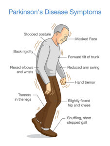
A new study describes how a spinal cord implant provides help to walk for Parkinson’s disease patients. The original study was published in Nature: . CNN published a simplified article that describes how a Parkinson’s patient with gait problems could walk again, when a spinal cord implant was inserted. This patient’s name is Marc Gauthier from a town near Bordeaux, France. He was 36 years old when he was diagnosed with Parkinson’s disease. For many years he could control his Parkinson’s disease symptoms with Dopamine replacement therapy.

Insertion of deep brain stimulator

But by 2004 the medication did not control his gait anymore. His doctors inserted a deep brain stimulator, which helped with tremors and muscle stiffness. But as the disease progressed, he developed a severe walking disorder. The drugs and the deep brain stimulator no longer helped his walking problem. His muscles stiffened up, and often he fell 4-times per day. He had to give up his work as an architect. Finally, in 2021 his doctors inserted a nerve stimulator with electrodes connecting to the lower spinal cord.

Spinal cord stimulation

Researchers from France, Switzerland and other parts of the world determined which part of the spinal cord were in need of electrical stimulation to improve his gait and balance problems. Dr. Eduardo Moraud is an author of the study and researcher at Lausanne University Hospital. He said: “The stimulation here is focused on the spinal cord. We target the region of the spinal cord that will control all the leg movements.” The team managed to develop an electric stimulator for implantation under the skin over the abdomen with electrodes going to the lower spinal cord. This way his physicians stimulated muscle groups and also relaxed them in sequence so that his gait improved and steadied. His balance stabilized as well and he could walk stairs again.

Future routine surgery to implant spinal stimulator

Marc Gauthier’s case is a first in approaching Parkinson’s disease with a spinal stimulator. The researchers stressed that they will improve the technology and learn from other patients how to individualize this technique. But eventually the spinal cord stimulator could become a routine approach for end stage Parkinson’s disease. Dr. Moraud said: “Addressing deficits of gait and balance in Parkinson’s disease is extremely challenging. These deficits can be very heterogenous. They can be variable across patients. They can affect walking but also symmetry, balance, posture. The neuroprosthetic approach that we have developed here allows for the first time to target and address these problems individually in a highly specific manner for each patient. It operates in real time, and importantly, it is complementary to other existing therapies.”

Marc Gauthier’s progress since his spinal cord stimulator

The researchers identified hotspots in the lower spinal cord with connection to the gait and balance problem of the patient. The surgeon connected the neuroprosthesis to these hotspots to stabilize gait, balance and muscle strength. Following the surgical procedure Mr. Gauthier had to undergo a few months of rehabilitation with the neurostimulator to practice walking, develop strength and coordination between the right and left leg. His body was no longer stiffening or freezing in place. He could now take a 3-mile lakeside stroll without stopping. He can now manage stairs going up or coming down smoothly. In the past his gait would suddenly freeze or he lost hist balance. None of this

Is happening now. He simply is walking normally.

The final word of Dr. Moraud

“The stimulation here is focused on the spinal cord. We target the region of the spinal cord that will control all the leg movements.” Inserting a neurostimulator is not a cure, simply a procedure to help the patient lead a normal life.

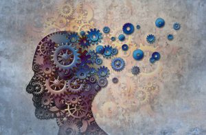
Help to Walk for Parkinson’s Disease Patients

Help to Walk for Parkinson’s Disease Patients

Conclusion

A new surgical procedure, namely inserting a neurostimulator under the abdominal wall, is the latest approach to treat Parkinson’s disease. This is help to walk for Parkinson’s disease patients. The case, which I described here is the first case where doctors performed this new procedure. They identified hot spots in the lower spinal cord and connected them to the neurostimulator with electrodes. After a lengthy rehabilitation period the patient learned how to use the stimulator for optimal walking, balancing, and going stairs up and down. There are no more falls or gait problems and no freezing. At the end the patient can walk normally and even manage a walk of 3 miles. Researchers anticipate that this method will become an accepted treatment modality for gait problems of Parkinson’s disease patients.

Oct
30
2021

Acetaminophen Damages the Fetus

Many women take acetaminophen when pregnant, but acetaminophen damages the fetus. It is important to realize that acetaminophen is a common over-the-counter pain reliever. In addition, it is also often combined with codeine as headache pills. Acetaminophen goes under these brand names: Tylenol, Tylenol Arthritis Pain, Tylenol Ext and Little Fevers Children’s Fever/Pain Reliever. The international name of acetaminophen is the name “paracetamol”.

An international group of 13 scientists are calling health care professionals to limit the use of acetaminophen in pregnant women. CNN reported about this under this link.

These scientists published an article in the medical journal Nature on the dangers of paracetamol use in pregnancy.  Specifically, they said that acetaminophen can alter fetal development, which includes reproductive, neurodevelopmental and urogenital disorders.

History of acetaminophen

The chemical name for acetaminophen is N-acetyl-p-aminophenol. Acetaminophen is simply a shortened version of that chemical name. It was introduced in the US in 1955 as Tylenol® and in the United Kingdom in 1956 under the brand name Panadol®. In particular, acetaminophen was recommended to control fevers and to help with pain control.

In the 1960’s the Swiss watch industry provided workers who complained of headaches freely with acetaminophen. With this in mind, within a few years studies showed that many of these women who took a lot of this medication developed kidney problems. This led to an increase of the creatinine level in the blood. The kidney damage from acetaminophen was dubbed “phenacetin kidneys”. Outside of the US acetaminophen has the name phenacetin. Many of these patients subsequently had to receive dialysis and later kidney transplants.

Acetaminophen toxicity

The recommended dose of acetaminophen is 650 mg to 1000 mg 4-6 times daily, not to exceed 4 grams/day. The therapeutic window for this drug is very narrow, because 7.5 grams per day to 10 grams per day are already toxic. For children the dose is 15 mg/kg every 6 hours to a maximum of 60 mg/kg per day.

The other known toxicity concerns liver function. This article about Tylenol toxicity explains this in more detail.

Many people do not know about the limit for the over-the-counter acetaminophen and take too much for a fever or a painful condition. Just because a drug is available over the counter does not mean that it is harmless. If you don’t watch for toxic levels, you could end up dead or find yourself waiting for a liver transplant.

New evidence that acetaminophen damages the fetus

91 scientists from Australia, Brazil, Canada, Europe, Israel, Scotland, the UK and US have signed a declaration. In it they ask pregnant women not to take acetaminophen “unless its use is medically indicated”. Among the reasons for the declaration is that acetaminophen certainly can cause neural tube defects and cardiovascular disorders in fetuses. Pediatrician Dr. Leonardo Trasande, director of environmental pediatrics at NYU Langone Health, did not partake in this research. He has done safety studies on acetaminophen and pointed out the similarity in chemical structure between acetaminophen and phthalates. Like phthalates acetaminophen disrupts the reproductive development in animals and humans.

Evidence for neurodevelopmental disruption

Dr. Shanna Swan, a professor of environmental medicine at the Icahn School of Medicine at Mount Sinai in New York said: “There’s enough evidence to find increased risk of undescended testicles and a shortening of the anogenital distance, which is a predictive of later decreased sperm count and decreased fertility. We also see impaired ovarian function which has consequences for later fertility, although females have been less studied.”

The conditions that relate to acetaminophen toxicity were attention deficit hyperactivity disorder (ADHD), behavior abnormalities and autism spectrum disorders. In addition, language delays, conduct disorders and decreased IQ were due to neurodevelopment disruption from acetaminophen toxicity.

Mechanism of action of acetaminophen and side effects

Acetaminophen has been on the market for over 60 years. But scientists still don’t know exactly how it acts in the body to help control pain and reduce fever. There is a consensus that acetaminophen acts on the central nervous system inhibiting the synthesis of prostaglandins. These biological compounds have a leading role in causing fever, pain and inflammation. But the scientific proof for this consensus is still outstanding.

Side effects

There are a multitude of side effects that can occur with the use of acetaminophen. Common side effects are hives, itching, swelling of the mouth and throat and tingling in the mouth or throat. Other side effects are swelling in the face or hands, breathing difficulties or chest tightness. Acetaminophen can cause a loss of appetite, nausea and vomiting. Severe stomach pain can be another symptom of acetaminophen side effects. As you can see from the above link there are many more known side effects of acetaminophen.

Acetaminophen Damages the Fetus

Acetaminophen Damages the Fetus

Conclusion

Acetaminophen (=paracetamol, phenacetin) is a popular over-the-counter fever and pain remedy. But a narrow therapeutic width can cause serious overdoses where both the liver and the kidneys suffer irreparable damage. When people unknowingly take too much acetaminophen, they enter into the toxic range. This can cause disability and death. Kidney damage from acetaminophen became known as “phenacetin kidneys” already in the 1960’s in female workers of the watch industry in Switzerland. They developed headaches from constantly working with magnifying glasses and had free access to acetaminophen provided by the employer. Later, in North America liver disease developed when patients overdosed with over-the-counter acetaminophen for fever and pain control.

Interruption of fetal development from exposure of the fetus to acetaminophen 

At the present time the focus is on newer findings of researchers. They noticed that exposure of pregnant women to acetaminophen damages the fetus. This results in undescended testicles and a shortening of the anogenital distance which is a predictive of later decreased sperm count and decreased fertility. These are findings for males. Findings in females are less studied at this point in time. Dr. Leonardo Trasande pointed out the similarity in chemical structure between acetaminophen and phthalates. Like phthalates acetaminophen disrupts the reproductive development in animals and humans. Patients should take acetaminophen only under supervision with doses that are safe. The old notion that acetaminophen would be safe in pregnancy is no longer true in light of the new medical findings. Any pregnant woman should discuss with her physician what she can safely take.

Incoming search terms:

Jun
26
2021

Being A Morning Person Can Prevent Depression

A British study concluded that being a morning person can prevent depression. It was reviewed also in CNN. The study used sleep data from 85,000 UK participants in the Great Britain Biobank Study. They wore wrist activity monitors that provided sleep data. Researchers compared the sleep data with the self-reported mood changes. They found that if the sleep pattern is misaligned with the circadian rhythm, those who are night owls are at a higher risk of developing mood disorders. The controls were those who were morning persons. They were not affected by the misalignment effect.

Normal sleep pattern

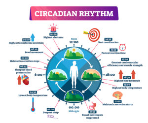
Natural sleep habit or the circadian rhythm starts between 10 PM and 11 PM and continues for 7 to 8 hours. Your hormones are replenished during your sleep. This helps your body’s hormones and the immune system to restore itself overnight.

“The health problems associated with being a night owl are likely a result of being a night owl living in a morning person’s world, which leads to disruption in their body’s circadian rhythms”. This is what sleep specialist Kristen Knutson said. She is an associate professor of neurology and preventive medicine at Northwestern University Feinberg School of Medicine.

Reclaim your natural sleep habit

The key is to learn to live within your circadian rhythm pattern. Caffeine is a powerful stimulant and will stop you from falling asleep. For this reason, it is best to avoid caffeine-containing beverages. If you cannot live without your favourite cup of java, switch to the decaffeinated version. Stop using LED lights (from TV, computers, tablets or smartphones) 2 hours prior to your bedtime. Use dark curtains and enjoy a comfortable bed. Maybe read that thriller, earlier in the day instead of making it your bedtime story. And do yourself a favour: you do not need the eleven o’clock news on TV!  They will probably stop you from falling asleep. Go to sleep between 10PM and 11PM.

Children can have problems with sleep disturbances and depression

Another study published March 22, 2021 in the Jama Network showed that depression had an association with sleep disturbances in youth and children.

A meta-analysis of 16 publications looked at depression and disturbed sleep. It showed that depression was 1.5-fold higher in sleep disturbed youths/children compared to controls with a normal sleep pattern.

Other studies re. being a morning person can prevent depression

Another study with Dr. Knutson as the lead author appeared in 2018. The authors found that various health conditions, mood disorders and mortality were on the increase the more the sleep rhythm deviated from the circadian rhythm. Morning persons were protected from this effect. But night shift workers and night owls were at a higher risk of disease. Specifically, they found the following associations for evening types.

  • Evening types compared to morning types had a 1.94-fold risk of psychological disorders
  • The risk of diabetes was 1.30-fold for evening types
  • Neurological disorders had a 1.25-fold risk in evening types
  • Gastrointestinal/abdominal disorders occurred 1.23-fold more often in evening types
  • respiratory disorders were 1.22-fold more common in evening types
  • Evening types had a 10% increased risk of all-cause mortality

The researchers concluded that externally imposed timing of work and social activities has potentially serious health consequences.

Circadian misalignment responsible for disease

Dr. Knutson also said: “Circadian misalignment could also lead to inadequate sleep duration and quality, which could also impair mood and exacerbate mood disorders.”

In other words, circadian misalignment to circadian rhythm problems. This can cause mood disturbances, but eventually lead to the diseases listed above.

Evidence of health risks in night shifts workers

The medical literature is full of examples that the health of night shift workers is significantly affected by circadian misalignment. Here are a few examples.

1.Here is a random selection to illustrate the health risks of night shifts workers. A study from 2015 examined the sleep patterns of 315 shift nurses and health care workers in Iranian teaching hospitals. They found that 83.2% suffered from poor sleep and half of them had moderate to excessive sleepiness when they were awake.

2.This South Korean study examined 244 male workers, aged 20 to 39 in a manufacturing plant. Researchers compared blood tests from daytime workers to blood tests from night shift workers. They also obtained inflammatory markers like the C-reactive protein and leukocyte counts. Night shift workers had significantly higher values. The investigators concluded that shift workers have increased inflammatory markers. This is a sign of a higher risk of developing cardiovascular disease in the future.

Higher mortality and higher cancer risk in nighttime workers

3. A Swedish study found that white-collar shift workers had a 2.6-fold higher mortality over a control group of daytime white-collar workers.

4. Another study compared night workers in the age group of 45 to 54 with daytime workers and found a 1.47-fold higher mortality rate in the night shift workers.

5.In a study from China 25,377 participants were part of a study that investigated cancer risk in males with more than 20 years of night shift work. They had a 2.03-fold increased risk to develop cancer compared to males working day shifts. Women with night shift work in this study showed no effect with regard to cancer development.

Healthy telomeres with healthy sleep pattern

It is true that you can suffer multiple health problems, as all of your hormones depend on the resetting during your deepest sleep between 2AM and 4AM triggered by the nighttime melatonin response. Even your telomeres, the caps of chromosomes in every cell get shortened from too much stress and too little sleep.

One example of such a study comes from Milan, Italy. https://oem.bmj.com/content/75/Suppl_2/A480.1

In this 2018 study researchers compared 46 nurses who had worked in night shifts with 51 nurses working day shifts. Among the night shift workers breast cancer was common, but not among day workers.

Shortened telomeres, hypomethylation of BRC1 gene and p53 gene

In the night shift nurses from Milan there was hypomethylation of the breast cancer gene BRC1. There was also hypomethylation of the general cancer gene p53. At the same time significant telomere shortening occurred in night shift nurses who had worked night shifts for more than 15 years. This likely all worked together in causing night nurses to develop breast cancer more frequently.

Shortened telomeres mean a shortened life span. The reason for this is that people with shortened telomeres develop heart attacks, strokes and cancer. This is what shortens the life span. How do we avoid this risk? Go back to healthy sleep habits. As mentioned above it is best to start going to sleep between 10PM and 11PM and sleep for 7 to 8 hours. Night owls delay going to sleep by 2 to 3 hours.

Being A Morning Person Can Prevent Depression

Being A Morning Person Can Prevent Depression (click image to enlarge)

Conclusion

A publication in Molecular Psychiatry demonstrated that evening person (night owls) are more likely to develop depression. This is in comparison to morning persons. As discussed, other researchers showed that evening persons also can develop diabetes and neurological disorders. In addition, respiratory disorders and gastrointestinal disorders are more common in night owls as well. When it comes to mortality, evening persons have a 10% increase of mortality over morning persons. There is a large body of literature regarding diseases of night shift workers. Night Shift work is perhaps the most extreme example of a circadian misalignment. It leads to poor sleep, inflammation in the body, increased cancer risk and higher mortality compared to day shift workers.

Prevent telomere shortening

Even the telomeres get shortened in night owls and night shift workers. We can prevent problems like these by going to bed in time and sleeping according to our circadian rhythm. If you had a poor night’s sleep, make up for it with the help of an afternoon nap. Do not sleep all afternoon though; half an hour or one hour will be enough. Even a short, restful nap after coming home from work can restore your feeling of wellness.

Jan
09
2021

Melatonin Is More Than a Sleeping Aid

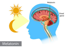
Notably, the January 2021 issue of the Life Extension magazine informs you that melatonin is more than a sleeping aid. It contains an interview between Dr. Roman Rozencwaig and a Life Extension (LE) magazine reporter. It must be remembered that Dr. Rozencwaig dedicated much of his career to the healing effects of melatonin. Another keypoint is that in 1987 Dr. Rozencwaig published a paper together with two other researchers. Specifically, it showed that melatonin production by the pineal gland declines in older age. Markedly, they stated that this is the reason why people age and why diseases of aging develop. Another key point is that Dr. Rozencwaig also stated that taking oral melatonin can promote a healthier life.

Melatonin deficiency causing aging and various illnesses

With the aging process the pineal gland calcifies and melatonin production is steadily declining. Surely, along with this is a deterioration of the circadian hormone rhythm. Meanwhile, the neuroendocrine system in the brain gets disorganized. Accordingly, this causes various diseases to occur. To emphasize, Dr. Rozencwaig says that a proper balance between melatonin and neurotransmitters is what we need to maintain health and longevity. As a result, a daily intake of melatonin supports healthy aging and longevity.

The many clinical effects of melatonin

Oral melatonin tablets help you to fall asleep easier, particularly the population that is older than 60 years.

But besides that, melatonin has many other clinical effects.

  • Melatonin improves immunity, which improves resistance against infections. It helps also in cancer prevention
  • Melatonin maintains the circadian hormone rhythm by synchronizing pituitary and hypothalamic hormone production
  • It protects the brain and may prevent Parkinson’s disease, Alzheimer’s disease, multiple sclerosis, autism, and others
  • Melatonin modulates anti-inflammatory cytokinins in different diseases

Dr. Rozencwaig mentioned that melatonin slows down the aging process. There are multiple intertwining reasons for this. 

Melatonin’s actions against the aging process 

  • Melatonin regulates gene expression. This means that some signs and symptoms of aging can be reversed through genetic switches
  • Because melatonin regulates the immune response, the body is more protected against viral, bacterial and parasitic infections
  • Melatonin helps to overcome chronic inflammation that produces cytokines
  • Melatonin is also liver-protective through stimulation of an enzyme (AMPK). This enzyme regulates cellular metabolism.
  • There are other processes that melatonin is involved in: energy metabolism by protection and restoration of mitochondria.
  • Melatonin protects against osteoporosis by balancing and regulating bone formation versus bone loss.

More actions of melatonin

  • An important function of melatonin is the stimulation of antioxidant enzymes like glutathione peroxidase and superoxide dismutase (SOD)
  • Melatonin regulates sirtuins, which are proteins that maintain cellular health. They protect you from obesity, type 2 diabetes, cancer, heart attacks and strokes, dementia and more
  • As already mentioned, melatonin is a neuroprotective agent and may prevent Alzheimer’s and dementia
  • Melatonin stimulates apoptosis of cancer cells.
  • Oral health and melatonin are related. Melatonin suppresses herpes infections and periodontal disease. Melatonin prevents oral cancers to a certain degree. In addition, dental implants survive better when melatonin is present in saliva.

Prevention of cognitive decline

Dr. Rozencwaig mentioned that melatonin stops much of the cognitive decline of aging. To achieve this the following processes take place.

  1. Melatonin improves the sleeping pattern and increases the amount of REM sleep.
  2. During sleep melatonin removes toxic amyloid and tau proteins. We know that with Alzheimer’s disease these are the proteins that accumulate in the brain.
  3. Melatonin improves myelination of white matter in the brain. This prevents brain atrophy of old age.
  4. The brain is metabolically very active and produces toxic free radicals. But melatonin is a strong antioxidant dealing with free radicals. Melatonin can cross the blood brain barrier and stimulates enzyme production to eliminate toxic reactive oxygen species.
  5. Chronic inflammation also increases with age, but melatonin deals with this condition in the brain.
  6. Here are 3 subtypes of melatonin receptors. The body integrates the multitude of actions of melatonin with the help of these receptors.
Melatonin Is More Than a Sleeping Aid

Melatonin Is More Than a Sleeping Aid

Conclusion

Melatonin is a powerful antioxidant that has many other useful protective qualities as explained. The body integrates various functions like anti-aging, anti-free radical activity, neuroprotection in the brain and more. Melatonin even synchronizes pituitary and hypothalamic hormone production. This helps to integrate the effect of melatonin, which benefits the body in many ways. Melatonin prevents Parkinson’s and Alzheimer’s disease, multiple sclerosis, autism, obesity, type 2 diabetes, cancer, heart attacks, strokes and dementia. Melatonin production deteriorates from the age of about 60 onwards. It is important to supplement with melatonin at nighttime from that age on. Usually, you only need small amounts of melatonin, between 1mg and 3 mg at bedtime. This prevents most of the serious diseases of old age, stimulates your immune system and lets you age gracefully.

Incoming search terms:

Jan
19
2019

Alzheimer’s disease is treatable with hormones

Dr. Thierry Hertoghe, an endocrinologist from Belgium, stated that Alzheimer’s disease is treatable with hormones. This talk was part the 26th Anti-Aging Conference of the American Academy of Anti-Aging Medicine in Las Vegas (from December 13 to 15, 2018).

First of all, Dr. Hertoghe treated many Alzheimer’s patients himself and noted that they often have multiple hormone deficiencies. Secondly, common deficiencies affect thyroid hormones, human growth hormone, estradiol for women and testosterone for men. But even vasopressin and oxytocin are hormones that may be lacking. Third,  after doing thorough blood tests to assess hormone levels, Dr. Hertoghe replaced what hormones were missing. Finally, many Alzheimer’s patients got their energy, muscle strength and memory back.

In the following I am summarizing what Dr. Hertoghe told the audience about the various hormones. Alzheimer’s disease is treatable with hormones. Later I provide the hormone doses that Dr. Hertoghe uses for replacement.

Progressive memory loss

Generally, patients who develop Alzheimer’s disease start losing short-term memory first, but in time they will also lose long-term memory. Often this disease process starts in the 60’s as age-associated cognitive impairment. In the 70’s it may progress further to mild cognitive impairment, only to take off in the 80’s as Alzheimer’s disease. The astute clinician may order some screening blood tests in the 60’s and 70’s. In a male low testosterone, low DHEAS and low thyroid hormones may be present. Certainly, blood tests will show this readily. Frequently, in women low estradiol, low thyroid and low DHEAS may also be present. The reason this is important is that simple hormone replacement can return a person back to normal. Yes, this is right: hormone replacement can bring a person with age-associated cognitive impairment or mild cognitive impairment back to normal! In other words, Alzheimer’s disease is treatable with hormones.

Hormones important to monitor with Alzheimer’s disease

There are 6 hormones that are important for memory restoration in Alzheimer’s patients: IGF-1 (and growth hormone), thyroid hormones, estrogen and testosterone, vasopressin (and oxytocin) and pregnenolone. However, as Alzheimer’s patients often have sleep problems, another important hormone is melatonin.

Oxytocin to calm down aggressive Alzheimer’s patients

Notably, Dr. Hertoghe found that Alzheimer’s patients often are restless and can be aggressive. This makes it difficult to care for them in a home. Oxytocin is the hormone of trust, affection, sociability and concerns about others. It calms down aggressiveness. But with oxytocin treatment the Alzheimer’s patient feels better, becomes friendly, cooperative and warm-hearted.

As an illustration Dr. Hertoghe gave an example of one of his 80-year old patients with aggressive Alzheimer’s disease. She became unmanageable for her non-married son and other contacts. 5 IU of oxytocin sublingually changed this woman into a friendly, compassionate, warm-hearted woman, and the aggressiveness disappeared completely.

Insomnia in Alzheimer’s patients

About 45% of Alzheimer’s patients develop “sundowning”. When the sun goes down they start getting hyperactive, develop unacceptable behaviors and they become restless. Research papers showed that blood melatonin levels are low in these patients. Indeed, this is why they respond very well to small amounts of melatonin at bedtime. As a conclusion, within only a few days of starting this, their sundowning disappears, and they become easier to look after.

Dr. Hertoghe provided material from several research papers that showed that Alzheimer’s patients are often deficient for melatonin. Replacement with varying doses of melatonin solved even more complicated insomnia problems.

Melatonin is a powerful anti-oxidant. Interesting animal experiments have shown that melatonin has memory-enhancing properties. Researchers believe that melatonin improves the extracellular senile plaques with amyloid-beta peptide accumulation (first of 2 Alzheimer’s lesions). In addition melatonin also decreases the intracellular neurofibrillary degeneration tangles, the second of the two specific Alzheimer’s lesions.

IGF-1 and human growth hormone

Several studies have shown that Alzheimer’s patients have a significant drop in IGF-1 levels and growth hormone levels. This affects their short-term and long-term memory. Serum IGF-1 has an inverse correlation with cognitive impairment. Dr. Hertoghe said that IGF-1 treatment in Alzheimer’s patients increases their brain volume, increases the functional network of neurons in the brain and increases memory.

Brain atrophy in Alzheimer’s patients from chronically depleted IGF-1

Dr. Hertoghe showed a slide of a normal brain with a view from the outside and a cross section view of the brain. The same slide contained the view of an Alzheimer’s patient’s brain. It showed brain atrophy resulting in a much smaller brain and the cross section displayed an increase of the hollow spaces (e.g. the third and forth ventricle). He stressed that in his view the brain shrinkage of Alzheimer’s patients is due to prolonged low levels of IGF-1. This in turn is due to a lack of production of human growth hormone.

With IGF-1 treatment the serum IGF-1 was increasing and the cognitive function in older adults recovered. Dr. Hertoghe provided many literature citations to support this, which I will not repeat here.

Case report of a male patient with Alzheimer’s disease

Dr. Hertoghe presented one of his patients with Alzheimer’s. Lab tests showed that he had deficiencies of thyroid hormones, DHEA and testosterone. But despite replacement of these hormones he remained severely affected with Alzheimer’s. He did not remember his own name, could not go to the toilet on his own, spoke only a few words and suffered from severe fatigue. He received 4 injections around his eyes with IGF-1 and mesotherapy from his doctor (described below) with human growth hormone and IGF-1. Within a few weeks he had a complete reversal of his cognitive decline. He could return to his professional driving career doing halftime work with a delivery van in the city. He could read a newspaper and understood what he was reading. Alzheimer’s disease is treatable with hormones.

Thyroid hormones

According to Dr. Hertoghe thyroid hormones help to establish short-term and long-term memory and treat the apathetic depression in Alzheimer’s patients. Many Alzheimer’s patients are hypothyroid.With this deficiency they have swollen lower eyelids, a puffy face and paleness of the face. In a 1990 study a group of Alzheimer’s patients had 26% lower T3 levels when compared to normal controls. Many patients with hypothyroidism have memory loss, before their deficiency is corrected. Dr. Hertoghe stated that 13% of all dementia cases are reversible by proper thyroid hormone treatment.

Estradiol can improve long-term memory loss

Research showed that estradiol could improve long-term memory in dementia and Alzheimer’s disease cases. Many female Alzheimer’s patients are deficient in estrogens. If they do, they have dry eyes, a pale face and thin, dull hair. In a 2005 study 33 control women were compared to 48 women with Alzheimer’s disease. The estradiol levels in the Alzheimer’s disease group showed significant depletion compared to the normal control group. There was no significant difference found with regard to progesterone, testosterone and LH&HSH levels. Another study showed that in cerebrospinal fluid of women with Alzheimer’s disease the estradiol level was significantly reduced while the beta-amyloid levels were significantly increased.

Dr. Hertoghe reviewed several studies that showed that symptoms of Alzheimer’s disease disappeared with estradiol supplementation. Both memory and mood responded to the treatments.

Men with Alzheimer’s disease are often testosterone deficient

Testosterone is important for long-term memory. Men in andropause report erectile dysfunction, general weakness and memory loss. The physician needs to be aware that the patient may be starting to develop Alzheimer’s disease. Dr. Hertoghe showed a slide based on a publication, which stressed that testosterone enhances memory. It increases brain blood flow and thickens the myelin sheets. Testosterone increases dendrite and synapses and in addition decreases amyloid beta-peptide production. Neurotoxicity is also reduced. The end result is improvement of Alzheimer’s in males with testosterone replacement.

Pregnenolone improves short-term memory

Pregnenolone gets synthesized in the brain, spinal cord and peripheral nerves. Dr. Hertoghe said that pregnenolone is a neurostimulating “neurosteroid”. Pregnenolone concentrations in brain tissue are about 25- to 35-fold higher than in the blood stream. Some cases of Alzheimer’s disease can come from a lack of pregnenolone and pregnenolone sulfate. Patients who have Alzheimer’s because of a lack of pregnenolone have blood levels that are 2.5-fold lower than pregnenolone levels in normal controls. When these patients are treated with pregnenolone, their memory improves. The mechanism of the effect of pregnenolone is by increasing acetylcholine by more than 50% in the hippocampus. It also protects the hippocampus from glutamate and amyloid beta. Pregnenolone improves short-term memory over a period of 3 to 4 months of treatment.

Vasopressin improves short-term and long-term memory loss

Postmortem studies on Alzheimer’s patients showed that there is decreased vasopressin in the brain cortex. In patients with alcoholic dementia (Korsakoff psychosis after recovery) there was decreased vasopressin in the cerebrospinal fluid. Often patients with diabetes insipidus have decreased vasopressin and are in danger of developing dementia. If not treated, they develop short-term and long-term memory loss. When treated with vasopressin or Desmopressin their memory recovers within 4 hours of starting therapy. Younger patients (50 to 73) do better with memory recovery than older patients (74 to 91).

Treatment details of hormone replacement for Alzheimer’s disease

Before hormone treatments are given to a patient it is important to do a battery of blood tests. This will help the physician to identify the missing hormones in a particular patient. Each of the missing hormones are then administered separately.

Oxytocin

This hormone can be given sublingually or intranasally. Sublingually 5-10 IU are given daily. With the sublingual approach 1 or 2 sprays are given daily. Each spray contains 8 IU of oxytocin. Improvement is visible within 2 to 5 days. A full recovery takes 2 to 3 months.

Melatonin

Most patients in the higher age group do no longer produce their own melatonin. With the oral route 1-3 mg are given every night before going to bed. An alternative is to use sublingual tables 0.5mg to 1.0mg at bedtime. The first improvement can be seen 2-5 days after the start of replacing melatonin, the full impact takes about 2-3 months from the start of the treatment.

IGF-1 and human growth hormone

Replacement of IGF-1 can be done by injecting IGF-1 or human growth hormone (HGH). HGH stimulates the liver to produce IGF-1. IGF-1 is somewhat cheaper than HGH. When IGF-1 is used, 0.3mg to 1mg is injected at bedtime. Progress is slow; the first improvement is visible at 2-4 months, it takes up to 24 to 36 months for a full recovery.

For severe memory impairment with Alzheimer’s, the doctor does a double treatment approach with both IGF-1 and HGH: first subcutaneous IGF-1 injections around the eyes 4 times per day (0.01mg each). Secondly, at the doctor’s office the doctor administers mesotherapy injections with 1mg of HGH and 1mg of IGF-1 and vasodilators 3 times per week. Two weeks later the doctor administers another course of mesotherapy. He may repeat this twice in 14-day intervals. Now the interval increases to monthly therapy for 3 months and finally every 3 to 4 months. The patient can use IGF-1 nose drops instead of subcutaneous IGF-1 injections.

Thyroid hormones

Dr. Hertoghe prefers desiccated animal thyroid hormone replacement as the T3/T4 ratio is best matched to what the ratio is in humans. Depending on the severity of thyroid hormone deficiency the patient takes 30-150mg of thyroid hormone every morning. Dr. Hertoghe starts with a low dose and slowly increases the dosage. Clinical progress is very slow. It takes until the second month before the first improvement takes place. Full improvement can take 8-12 months.

Estradiol

Replacement of estradiol in postmenopausal women with Alzheimer’s disease received ether more than 0.1mg per day or 0.625mg of conjugated equine estrogen daily. In both cases there were improvements of their memory and improvement on the Hamilton depression scale.

Dr. Hertoghe’s preferred way to treat postmenopausal women with Alzheimer’s disease is as follows. The first 25 days of each month he gives them 1-2mg of oral estradiol valerate each day and 100mg of micronized progesterone. If they prefer an estrogen cream, he gives them 1-3mg per day transdermal estradiol and 100mg micronized progesterone capsules.

The first improvement is visible after 2-4 months; there is further improvement the next 8-12 months.

Testosterone

There are two methods of how to do hormone replacement with testosterone, either by injection or as transdermal cream. The injection treatment uses 250mg of testosterone enanthate or cypionate every 2 -3 weeks. The patinet can also self-administer testosterone enanthate (50mg twice per week) for a more even blood level of testosterone. The transdermal approach involves 100-250mg transdermal, nanoliposomal testosterone daily.

The memory will improve 2-4 months into replacement therapy. The full improvement takes 8-12 months.

Pregnenolone

The replacement therapy is 100mg per day in the morning for the first 4 months. Then there is a dosage reduction to 50mg daily. Studies have shown that 30mg of pregnenolone is not enough to treat memory loss. Short-term memory improved after 3 to 4 months in about 75% of patients.

Vasopressin

The best vasopressin preparation to use is bio-identical vasopressin. It comes as 1 nasal spray with 10IU of vasopressin. Upon awakening the patient or caregiver applies 1-2 sprays into the nose. The patient receives the second dose 10 minutes before lunch by nasal spray.

Apart from hormones, lifestyle changes are also recommendable.

Alzheimer’s disease is treatable with hormones

Alzheimer’s disease is treatable with hormones

Conclusion

Who would have thought that Alzheimer’s disease could have anything to do with hormones? Dr. Hertoghe, the endocrinologist from Belgium did many hormone tests on Alzheimer’s patients and concluded that various degrees of hormone deficiencies can indeed cause Alzheimer’s disease. But what is more is that you can replace the missing hormones and see complete cures in patients with Alzheimer’s disease. Alzheimer’s disease is treatable with hormones. This is something conventional medicine can only dream of. At this point this hormonal approach is not yet mainstream medicine; but it would not be a surprise to me, if in 10 or 20 years interested physicians do this type of therapy routinely in their practice. When hormones are missing, replace them. When the memory is fading, think about testing for missing hormones! It will make a difference in the quality of life for the patient as well as for his family.

Oct
20
2018

Leg Paralysis Can Be Treated

Several publications show that leg paralysis can be treated. This goes against the clinical experiences throughout the world. The common school of thought was that spinal cord injuries from a severe accident would lead to permanent damage. New research has proven that this is not so.

The publication to show that leg paralysis can be treated

On Sept. 24, 2018 the New England Journal of Medicine reported about several cases where completely paralyzed people were able to walk again with the help of a walker. A surgeon implanted a spinal cord stimulation device under the skin of the abdominal wall. From there electrodes were going under the skin into the lower lumbar spine and upper sacral area close to the spinal cord. This allowed the muscles of the lower body half to receive the same nerve impulses that the muscles above the injury received. With extensive physiotherapy treatments the body was able to relearn the muscle contractions of the legs and feet.

Relearning how to walk

The next step was to relearn the steps of walking. There was a group of 14 patients with spinal cord injuries who had implantation of a spinal cord stimulation device. They were eventually able to walk again with a walker. The walker was necessary to stabilize their gait. One male took a chance and did not use a walker. He fractured his hip after a fall. But eventually he was making a full recovery and is able to walk now with a walker.

Details of a case of full leg paralysis

Kelly Thomas was driving in a car and lost control. The car ended up at a tree, severely deformed. She was unconscious for several weeks and needed treatment in the hospital. She was 19 and paralyzed from the chest down. Kelly is 24 now and she is able to walk again with the help of a walker. A surgeon had implanted a stimulation device in her abdominal wall with electrodes going to her lower back. The stimulator is passing on the signals coming down from the healthy spine. Electrical signals from the healthy spinal cord make their way to below the severed spinal cord. This though was only the first step.

The second step, a lengthy physiotherapy program

The second step was a lengthy physiotherapy program. All of the previous memories of learning to walk as a child are no longer there in a paralyzed person. The body has to relearn muscle contractions, coordination of muscles, moving of a foot or lower leg. Then all of these sub movements have to blend together into a smooth movement associated with walking. A walker helps to stabilize the gait. This link described more details regarding Kelly’s recovery and how hard she had to work on the physiotherapy part to finally achieve her walking. Leg paralysis can be treated.

Other studies showing people rising from wheelchairs

Here are two other studies that show how other people were able to rise from their wheelchairs.

2015 study

A 2015 study explains how the researchers were able to make one patient walk again. They used a recording of the brain currents (EEG) and pass that information on to below the spinal cord injury. This involved a lengthy learning procedure followed by many physiotherapy treatments. In the beginning it was important to have the patient suspended from the ceiling to prevent falls. Subsequently the patient could walk unsuspended.

2016 study

In a 2016 study eight patients were treated with the system described in the previous paper. Brain-machine interfaces recorded and transmitted the electrical brain activity to below the spinal cord injury. An intense 12-month physiotherapy program enabled the patient to regain her capability to walk. Only 50% of the participants were able to complete walking. There were some drawbacks of this procedure. Thoughts were interfering with brain wave recording. Also, a lack of focusing on the walking process could make it impossible for the person to walk.

Discussion re. leg paralysis can be treated

Walking again after a spinal paralysis is the dream of 1.275 million people with paralysis in the US.  Since these recent scientific findings one can truly say “leg paralysis can be treated”. There are about 8000 that would like to participate in a program, which Kelly Thomas has successfully completed. Her procedure seems to be the scientifically more robust program, although it is invasive considering that a surgeon has to implant the spinal cord stimulation device. The implanted device funnels the brain signals from above the severed spinal cord to below the injured cord. From there the electrical signals travel via the regular nerves into the muscles of the lower extremities. Extensive work with a physiotherapist is still necessary to complete the ability to walk again. For tissue defects, extracellular matrix treatment helps. For leg paralysis think spinal cord stimulation device implantation and physiotherapy treatments.

Leg Paralysis Can Be Treated

Leg Paralysis Can Be Treated

Conclusion

Lately great strides forward made it possible to help help people with paralysis enabling them to walk again. The most promising system is the one involving Kelly Thomas presented here. Briefly, following a serious car injury with a spinal cord crushing injury she received a spinal cord stimulation device. The stimulation device sends the electrical encoded muscle commands to below the scar of the spinal cord injury. The electrical impulses from above the spinal cord scar transmit smoothly to below the scar. The body does the rest.

Lots of physiotherapy

But the body needs a lot of coaxing to relearn the old body movements that connect with walking. A lot of that knowledge receded into the background following the spinal cord injury. However, extensive and prolonged physiotherapy treatments can achieve this. The Spinal Cord Injury Research Centers throughout the US have done a tremendous job researching this area. This resulted in new ways how to make paraplegic people walk again.

Sep
07
2018

Two Proteins Responsible For Getting Epilepsy

Epilepsy is sometimes difficult to control, but research has now identified two proteins responsible for getting epilepsy. According to the researchers this finding has the potential of turning our current knowledge about epilepsy upside down.

How the brain works and seizures originate

The brain is an accumulation of a myriad of nerve cells that are connected with an equal number of nerve fibers.

The communication from one end of the brain to the other end occurs via electrical signals. They have their origin in the nerve cells and travel through the nerve fibers at an astonishingly fast rate.

The nerve cells of the brain also have a connection to the muscles, organs and skin. This is achieved with the help of nerve fibers. They travel through the spinal cord and peripheral nerves to reach the end organs. Again the transmission of these communication signals are through electrical impulses. Usually these signals are perfectly balanced by inhibitory nerve cells, whose job it is in the spinal cord and throughout the brain to keep these electrical signals under control.

Unfortunately some people are not so lucky. They have a “low seizure threshold” and a fever or stress can trigger a seizure or epilepsy. This is a state of the brain where activated parts are overruling the inhibitory parts and an epileptic seizure or partial seizure results. In the case of a grand mal seizure the person may be unconscious for a brief period of time while the muscles have shaking spells. It may start with involuntary muscle twitching around the eyes, the patient becomes unconscious and may fall down onto the ground. The patient may bite the tongue, stop breathing, shake arms, then the trunk muscles and finally both legs. Around 1 in 100 people get epilepsy, and it is mostly children and people above the age of 65 who get it.

New research regarding two proteins responsible for getting epilepsy

Rochelle Hines led a team of neuroscientists from the University of Nevada, Las Vegas in this research. They investigated two proteins involved in inhibiting the central nervous system. In the past it was thought that an overstimulation of nerve cells would have caused electrical overstimulation of the central nervous system causing epilepsy. Now this new research showed that it is two proteins that are missing. Normally these proteins suppress the electrical nerve activity that leads to epilepsy.

First of all, gephyrin is a protein that regulates GABA receptors. GABA is a brain hormone that calms the brain. Another protein, called collybistin is a regulator of the localization of gephyrin.

Different approach to study epilepsy will find better medications

Rochelle Hines and her team found two key proteins, gephyrin and another protein, the alpha-2 subunit of the GABA receptor. They found that they have to interact with each other in order to calm the brain. If this does not occur, seizures (epilepsy) can occur. The emphasis of this research was on finding ways to stimulate the inhibition of the GABA receptor system. In the past the emphasis was to generally calm down the entire brain with medications. This caused a lot of side effects because the traditional anti-seizure medications do not target a specific area of the brain. Now scientists can work with the two key inhibitory proteins, gephyrin and the alpha-2 subunit of the GABA receptor. At this point a new medication is not yet available, but certainly when the development of it is complete, it will be more specific and have fewer side effects.

Some citations from the lead researcher, Rochelle Hines

“Regulating this ‘compartment’ of proteins in the brain that controls cell signalling may lead to better therapies for stopping or preventing seizures. If we can better understand the activity of the brain pattern, we can understand how it might go wrong in a disorder like epilepsy, where brain activity becomes uncontrolled. And if we can understand what is important for this control, we can come up with better strategies for treating and improving the quality of life for people with epileptic seizures and maybe other types of disorders as well, such as anxiety or sleep disorders.”

It is interesting to me that seizures and anxiety maybe the same process in the brain. While with anxiety the brain is irritated, it is still controlled. In contrast in the case of epilepsy the control of the GABA system is gone and the brain excitation is uncontrolled. Medication that will increase the proteins gephyrin and the alpha-2 subunit of the GABA receptor is badly needed.

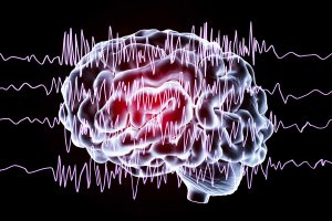
Two Proteins Responsible For Getting Epilepsy

Two Proteins Responsible For Getting Epilepsy

Conclusion

Epilepsy and seizure disorders appear to now be due to a lack of inhibition in the central nervous system. Two proteins that work on the inhibitory GABA receptors hold the key. They are gephyrin and the alpha-2 subunit of the GABA receptor. There are mouse strains that are deficient for these proteins, and they come down with epilepsy. The human brain has the same proteins that are necessary to inhibit the brain. In anxiety disorders it seems like there is a mild loss of these inhibitory proteins. In contrast with epilepsy the deficiency of these hormones is more severe. Researchers are now concentrating on developing new drugs or modifying existing drugs to stimulate the production of these inhibitory proteins. The hope is that these medications will have fewer side effects, because they will be more receptor specific.

Incoming search terms: