Feb
27
2016

Orthopedics Without A Knife

Dr. Fields gave a talk in Las Vegas about orthopedics without a knife. His talk took place at the 23rd Annual World Congress on Anti-Aging Medicine on Dec. 12, 2015 in Las Vegas. Dr. Fields gave a talk entitled “Regenerative orthopedics – non-surgical repair with stem cells/PRP/prolotherapy”. In essence the talk was about alternative treatments to surgeries in orthopedic medicine.

Dr. Peter Fields, MD, DC is a board certified medical physician and chiropractor. He is also the director of the Pacific Prolotherapy & Medical Wellness Center in Santa Monica, CA.

Introduction

Joints, muscles, tendons, ligaments and joint capsules control the movements in joints. Due to injuries and wear and tear these body parts can have a lack of function, which will lead to pain and disorders. The result can be weak, torn or damaged ligaments and tendons, arthritic changes, excessive joint motion, increased pressure, and a decrease in range of motion.

This is the common treatment cycle in medicine

Joint pain prompts you to see the doctor. You are told it is arthritis, and you get non-steroidal anti-inflammatories (NSAID’s). You come back with more pain, and you’ll get a stronger NSAID prescription. Eventually a cortisone injection is given, which helps for a few months, but then the pain reoccurs. The doctor arranges for an MRI scan. A referral to an orthopedic surgeon is likely to be the next step, and an arthroscopy (pinhole surgery) is arranged. In that case, if this does not resolve the pain, surgery like a knee replacement or hip replacement is suggested.

Common sayings when traditional medicine has nothing to offer

You may have heard some of these common sayings before. “Nothing more we can do about it!” -“I suggest you learn to live with it”- “You should never play that sport again!”- “Take these pain medications” and “The only alternative is surgery!”

The problem is, that none of these pieces of advice are really helpful. This type of approach does not treat the cause; it is directed against symptoms.

How to treat the cause?

Prolotherapy

Prolotherapy is a natural, non-surgical method to assist the body to heal torn soft tissues. It works in cases like torn ligaments, damaged tendons, cartilage, menisci or a torn labrum in the shoulder. Hyperosmolar dextrose solution is injected into the injured area. This stimulates the body’s healing forces and the body repairs what is damaged. More information is found here. In essence, prolotherapy fixes the cause, not just the effect; it heals, and it is permanent. Prolotherapy strengthens tissues, relieves pain and increases the range of motion in joints. There is 80 to 85% full pain relief and more than 80% improvement in range of motion. Prolotherapy promotes the healing of torn or damaged ligaments and tendons.

Conditions suitable for treatment with prolotherapy

Suitable conditions for treatment with prolotherapy are sports injuries, muscle tears, arthritis, tendinitis, bursitis, sciatica, TMJ problems, and fibromyalgia. Common areas treated with prolotherapy are the hip, knee, shoulder, ankle, neck, lower back and elbow. Dr. Fields showed MRI scans before and after prolotherapy treatments of ligament injuries within the knee and of shoulder ligament tears before and after treatment. Normally the physician expected these injuries to require surgery. But all that was done was one or two injections (prolotherapy treatments) with reactivation of the affected joint. There were astonishing results shown with MRI’s before and after herniated disc injuries and how they healed in a relatively short time following prolotherapy.

PRP prolotherapy

Platelet rich plasma (PRP) is a tool from regenerative medicine to amplify the healing response in connection with stem cell therapies .  The lab technician takes blood from the patient and subsequently spins it down in a centrifuge. The platelet rich fraction (PRP) contains all of the growth factors, which have the healing power of the blood. The physicians combines this with prolotherapy to make healing even more successful. This is particularly useful for labral tears in shoulders, meniscus tears in knees and other localized injuries.

Stem cell prolotherapy

Stem cell therapy has been the gold standard for repairing more serious problems. Dr. Fields combines stem cell therapy with prolotherapy to treat more serious injuries like end stage arthritis.  This is the case when bone rubs on bone, where conventional orthopedic medicine would offer a joint replacement in the hip or knee. Stem cell prolotherapy can repair any joint that has cartilage damage. A severe meniscus tear in a knee or a severe labrum tear in a shoulder would also be situations where stem cell prolotherapy is superior to surgery or to just using prolotherapy alone.

Here is a description of the procedure

Before the patient’s procedure the physician first harvests bone marrow stem cells by way of a pelvic bone aspirate; secondly the physician obtains mesenchymal stem cells from fatty tissue by aspiration of abdominal fat. A cell separator provides the stem cell fractions. The physician combines both types of stem cells, the bone marrow stem cells and the mesenchymal stem cells from fat as each one has its own strengths. These two stem cell types are more effective in combination to repair whatever tissue needs repair. Thirdly, the lab technician will draw blood from the patient to obtain PRP, which contains the growth factors needed to activate the stem cells to do their job of healing. The last step is that the physician now combines hyperosmolar dextrose (the prolotherapy part) with the stem cell preparation and mixed in PRP and injects this mixture into the injured area.

Conditions that respond to stem cell prolotherapy

This procedure has superior healing power. Before and after MRI scans of all of the major body regions showed impressive results. Several video recorded testimonials  complemented the MRI scans. It is surprising how quickly and completely fairly severe injuries can heal using stem cell prolotherapy. One particularly nasty condition is osteonecrosis of the hip, which can occur as a side effect of chronic cortisone treatment for arthritis, asthma or chronic obstructive lung disease. One or two stem cell prolotherapy treatments will heal this condition because the stem cells build up brand new bone and get rid of the old necrotic bone from the osteonecrosis. Conventional medicine has no answer for this condition. Regenerative orthopedics is successful by using stem cell prolotherapy.

What are the advantages of regenerative orthopedics?

Regenerative orthopedics reduces pain very quickly and it improves function rapidly. Healing occurs naturally, and it strengthens the tissues involved. Particularly complicated lower back pains or lower neck pains (due to degenerative disc disease, facet joint osteoarthritis, spondylolisthesis and significant foraminal stenosis) respond really well to stem cell prolotherapy, getting rid of chronic pain. The speaker showed before and after MRI scans. He also shared testimonials from patients about the various procedures.

End result following stem cell prolotherapy versus conventional surgery

This is quite in contrast to what conventional orthopedics has to offer: discectomy with fusion surgery, where the patient often has scar pain later. With a laminectomy to treat a foraminal stenosis the patient may have limited improvement of the chronic back pain for a couple of months, only to experience new back pain from a subsequent spinal stenosis as a late complication from the prior surgery. The end result with conventional orthopedics is disability, pain and suffering; the end result with regenerative orthopedics is a patient that is well, active, pain free and thankful.

Orthopedics Without A Knife

Orthopedics Without A Knife

Conclusion

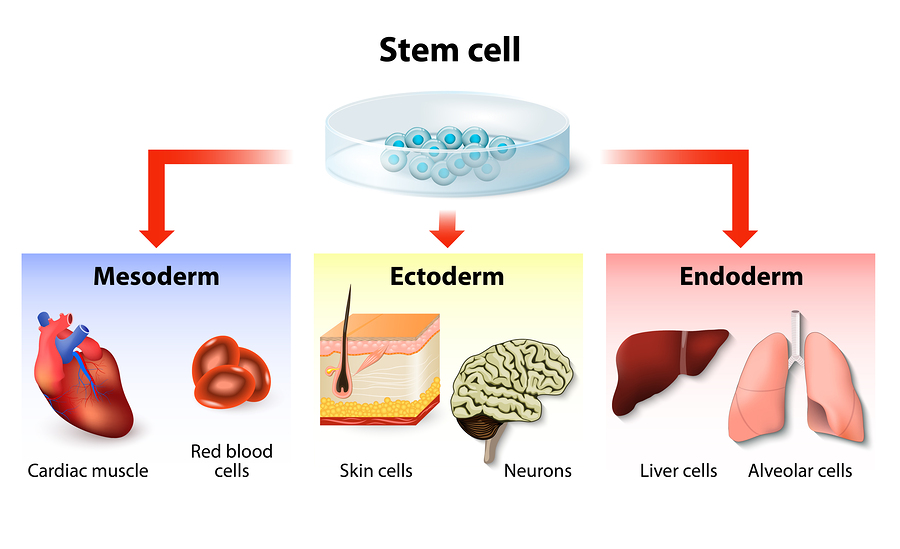
There is a form of orthopedics without a knife: regenerative orthopedics. The tools are prolotherapy for minor musculoskeletal problems. Some very conservatively minded physicians still scoff at this, but wrongly so. PRP prolotherapy is suitable for more severe injuries that require more healing power. Stem cell prolotherapy is what the physician uses for the severe cases. All of the healing power (minus the knife) is put to use. Two types of stem cells initiate healing where there is a need for it. The stem cells transform into the cell types that do the repair.

Two types of stem cells needed sometimes

Research has shown in the past that the mesenchymal stem cells alone will not heal cartilage of joints very well, but in combination with bone marrow derived stem cells this heals quite well and efficiently. Healing osteonecrosis and complicated lower neck and lower back problems borders to miraculous healing. Regenerative orthopedics is definitely something to remember should you get into trouble down the road. There are alternatives to the knife!

Incoming search terms:

Oct
10
2015

Tissue Repair With Extra Cellular Matrix

Are you ready to learn about futuristic medicine consisting of tissue repair with extra cellular matrix? On September 5, 2015 I watched an interesting documentary on Discovery Channel while working out on the treadmill in the gym. This gave me the idea that this would be good material for a blog. After a little research on the Internet I found the full extra cellular matrix story, which you can read about below.

An amputated finger grows back

Lee Spievak, a man who loves flying model aircraft had an injury to his his right middle finger. A rotating model airplane propeller chopped off the end of his right middle finger. His surgeon felt that there was nothing much that could be done. But his brother who works in regenerative medicine knew about a powder made from pig’s bladder tissue, which Dr Stephen Badylak from the University of Pittsburgh, had pioneered. His brother sent a sample of powder (extra cellular matrix, ECM) to Lee Spievak who sprinkled some on the open wound (the stump).

New tissue forming with extra cellular matrix powder

Within two applications he saw that new tissue was forming. In a matter of 4 weeks it sealed up, the wound and a new finger grew to the same length as before. In the course of 4 months his nail, skin, his feeling and even his fingerprint were back to normal.

This story happened in Cincinnati in 2005. In this news story it is explained why the ECM powder worked so well: it prevented the wound from closing and it stimulated the body to heal.

A large thigh muscle defect grows back

Marine Sergeant Ron Strang was severely wounded by a roadside bomb in Afghanistan where a large part of his left quadriceps muscle (left thigh) was ripped off. After several surgeries the surgeons decided that Ron was a good candidate for part of a trial that is ongoing involving about 80 Veterans with similar injuries. Dr. Steven Badylak from the University of Pittsburgh suggested with the next surgery to put extra cellular matrix from pig bladder into the remaining quadriceps muscle to see whether it would regrow part of it.

Surgery with addition of extra cellular matrix from pig’s bladder

The surgery followed by physical exercise was so successful that Sergeant Strang is now able to run and do all the activities he wants. There is still a scar, but in comparison to the initial injury where a big chunk of muscle was missing, the remaining scar was insignificant.

Dr. Badylak explains in the video of the link that the insertion of the sheet of extracellular matrix immediately recruits the patient’s own stem cells, which makes new muscle cells, new nerve tissue, new skin, whatever the body needs to heal what’s missing in the injured area.

Dog gut growing into a dog aorta

Dr. Badylak from the University of Pittsburgh had a veterinary medicine degree before he studied medicine and became a surgeon. From the beginning his interest was in regenerative medicine.

After he saw the success with Lee Spievak’s finger regeneration, he thought that there must be a way to regenerate other tissues. He started doing experiments on dogs where he removed part the arch of the aorta and replaced it with a piece of gut from the same dog to see whether the dog would survive and whether the gut would be strong enough to withstand the pressure from the outflowing blood in the aorta. He figured that the tubular structure of the gut would be a better template than the synthetic aorta pieces that are still in use by thoracic surgeons. To his surprise the first dog (his own dog named Rocky) survived and did well.

Dog experiments to understand how extra cellular matrix works

He accumulated data on a total of 15 dogs. All of them survived and did well. He could not understand what had happened, so he reexamined one of these dogs where he got histological samples and analyzed them under the microscope to see what was going on. What he expected was the typical findings of the gut transplant, but instead he found a new aorta with all of the histological findings of aortic tissue. There was a transformation of a piece of gut into aortic tissue!

Next Dr. Badylak repeated the surgical procedure, but this time he inserted a piece of gut from a cat, removed the lining of it (the mucosa) and the muscle layer, (the muscularis),. The remainder was only the extra cellular matrix, a thin tubular structure of ECM.

Aorta scaffolding made of extra cellular matrix survives in dogs

When he was done, he was wondering whether the body would reject the catgut ECM. After all, it came from another species. Normally with whole organ transplants one can expect rejection of the foreign tissue. None of that happened. The experiment went flawlessly: the transplant survived like all the others and again the ECM had turned into dog aorta. It was possible to integrate the extra cellular matrix into the aorta without any scar formation! None of this fitted any conventional medicine model; it was the blueprint for the regenerative medicine model.

Dr. Badylak recognized that this was a huge step forward, and he would need easy access to ECM material. He got it from the pig slaughterhouses dotting the Indiana countryside surrounding Purdue. There would never be a shortage of tissue for preparing the scaffolding of the ECM for various applications.

Repair of tissue defects with extra cellular matrix in various body regions successful

By now the surgeon had proven that the gut or ECM transplant was switching off an inflammatory reaction, which suppressed scar formation, and simultaneously promoted regeneration. But the missing puzzle still was how the body generated the aortic tissue.

Dr. Badylak tested whether the procedure would work for large veins, smaller arteries and Achilles tendons.  He did this all in dogs and using pig’s ECM. The answer was it worked all beautifully with no scarring and perfect healing results. Control dogs did not get the ECM, but were only operated on and then repaired conventionally in their Achilles tendons.  They developed a limp from scar tissue. This is what often happens in humans as well with conventional surgery. But none of the dogs that had 3 cm cuts and then received a treatment with pig’s ECM developed a limp or scarring. They healed perfectly.

Large company supports Dr. Badylak’s work

In 1992 DePuy licensed Badylak’s ECM-derived “biologic scaffolds” for all orthopedic applications. DePuy is a big company that makes supplies for hip and knee replacements and much more. This was an ideal support for Dr. Badylak’s work.

In 1999 the FDA approved pig’s bladder ECM for human applications. This included the use of pig’s ECM for shoulder rotator cuff tears in patients. The FDA also approved it for abdominal hernias and for esophageal reflux damage. In addition the FDA approved it to induce the regrowth of the outer lining of the brain following brain surgery.

He could now continue his research and find out what the missing puzzle was.  How did the body use the pig’s ECM and repair tissues?

Stem cell recruitment by ECM

Dr. Badylak was visiting a colleague of his in Los Angeles, Dr. John Itamur who had previously repaired a rotator cuff tear on a patient 8 weeks earlier using porcine ECM. The same patient had an unrelated shoulder injury. This required surgery just adjacent to the previously repaired rotator cuff. The surgeon decided to take a small biopsy to see how the healing tissue looked. This was when Dr. Badylak came for a visit. The microscope showed a surprise: the scaffolding had disappeared as expected. But there were a lot of new cells there. They did not look like inflammatory cells, muscle cells or nerve cells; they were stem cells. Dr. Badylak read several papers that told him that ECM breaks down into so-called crypteins. These peptides have powerful stem cell recruiting properties.

Experiment show how extra cellular matrix recruits stem cells 

In 2003 he started groundbreaking experiments in mice that proved this theory to be correct. He X-rayed a group of mice to kill all of their bone marrow stem cells. Then he injected stem cells tagged with a fluorescent marker. They repopulated the bone marrow with these tagged stem cells from the same strain of mice. Now he removed a piece from the Achilles tendon and repaired the defect with pig ECM. Stem cells tagged with a fluorescent marker were flooding the Achilles tendon repair area. Even months after the Achilles tendon repair with ECM the new Achilles tendon was still filled with some of these tagged cells showing that some of them had matured into regenerated tissue.

Video showing would healing with extra cellular matrix and the final outcome of dog Rocky

Here is a link that contains a video about Sergeant Strang and his severe leg injury (repair of a rectus muscle tear). You may wonder how Rocky, the initial dog did who had an aortic segment replaced by a piece of gut. Rocky lived for another 8 years and was healthy until the very end.

Tissue Repair With Extra Cellular Matrix

Tissue Repair With Extra Cellular Matrix

Conclusion

You saw how the observation of a healing finger turned into experiments on dogs. Aortic defects and Achilles tendon defects healed without scarring. You learnt how pig’s or cat’s ECM were in use as scaffolds and that the body absorbed this. They recruit stem cells from the host’s body that subsequently do the healing. The exciting news about ECM is that it promotes healing, recruits stem cells, but also suppresses inflammation and scar formation.

We already hear that ECM is used in hernia repairs, rotator cuff repairs for shoulder injuries, and also in hair transplants, where Acell material is mixed in to improve the transplant success.

It is being used in lower esophagus surgery in cancer cases and with reflux esophagitis.

What will be the next application for ECM? We do not know everything, but it is a promising step into the future of regenerative medicine!