Mar
01
2004

Inflammatory Marker Linked To Blindness

This outline is about “inflammatory marker linked to blindness”. Up to now age-related blindness or “age-related macular degeneration” (AMD) as it is medically called, has been a mystery. Notably, the retina is the light-sensitive area of the eye similar to the film in a camera. Specifically, the “macula” is that part of the retina that has the highest visual acuity. It is important to realize that several studies have been conducted lately regarding age-related blindness. Most compelling evidence sheds more light on this important health hazard of old age. One day these studies might even lead to a cure or powerful preventative measures to avoid AMD from ever developing.

Macular degeneration related to C-reactive protein

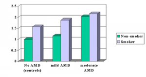
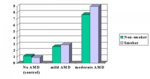
Particularly, one such study is the one by Dr. Johanna M. Seddon and co-workers published in the Feb. 11, 2004 issue of the Journal of the American Medical Association. Almost 1000 patients with various degrees of age-related degrees of blindness from the Age-Related Eye Disease Study (AREDS) were classified by the degree of their macular degeneration. As an illustration, I have produced the bar graphs below based on these studies.

Details of AMD in relation to CRP

For one thing, the researchers defined four groups, namely those with no AMD who served as controls. The second group were those with mild AMD, the third group those with moderate AMD. And the fourth group were those with severe AMD who were legally blind. Specifically, they suspected that an inflammatory marker in the blood stream of these patients, called C-reactive protein (CRP), might be present in the more severe cases of blindness when compared to the control group who did not have any inflammatory changes in the macula. Indeed, the bar graphs below show exactly what the test results indicated. Another key point, they also found that smokers (blue bars) tended to have slightly worse blood tests in terms of CRP (more inflammatory substances circulating in the system) within the same severity category of the age-related eye changes.

CRP (mg/L) Levels in Various Degrees of Severity of Age-related Macular Degeneration (AMD)

Inflammatory Marker Linked To Blindness

Inflammatory Marker Linked To Blindness

Risk of AMD depends on value of CRP

The investigators studied the risk for the highest percentile of the CRP tests within various subgroups of AMD. They found several differences as shown in the next table. First there was a low probability to develop AMD in a person with a normal looking macula. The investigators took this risk as the 1.0 point for comparison. In contrast a person with a normal looking macula who smokes has a 1.5-fold risk of developing AMD later. Patients with a moderate degree of AMD have about a 2-fold risk of getting a severe degree of AMD. This is true for smokers and non-smokers. Once the inflammatory cycle has started, the process of causing a moderate degree of AMD is so strong. This means that the effect of smoking will not add that much in comparison.

This is the first study of this kind that established that CRP is useful as a screening for the risk to develop AMD. Physicians already use CRP  as a test for monitoring progress in rheumatoid arthritis or to monitor for the risk of developing a heart attack or stroke.

AMD risk studied by another research group

Another study by Dr. Johanna M. Seddon and co-workers was published recently in the Archives of Ophthalmology. 261 people aged 60 years and older with established AMD were followed for 4.6 years and checked for deterioration. 101 patients had deterioration of their AMD.

Risk of Developing Age-Related Macular Degeneration (AMD) in Highest CRP Percentile
 Inflammatory Marker Linked To Blindness1

Omega-3 fatty acids protect against AMD

The authors analyzed the patients’ diet habits and found that increased fat intake was a high risk factor for deteriorating AMD. Both vegetable and animal fat had a 2-to 3-fold increased risk for deterioration of the AMD to a more severe stage (legal blindness). Fish, omega-3 fatty acid and nuts had a protective effect, but only when omega-6 fatty acid (linoleic acid) intake was low in the same group. The studies showed that the risk of age-related blindness was reduced by 40% when patients ate nuts at least once per week. The authors concluded that a “fat conscious diet” would be good for “maintaining good eye health” and at the same time be beneficial for prevention of heart attacks and strokes.

The authors will do further studies to investigate potential ways of helping patients with AMD and to understand the mechanisms of the disease process better.

References

1. JAMA 2004;291:704-710  2. Arch Ophthalmol – 01-DEC-2003; 121(12): 1728-37
Feb
01
2004

Cinnamon A Natural Insulin Booster For Diabetics

In a recent edition of the medical journal Diabetes Care an interesting article appeared regarding the healing effects of the spice cinnamon. A medical research team in Pakistan (Dr. Khan et al.) in collaberation with a U.S. research team divided a group of 60 comparable diabetics (males and females) in the age range of 45 to 55 and fed one half different concentrations of cinnamon while the other half served as a placebo control. There were three different concentrations of capsules of cinnamon given: 1g, 3 g and 6 g. The placebo control group got capsules with inert material. Here are the results:

The placebo control group showed no change in blood values. The effect documented in this table was achieved after 40 days of cinnamon exposure and was “washed out” after 20 days. Other experiments had found that the substance MHCP (methylhydroxychalcone polymer) is the active ingredient in cinnamon that stimulates insulin and also acts on insulin receptors similar to insulin.

Cinnamon A Natural Insulin Booster For Diabetics

Cinnamon A Natural Insulin Booster For Diabetics

Dr. Richard A. Anderson and his colleagues at the Human Nutrition Research Center of the U.S. Department of Agriculture had already published a number of medical papers on the effects of cinnamon. He was the co-author of this study from the Department of Human Nutrition, NWFP Agricultural University of Peshawar, Pakistan.

Effect of cinnamon on blood values of diabetics
Blood component
investigated:
% Reduction
of blood test:
Blood sugar
level

18-29%
Triglycerides (blood
fat value)
23-30%
LDL cholesterol
(damaging cholesterol)
7-27%
Total cholesterol 12-26%
HDL cholesterol
(protective cholesterol)
unchanged

The interesting observation here is that several cardiovascular risk factors (blood sugar, triglycerides and LDL cholesterol) are simultaneously being reduced with something as simple as cinnamon powder. The authors stated that the cinnamon oil is not effective, only the cinnamon powder or a cinnamon stick dipped into tea (the water soluble component of cinnamon or MHCP). Dr. Anderson also warned not to make the mistake to eat more cinnamon buns or apple pie as there would be unhealthy amounts of sugar, starch and fat added. He suggested that the best to do instead would be to simply sprinkle cinnamon powder over whatever you are presently eating, as this will reduce the risk of getting diabetes or will reduce the risk of a heart attack in diabetics.

This article based on: “Cinnamon improves glucose and lipids of people with type 2 diabetes.” Diabetes Care – 01-DEC-2003; 26(12): 3215-8.

Here is a link to diabetes mellitus (type 2 diabetes).

Last edited December 8, 2012


Incoming search terms:

Dec
01
2003

Fat Cells Secrete Hormones That Raise Blood Pressure

Fat cells are known to secrete a number of substances that affect the lining of the arteries and that are also known to be associated with the metabolic syndrome. One of the observations that physicians were aware of for some time is that aldosterone, a hormone from the adrenal glands, is often elevated in patients with high blood pressure and obesity or people who are overweight.

Dr. Ehrhart-Bornstein and her group from the University Medical Center, Heinrich Heine University of Düsseldorf in Germany investigated this interaction between fat cell metabolites and the cells of the adrenal cortex in more detail. They used a tissue culture model with human adrenocortical cells (NCI-H295R). To their surprise they found two separate hormone factors that were produced by fat cells and that showed in the tissue culture system a 7-fold increase in aldosterone hormone release. As aldosterone is a mineralocorticoid hormone they called these new releasing hormones mineralocorticoid-releasing factors. Further characterization of these factors demonstrated that one was of a higher molecular structure and was heat-sensitive, the other one was smaller in size and was more heat resistant. Each factor alone lost much of the aldosterone releasing activity, but when recombined they had 93% of the original action. Synthesis of messenger RNA inside the adrenocortical cells was stimulated by a factor of 10-fold from the action of the mineralocorticoid-releasing factors. Other hormones were also somewhat stimulated such as release of cortisol by a 3-fold increase and DHEA by a 1.5-fold increase. Other known substances from fat cells were entirely ineffective in this testing system.

Fat Cells Secrete Hormones That Raise Blood Pressure

Adipose cells secreting aldosterone releasing factor

When asked how this new research might fit in with the observation that loss of fat through calorie restriction has a beneficial effect on high blood pressure, the authors commented that with less fat storage in fat cells during weight loss the production of mineralocorticoid-releasing factors would go down significantly and aldosterone would be released at a much lower rate thus decreasing blood pressure through the aldosterone/angiotensin/renin mechanism.

Nov. 12, 2003 paper on which this write-up is based: http://www.ncbi.nlm.nih.gov/pmc/articles/PMC283571/

Last edited October 26, 2014

Oct
02
2003

Heart Scan Saves Lives In Diabetics

A simple new nuclear perfusion study of the heart when applied to healthy appearing diabetics (adult onset or “type 2 diabetics”) showed silent hardening of the coronary arteries in 21.6%. This large study of an American medical team was recently presented at the 18th Congress of the International Diabetes Federation in Paris/France. Dr. F. Wackers, professor of medicine from Yale University school of medicine and one of the lead investigators, explained that 1,124 patients with diabetes in the age range of 55 to 75 years who were all thought to not have any heart blood vessel disease, either had nuclear perfusion studies performed and a control group did not.

As indicated above to the surprise of the investigators 113 patients of 522 (=21.6%) had positive heart scans showing perfusion difficulties of the heart muscle. Further testing with other methods revealed that 73% indeed had perfusion defects and 27% had other heart disease, electrocardiogram abnormalities and other heart dysfunctions. Conventional assessment tools such as a smoking history, determination of degree of obesity, blood pressure,kidney disease , high blood lipid levels, high C-reactive protein levels, the diabetes test hemoglobin A1C or homocysteine levels in the blood were also assessed. However, these conventional tests did not help in predicting that these patients would have developed perfusion defects in their heart muscle. This was due to hidden narrowing of the heart blood vessels (=coronary arteries) and this affected the supply of nutrients and oxygen to the heart even though these patients were completely symptom free at the beginning of the trial.

Heart Scan Saves Lives In Diabetics

Heart vessels and nuclear scan

Dr. Vivian Fonsega, a professor of medicine and pharmacology at Tulane University in New Orleans and co-researcher of the team, added that after a follow-up of 1 year those who had normal initial nuclear perfusion studies of the heart only 1% developed serious heart disease. These control patients who have now been followed for 3 years overall remained very healthy. In other words a normal (called “negative”) nuclear perfusion test in diabetics predicts a better longterm outcome than a positive perfusion test.

With this heart scan the cardiologist can identfy the high risk group among diabetics and can subsequently concetrate on doing something actively about the identified diseased heart blood vessel(=”coronary artery”) disease. Identified narrowing in the coronary arteries (“stenotic arterial lesions”) can be overcome by prying them open and placing heart stents across the affected section utilizing catheters (angiography). In other cases heart bypass surgery can be done by the heart surgeon to improve the perfusion of the heart muscle. The researchers stressed that those diabetics at risk can be identified with this test and the life expectancy of this high risk group of patients can be significantly prolonged. The study will continue for several more years so that the longterm results of any intervention can be measured when compared to controls.

Based on The Medical Post (Sept. 23, 2003 ): p. 55.

Here is a link to a chapter on diabetes and here is a link to heart attacks.

Last edited December 9, 2012

Aug
01
2003

Modify Risk Factors For Erectile Dysfunction (ED) In Elderly Men

Erectile dysfunction (ED, impotence) is a subject that is difficult to research because of its personal nature. Very few good studies are available regarding the question as to how common it would be among older men.

A team of medical experts under Dr. Constance G. Bacon from the Harvard School of Public Health and other institutions have investigated this problem in men older than 50 years and published the results in the August 5, 2003 issue of the Annals of Internal Medicine.

31,724 men aged 53 to 90 years were taking part in the Health Professionals Follow-up Study. Since 1986 they had been filling out detailed questionaires biennially. In 2000 detailed questions about sexual function were also included. Erectile dysfunction was defined as “having poor or very poor ability to have and maintain an erection sufficient for intercourse without treatment during the past 3 months”. The investigators found that about 1/3 of the men above the age of 50 had a sexual dysfunction. Such factors as orgasm, ability to have intercourse, sexual desire and overall sexual function were all affected more and more with every year after the age of 50. When this was further analyzed using multivariate analyses an interesting pattern of reasons for this emerged. The following factors were identified to be independent risk factors for the development of erectile dysfunction.

Modify Risk Factors For Erectile Dysfunction (ED) In Elderly Men

Modify Risk Factors For Erectile Dysfunction (ED) In Elderly Men

Each of the factors from this table is an independent risk factor and can be managed separately. For instance, the investigators found that a higher level of physical activity was associated with much less ED. The best group (men with no ED) was found among those who were always conscious about disease prevention and who had none of the conditions listed in this table or other chronic medical conditions. Leanness and physical activity were associated with good sexual functioning in this study.

Risk factors leading to erectile dysfunction (ED)
Symptoms: Comments:
increasing age
aging likely affects the blood supply to the swelling bodies of the penis; it also clamps down on testosterone production of the testicles
smoking accelerates aging and hardening of arteries
diabetes mellitus affects circulation and nerve impulse transmission
stroke
interferes with brain centers of arousal
antidepressant medication anticholinergic side-effect interferes with penile erection
beta-blocker medication reduction of libido (likely at the brain level from sympathetic nerve block)
alcohol consumption alcohol is a nerve poison that interferes with pudendus nerve function (lack of erections)
TV viewing time due to prolonged sitting there is a chronic lack of exercise that leads to nerve conduction and circulatory problems resulting in ED

This summary is based on a paper published in the medical journal of Annals of Internal Medicine 2003;139:161-168 by Dr. Constance G. Bacon and co-workers.

Here is a brief chapter on erectile dysfunction from Dr. Schilling’s web-based free Net Health Book.

Last edited October 26, 2014

Jul
01
2003

Obesity And Metabolic Syndrome

In the June 10, 2003 edition, following page24, of The Medical Post there was a minisymposium on obesity and the metabolic syndrome (also known as the “syndrome of hyperinsulinism”).

Four specialists had a discussion about this topic: Dr. Ehud Ur (endocrinologist, Dalhousie University, Halifax, N.S., Canada), Dr. Robert Dent (Director of the Weight Management Clinic, Ottawa Hospital, Ont.), Dr. Dominique Garrel (Director of Department of Nutrition and endocrinologist, University of Montreal, Quebec), and Dr. Arya Sharma (Prof. of Medicine, McMaster University, Hamilton, Ont.).

Introduction:

Obesity is now a health threat that about 25% of the North American population is suffering from. There is still a lot of discussion what the exact criteria should be, but the Third Report of the National Cholesterol Education Program Expert Panel on Detection, Evaluation, and Treatment of High Blood Cholesterol in Adults (ATP III) has simplified the detection of the metabolic syndrome.

Obesity And Metabolic Syndrome

Obesity And Metabolic Syndrome

The experts agree that when three or more of the criteria mentioned in this table are positive the person would be considered to have metabolic syndrome.

There is a wide age-related variety: in one study only 7% had metabolic syndrome in the age group of 20 to 29. The same study found 40% of study participants had the metabolic syndrome in the age group of 70 years and older. It is thought that too many calories coupled with too little activity over a longer period of time, perhaps coupled in some people with a genetic tendency to develop metabolic syndrome, leads to an accumulation of abdominal (so-called”visceral”) fat.

Because fat cells have their own hormone systems (leptins etc.) there is a change of metabolism including an elevation of the insulin level with associated loss of “insulin sensitivity”. So, the more obese a person becomes, the less effective insulin becomes in transporting blood sugar through cell walls. At the same time the liver metabolism is changing with the good cholesterol (HDL) being less produced and the bad cholesterol (LDL) being overproduced. The liver will produce a different mix of coagulation factors, which leads to a tendency to form clots in the veins of the legs and in the lungs. As the pancreatic capacity for insulin production gets exhausted over a period of time, the patient eventually develops type 2 diabetes mellitus. Due to the risk of the coronary arteries clogging up with the cholesterol changes and the accelerated hardening of arteries from diabetes, the risk for getting severe heart attacks in obese people with the metabolic syndrome when compared to a normal weight population is about 4-fold.

Elements leading to the diagnosis of “metabolic syndrome”
Finding: Comments:
abdominal obesity waist circumference more than 102 cm in men or more than 88 cm in women
elevated triglyceride level level of 150 mg/dl or higher
low HDL cholesterol level under 40 mg/dl in men or under 50 mg/dl in women
elevated blood pressure systolic or diastolic blood pressure exceeding 130/85 mm Hg
high fasting blood glucose level fasting glucose higher than 110 mg/dl

Treatment of metabolic syndrome:

The experts agreed that a reduction of only 5% to 10% of the body weight through a sensible combination of a mild exercise program (e.g. walking 30 to 45 minutes every day) and a calorie reduced food intake will make a significant difference in terms of normalization of the body chemistry. It is my estimate that perhaps 70% to 90% of all cases of obesity and metabolic syndrome can be treated this way.

However, the remaining cases should continue to see their physician and be followed like the doctor would follow someone who has high blood pressure. There are two types of medications available and they have nothing to do with the Phen-Fen diet pills from not too long ago that were found to cause pulmonary hypertension. These new diet pills are fairly safe and show weight loss results provided the patient co-operates with regard to a modified to low fat diet and some degree of regular exercise.

1. Sibutramine (brand name: Meridia) is a specific brain hormone inhibitor in the area where the appetite zone is located (serotonine and norepinephrine reuptake inhibitor). This medication helps the patient by experiencing satiety sooner so that the patient does not feel deprived despite less calorie consumption.

It is the medication of choice for those who tend to eat a lot. Like with other anti-depressants side-effects are a dry mouth, heart rate increases and sleep loss (insomnia).

2. Orlistat (brand name: Xenical) inhibits fat uptake at the level of the gastrointestinal wall (gastrointestinal lipase inhibitor). This leads to an inhibition of fat absorption by about 30%. The patient needs to keep the fat intake down to about 2 oz. (=60 gm) per day. If the patient consumes more fat, the side-effect of orlistat will be flatulence, abdominal cramps and diarrhea. If the patient is on a strict low fat diet, there would not be enough fat in the gut for the medication to be effective.

At this point it is not known how long the patient should be on such weight loss medication, if this was the chosen route. The experts felt that 1 year would be reasonable, but that the patient should be observed by the treating physician and it may be necessary after some intermission to go for another year of therapy all the way attempting to permanently change eating and exercise habits as an ongoing maintenance program.

Here is a link to another reference about the metabolic syndrome (syndrome of insulin resistance).

Last edited December 9, 2012


Jun
01
2003

Genetic Link Found For Bipolar Disorder

A staff psychiatrist at the Dalhousie Medical School in Halifax (Novia Scotia, Canada) has gathered 1100 DNA samples and psychiatric histories from patients with bipolar disorder and family members who do not have this psychiatric disease. Dr. Martin Alda, The Medical Post reports on page 46 of the May 20, 2003 edition, and his medical team were able to identify 4 areas of interest on chromosomes 15, 7, 6 and 21 where molecular markers for bipolar disease were located.

Two additional tools, namely responders to lithium (common bipolar disease stabilizer) and certain ethnic group differences, are being utilized as well. Dr. Alda has already found that unstable genes can be stabilized in the presence of lithium. By studying the genes involved in the expression of bipolar disorder and defining what triggers a depressive response and what triggers a manic episode, the researchers hope to unravel the mysteries that still surround this intriguing disease. Dr. Alda is also studying the connection of diabetes and biploar disease. Patients with biploar disease are 3 times more prone to diabetes than the general population. As these patients (bipolar patients with diabetes) are poor responders to lithium, there is a suggestion that perhaps the newly defined genetic loci are blocked in some way by the hormone changes in diabetics. Further investigations in this direction are planned by the research group.

Genetic Link Found For Bipolar Disorder

Genetic Link Found For Bipolar Disorder

Link to bipolar disorder: http://nethealthbook.com/mental-illness-mental-disorders/mood-disorders/bipolar-disorder/

Link to diabetes:

http://www.nethealthbook.com/articles/hormonalproblems_diabetesmellitus.php

Last edited October 26, 2014

May
01
2003

Allergies, Asthma And Diabetes All Helped By Fish Oil

Cod liver oil was what your grandmother told you to take. It turns out she was right as two studies from Manchester/England and Boston/US have shown. The common denominator are omega-3-fatty acids, which are found in fish oil, cod liver oil, mackerel, salmon and other fish, generally speaking all sea food that feasts on plankton.

1. A prospective study with a cohort of 1100 children from before their birth until their 5th birthday, which will be next year, is being conducted in Manchester/England.

A smaller pilot study with 37 children (4-year-olds from this cohort) was recently analyzed as reported in Denver by Dr. Clare Murray, a pediatric lung specialist from the University of Manchester. The investigators have done detailed diet analyses with the help of the parents. They found that children with severe asthma were taking in a lot less omega-3-fatty acids than a healthy control group. Further analysis showed that the asthmatic group took in a lot of the inflammation provoking omega-6-fatty acids, whereas the control group had a much better balance between these two unsaturated fatty acids. Apparently it is the ratio between omega-6 to omega-3 fatty acids that determines whether the prostaglandin metabolism is switched versus pro-inflammatory (ratio more than 3 to 1) or versus anti-inflammatory (ratio 3 to1 or less). This article can be found in the Medical Post, Vol39, No.17 (page 19), April 29, 2003.

2. Another study is mentioned on the same page of the Medical Post: Dr. Frank Hu from the Harvard School of Public Health is the lead author of a study published in Circulation: Journal of the American Heart Association. 5103 female nurses with established type 2 diabetes have been followed for about 18 years and their medical histories, life styles and eating habits were updated every two years.

Allergies, Asthma And Diabetes All Helped By Fish Oil

Allergies, Asthma And Diabetes All Helped By Fish Oil

In the beginning of the study every patient was free of heart disease and cancer. The big surprise was that eating fish 5 times per week diminished the risk for developing heart disease by 65%. Even the women in the study who ate fish once or twice per week had 40% less heart disease than those who did not eat fish. In addition, fish eaters survived those who were not fish eaters much better (lower mortality). Controls of women without diabetes who ate fish five times per week had also a reduction of heart disease by 35% compared to non fish eating controls. Dr. Hu stated that it is the omega-3 fatty acids in fish that are the active ingredient. They are known to reduce irregular heart beats (arrhythmias) that can lead to sudden death. Omega-3 fatty acids also reduce blood fat levels (triglycerides), clot formation and improve blood vessel function. He also noted that both genders have the same benefit (no difference between male and female), just that the study was done on female nurses.

Comments: For your information the table below shows what foods contain omega-3 and omega-6 fatty acids.

Omega-3 and omega-6 fatty acids in our food
Type of unsaturated fatty acid: Foods that contain this type of unsaturated fatty acid:
omega-3 fatty acid flaxseed oil, walnuts, macadamia nuts, fishoil, canola oil, mackerel, salmon, sardines, tuna and most cold water fish
omega-6-fatty acid corn oil, cotton seed oil, grape seed oil, safflower oil, soybean oil and sunflower oil

In the past 50 years the food industry has changed the ratio of omega-6 to omega-3 fatty acids in many common foods to the point that the ratios are now 12 to 1 and up to 25 to 1. It is cheaper to produce these foods in that manner as they often have a longer shelve life. Read food labels. Inform yourself about omega-3 fatty acids. Take 2 capsules of a high strength, molecularly distilled (to remove PCB’s, mercury and other heavy metals) fish oil once per day and include more fish in your meals. Avoid deep fried foods, as they contain omega-6 fatty acids.

Here are some links explaining this more:

Link about balanced nutrition.

More details about fat and fatty acids.

Last edited October 26, 2014

Jan
01
2003

Framingham Study…Obesity And Smoking Lead To Loss Of Life

The Netherlands Epidemiology and Demography Compression of Morbidity Research Group has published an important medical research paper in the Jan. 7, 2003 edition of the Annals of Internal Medicine (Ann Intern Med 2003;138:24-32).

The lead researcher, Dr. Anna Peeters, explained that the group has revisited the Framingham Heart Study 40 years later and analyzed survival statistics of the group of men and women who enrolled in this longterm study between 1948 and 1951. The population at the beginning of the study was aged between 30 and 49. The snap shot, after 40 years had elapsed, is the subject of this analysis. In order to make it easier to understand, I have tabulated the data as seen below.

This study shows that life style choices do matter: being overweight shortens your life by 3 years on average, being obese shortens it by 6 to 7 years.

Add the risk of smoking, and you end up shortening your life by 7 years in the case of being overweight (4 years more than without smoking) or more than 13 years, if you are obese.

Framingham Study...Obesity And Smoking Lead To Loss Of Life

Framingham Study…Obesity And Smoking Lead To Loss Of Life

This study was based on 3,457 participants who 40 years ago had a life expectancy of 85 years, if they were in the normal weight category and did not smoke.

The death rates were much higher than the researchers expected. The researchers from the Erasmus Medical Center in Rotterdam stated as a conclusion: ” just think about two things: Don’t get fat and don’t smoke”.

Years of life lost 40 years after Framingham Heart Study Was Started:
Non-smokers: Smokers:
  Male Female   Male Female
overweight *: 3.1 3.3 overweight*: 6.7 7.2
obese** : 5.8 7.1 obese ** : 13.7 13.3
* overweight:BMI25 to 29.9 ** obese:BMI = 30 and higher

Here are some links to my Internet based Healthbook regarding risks for heart attacks and strokes:

Heart disease: http://www.nethealthbook.com/articles/cardiovasculardisease_heartdisease.php

Strokes: http://www.nethealthbook.com/articles/cardiovasculardisease_strokeandcerebralaneurysm.php

Two things will lead to a normal weight (as you likely have heard before):

Proper nutrition…

http://www.nethealthbook.com/articles/nutrition.php

…and proper exercise (fitness):

http://www.nethealthbook.com/articles/fitness.php

Last edited December 10, 2012


Jan
01
2003

The Liverpool Eye Study…How Often The Eye Sight Of Patients With Diabetes Should Be Examined

In the January 18, 2003 issue of the Lancet (Lancet 2003; 361:195-200) Dr. Dr. Naveed Younis and his collegues published a study from the Royal Liverpool University Hospital, U.K. where diabetic patients underwent thorough eye examinations in regular intervals for 5 years. The question of this study was whether it mattered at what interval patients would be examined with regard to their eye-sight, so that perhaps blindness could be postponed or avoided through early interventions.

At the baseline of the study a special eye-photography method (three-field mydriatic photography) was used to document the blood vessel changes in the back of the eye. This helps the physician to assess whether or not there is a degree of retinopathy (blood vessel damage from diabetes) of the eyes or not. The investigators defined three groups:

For group one (no eye damage in the beginning of the study) there was a retinopathy rate of 0.3% at the end of the first year. Group two (moderate retinopathy in the beginning) showed a worsening of the retinopathy at the end of the first year of 5% (threatening blindness). Group three (significant retinopathy in the beginning) showed a rate of 15% of worsening retinopathy (threatening blindness) at the end of one year.

The Liverpool Eye Study...How Often The Eye Sight Of Patients With Diabetes Should Be Examined

The Liverpool Eye Study…How Often The Eye Sight Of Patients With Diabetes Should Be Examined

This shows that not every person with diabetes is equal with respect of having the threat of blindness. The medical investigators found that about 70% of patients fell into the group that did not have serious diabetic retinopathy. However, the other 30% would not do well with simply yearly eye examinations as it is recommended now. Instead the authors of the Liverpool study were able to make practical recommendations as follows:

After a baseline eye examination those who belong into group one would get an eye examination every 3 years. Those with prior moderate retinopathy (group2) on the initial baseline examination would get examinations at yearly intervals (until the degree of retinopathy worsens). Group 3 with significant retinopathy at the beginning would, however, be examined every 4 months (new recommendation) so that blindness hopefully could be avoided or significantly postponed through interventional therapies.

The following link is telling you about hardening of the artieries (arteriosclerosis) and how diabetes accelerates this process:

The following site is about the metabolic effects of diabetes on the body and in particular also about the danger of diabetic retinopathy:

Diabetes: http://www.nethealthbook.com/articles/hormonalproblems_diabetesmellitus.php

Last edited December 10, 2012