Oct
14
2017

A New Genetic Marker For Alzheimer’s

“A new genetic marker for Alzheimer’s”; so reported a study dated August 11, 2017. Most of all, they found that a genetic marker, TOMM40 was stronger than the established genetic marker APOE4. It seems like the older studies overlooked the importance of the new TOMM40 genetic marker. This new marker may have been present at the same time as APOE4.

Details of study regarding a new genetic marker for Alzheimer’s

The APOE4 is especially relevant for the formation of lipoproteins. APOE4 showed a strong association with the formation of amyloid plaque. This is located in the brain areas where Alzheimer’s disease developed. Therefore the thinking in the past was that APOE4 would be the culprit behind memory loss and Alzheimer’s disease. In contrast, the new study shows evidence that the TOMM40 genetic marker is the gene that actually orchestrates the development of Alzheimer’s disease. Thalida Em Arpawong is a postdoctoral fellow at the University of Southern California (USC) Dornsife College. She conducted research about the TOMM40 marker. Her supervisor was senior investigator Carol A. Prescott, who is a professor of psychology at the USC Dornsife College. She co-published the paper.

More info about the study involving a new genetic marker for Alzheimer’s

Professor Prescott used two verbal memory test results. They were the United States Health and Retirement Survey (HRS) and the English Longitudinal Study of Ageing (ELSA). In these tests immediate recall was compared to delayed recall 5 minutes later. Alzheimer’s patients have problems with short term memory recall.  In total the study examined 20,650 HRS participants and 11,391 ELSA participants. Their age was 50 years and above since this is the typical age for the onset of Alzheimer’s disease. Genetic data was part of the examination in 7,486 HRS participants and 6,898 ELSA participants. The scientists looked at 1.2 million genetic variations of the human genome to fit the memory loss. In conclusion, only one gene area, TOMM40 showed a strong association with decline in immediate and delayed memory recall.

Hence professor Carol A. Prescott summarized the findings: “The results from this study…raise the question of how many findings in other studies show an association with APOE4 that may in fact be due to TOMM40 or a combination of TOMM40 and APOE4.”

Possible future clues from a trial using TOMM40 marker

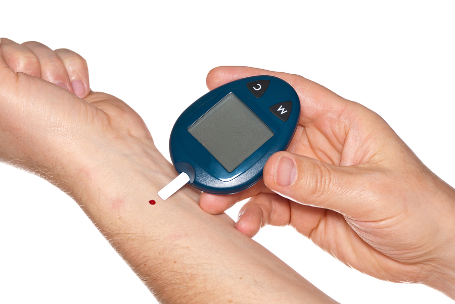
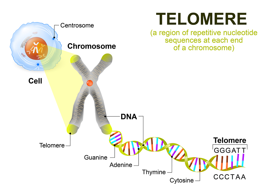
A review paper points out the start of a new trial, called TOMMOROW. The review paper points out that the location of APOE and TOMM40 are on chromosome 19 in very close proximity. Pioglitazone is a drug that controls diabetes. Patients tolerate it well. It is used in the TOMMORROW trial. As this review paper states the TOMM40 gene is responsible for the outer mitochondrion membrane. Consequently the paper states: the “outer mitochondrial membrane channel through which peptides and proteins travel into mitochondria to support mitochondrial function and biogenesis” is the key for understanding Alzheimer’s disease. Because pioglitazone is a drug that induces mitochondrial doubling the researchers hope that it will help Alzheimer’s patients.  It will probably be interesting to follow the phase 3 trial TOMMORROW, where research will observe the delay in onset of minimal cognitive impairment.

A New Genetic Marker For Alzheimer’s

A New Genetic Marker For Alzheimer’s

Conclusion

Research has found a new genetic marker for Alzheimer’s, TOMM40 that identifies a higher risk of getting Alzheimer’s disease. Its location is close to the marker APOE on chromosome 19. It appears that TOMM40 may be more reliable in identifying patients at risk for Alzheimer’s disease than the older APOE marker. As a result research has started a new phase 3 trial, called TOMMORROW. This will tell whether or not Pioglitazone, a diabetic drug maybe useful in delaying Alzheimer’s disease in high-risk patients.

ADDENDUM: This link shows that the TOMMORROW trial had to be shut down, because the drug Pioglitazone had no effect on slowing down the onset of Alzheimer’s disease.

Sep
30
2017

Parkinson’s Disease May Be Stopped

Parkinson’s disease is common in the US; new research shows that the use of an old anti-depression medication can stopParkinson’s disease The use of nortriptyline, a 50-year old antidepressant has shown to normalize a nerve cell protein. In rats nortriptyline dissolved toxic alpha-synuclein clusters in brain cells. These toxic protein clusters seem to be happening in the brain of Parkinson’s disease patients also. It is the protein by the name of alpha-synuclein that research first found in rats to cause the toxic protein clusters in nerve cells of the substantia nigra, a part of the brain stem.

But nortriptyline was able to normalize the concentration of the protein. In preliminary studies in humans the investigators found that there was a significant improvement of Parkinson’s disease with the use of nortriptyline.

Placebo controlled trial with nortriptyline

Now a research team from Michigan State University in Grand Rapids conducted a larger clinical placebo-controlled trial. The lead researcher Collier of the study group found that Parkinson’s patients who received treatment for depression with the tricyclic agent nortriptyline needed less dopamine, the main drug used to treat Parkinson’s disease. This indicated to the researchers that nortriptyline was preserving brain cells that were still making their own dopamine. In rat experiments they could show that it was the dissolving of toxic alpha-synuclein proteins by nortriptyline that was the key to therapeutic success.

Lisa Lapidus, a co-worker on the Michigan State University research team summed up their research: “What we’ve essentially shown is that we are dealing with a drug that the FDA approved already 50 years ago. Patients tolerate the medication relatively well. This could be a much simpler approach to treating the disease itself, not just the symptoms.”

Parkinson’s disease may be stopped also by old diabetes drug

Thomas Foltynie found that the diabetes drug exenatide helps patients with Parkinson’s disease. Dr. Foltynie is a professor of neurology at the University College London and co-author of the study.

Exenatide is an injection drug. When preliminary studies showed that this drug was effective in helping Parkinson’s disease patients lose their problems with walking and balance, a formal study followed.

Professor Foltynie designed a study where 60 people with Parkinson’s disease either got injections of exenatide or placebo injections. Patient exams followed regarding their musculoskeletal system and balance at baseline and every 12 weeks. A score system of 132 points assessed their Parkinson’s disease. After 48 weeks those who had been taking exenatide had a gain of 1 point on that scale while the placebo group dropped 3 points. After 48 weeks the drug administration (exenatide) finished. But after another 12 weeks another scoring and assessment of the Parkinson’s disease symptoms took place. The experimental group on exenatide scored 3.5 points higher than the placebo group. This suggests that exenatide is helping to treat the cause of Parkinson’s disease, not just the symptoms.

Parkinson’s disease may also stop through the use of caffeine

Parkinson’s disease was in the news again because of another study that involved breaking up misfolded alpha-synuclein through caffeine.

Misfolded alpha synuclein forms clumps inside dopamine producing cells in the substantia nigra of the brain stem. Misfolded alpha synuclein acts like a toxin to the dopamine producing cells and eventually these cells die off. This is the brain region that is responsible for making muscle movements smooth and stabilizes balance. The cells that have misfolded alpha synuclein clumps in them also go under the name of “Lewy bodies”.

Dr. Jeremy Lee from the University of Saskatchewan (Saskatoon, Saskatchewan, Canada) has isolated two compounds from coffee. They are called C8-6-I and C8-6-N. They can bind to alpha-synuclein and prevent clumping, which stops the toxic effects on dopamine producing nerve cells. Like with nortriptyline the caffeine effect is a curative approach to Parkinson’s disease.


Parkinson’s Disease May Be Stopped

Parkinson’s Disease May Be Stopped

Conclusion

There is a new therapeutic approach to Parkinson’s disease. Researchers have detected a protein called alpha-synuclein to cause toxic protein clusters in nerve cells of the substantia nigra, a part of the brain stem. When these cells die from the accumulation of these misfolded proteins, patients come down with Parkinson’s disease. But three different methods of treatment can improve Parkinson’s disease by dissolving the protein alpha-synuclein.

  1. Nortriptyline was able to normalize the concentration of the protein. In preliminary studies in humans the investigators found that there was a significant improvement of Parkinson’s disease with the use of nortriptyline.
  2. Exenatide, an injection drug for diabetes, has been described to help Parkinson’s patients get better.
  3. Caffeine can also dissolve misfolded alpha synuclein (two compounds from coffee called C8-6-I and C8-6-N). This helps patients with Parkinson’s disease to stabilize.

This is only the beginning of a new approach to Parkinson’s disease and an attempt to cure the disease by dissolving the underlying mechanism. So far the drugs that are in use for Parkinson’s disease are only attempting to stimulate dopamine producing nerve cells to produce more dopamine. But the underlying pathology of accumulating misfolded alpha-synuclein clumps is not yet in the treatment protocol. The new research is different, as it takes this into account in an attempt to prevent the condition.

Incoming search terms:

Sep
23
2017

Close Diabetes Control Prolongs Life


A 20-year study showed that close diabetes control prolongs life. A study divided 160 people with diabetes into two groups. The one group continued to get standard care. Yet the other group received a multi targeted, aggressive treatment protocol. As a result after 20 years the group with the intensive treatment protocol lived 7.9 years longer than the group with the standard treatment.

Dr. Oluf Pederson was the senior investigator of the physician team that followed the diabetes group. He said that they concentrated on a number of known adverse factors and treated them aggressively. These factors were first of all high blood glucose values and clotting risks, also high blood pressure and high triglycerides and in addition cholesterol values. Behavior modification was the therapeutic method to get people with risk factors to exercise more, adopt a healthy diet and stop smoking. Medication in select cases also played a role.

More details about the study

The intervention of intensive treatment lasted 8 years. After that the patients were still in a follow-up study for 13 years. At the beginning of the study patients were on average 55 years old and were borderline obese.

The investigation team screened for complications of diabetes. This included screening for kidney disease, heart disease and blindness. Dr. Joel Zonszein, the director of the New York Clinical Diabetes Center at Montefiore Medical Center said: ”These results are impressive and most patients do not receive the correct treatment, according to national surveys.”

Other studies about diabetes  

Foreign studies

Study from Croatia
  • Another study from Croatia involved 200 patients. It concentrated on patients who did not respond to metformin. Physicians used alternative treatment modalities, and they observed and measured blood sugars and hemoglobin A1C in the following 6 months. The study concluded that those patients who received aggressive treatment of their condition did better than those who did not receive the same vigorous approach.
Study from Japan
  • This Japanese study documented that female patients with type-2 diabetes developed kidney damage earlier than their male counterparts.  Consequently, the investigators pointed out how important it is to treat diabetes aggressively to avoid kidney damage.
Study from Singapore
  • This 2016 study from Singapore analyzed retroactively the impact of diabetes on the long-term survival after coronary bypass grafting (CABG).  5720 consecutive patients had their isolated first CABG surgery between 1982 and 1999. The mean follow-up was 13 years. 34.6% of the patients had diabetes, 51% had high blood pressure and 46.6% had elevated blood lipids. The initial mortality after the CABG surgery was 2.4% in the diabetic group and 1.8% in the non-diabetic group. 20-year survival rates following CABG surgery were 30.9% in diabetics and 49.2% in the non-diabetics, an 18.3% difference. The 20-year freedom from cardiac mortality rates was 56% in diabetics and 68.4% in non-diabetics. Other risk factors that led to cardiac mortality were the following: female gender (1.43-fold risk), diabetes (1.51-fold risk), previous heart attack (1.54-fold risk) and a low left ventricular ejection fraction of less than 35% (2.6-fold risk). The conclusion from this study was that long-term survival in diabetics following CABG surgery was much lower than that of non-diabetic controls. Hence the key to improving long-term survival for diabetics is to treat comorbidities like high blood pressure and elevated lipids aggressively as well as getting blood sugars and hemoglobin A1C values under control.

US studies

  • In this US study 558 youth (age less than 21) between February 2012 to July 2015 received follow-up. Between 40% and 50% of these diabetics needed insulin to improve their diabetes. Unfortunately their diabetes showed poor control, as their high hemoglobin A1C values indicated. Median HbA1C was 6.7%, 8.5%, 9.6%, and 9.7% in those with disease duration less than 1 year, 1-2 years, 2-3 years and less than 4  In other words, the longer the young patients had diabetes, the less seriously they took their treatment. Only 33% treated their high blood pressure and only 11% their elevated blood lipids. Microalbuminuria, an indicator of diabetic kidney disease, and non-alcoholic fatty liver disease were present in 5% to 6% of these young diabetic patients. The authors came to the conclusion that there were serious gaps in treating these young diabetics. Further follow-up data of the same group of patients in the coming years will provide further data. In conclusion, the new hemoglobin A1C ranges of 3.8% to 4.9% as the new normal range explains why these youths who do not treat their diabetes properly are at high risk to develop complications from their poorly controlled diabetes.
Heart attacks and erectile dysfunction
  • Heart attacks are more common among patients with uncontrolled diabetes. This US study classified diabetics according to the tightness of their diabetes control. Researchers found examining 606 men and 606 women with diabetes that they could reduce their risk of a heart attack, if they controlled smoking, glycated hemoglobin (hemoglobin A1C), systolic blood pressure, and total and high-density lipoprotein cholesterol. The control of all these risk factors could contribute to the prevention of heart attacks. 35% of men and 45% of women could prevent having a heart attack. A laxer control still would prevent 36% of heart attacks in men and 38% in women. A very aggressive diabetes control could prevent 51% of heart attacks in men and 61% in women. Most noteworthy: close diabetes control prolongs life.
  • Erectile dysfunction (ED) is a big problem among diabetic men. This study from Seattle shows the investigation of 136, 306 men with erectile dysfunction. 19, 236 of these men had diabetes prior to their ED problem. Over a two-year observation period diabetic men had much worse ED problems. As a result they needed to receive secondary line treatments  like penile suppositories or injectables. Others needed tertiary treatments like penile prostheses. In those whose diabetes control was good, oral agents as first-line therapies were usually sufficient.
More studies about risks and benefits of lifestyle
  • Middle-aged women with diabetes have a 4- to 5-fold higher risk for developing heart attacks while men do not show such a higher risk. It is probably particularly important for women to control diabetes when they are diagnosed with it to reduce the risk of coming down with a heart attack.
  • In 2011 Taylor from Newcastle University showed in a group of diabetes patients that he could cure diabetes permanently with an extremely low calorie diet. The trial was simple: he took overweight or obese patients with diabetes and put them on a starvation diet of 600-700 calories per day for 8 weeks. Consequently 43% of diabetic patients received a permanent cure of their diabetes. More info: http://nethealthbook.com/news/cure-diabetes-permanently/


Close Diabetes Control Prolongs Life

Close Diabetes Control Prolongs Life

Conclusion

The new hemoglobin A1C ranges that are desirable are between 3.8% to 4.9%. When diabetics bring their hemoglobin A1C level into this range, they do not get complications from their previously poorly controlled diabetes. Close diabetes control prolongs life. But as can be seen from a brief review of the literature physicians tend to be lax, patients are lax, and diabetes is often not well controlled. This leads to erectile dysfunction in males, to heart attacks and kidney failure in both sexes. Blindness and painful diabetic neuropathy are also common complications of poorly controlled diabetes. Amputations from clogged arteries are also among the complications. “Close diabetes control prolongs life” is the new mantra that everybody with diabetes needs to follow.

Lifestyle changes control diabetes and prolong life

As stated above Dr. Taylor from Great Britain has shown that a brief 600 to 700 calorie diet can cure 43% of diabetic patients permanently. Quit smoking, bring the glycated hemoglobin (hemoglobin A1C) into the normal range, control your systolic blood pressure as well as your total and high-density lipoprotein cholesterol. Do all these things, exercise regularly, and your diabetes will be well controlled. Remember: close diabetes control prolongs life!

Sep
02
2017

Resveratrol Effective In Humans

Resveratrol is a powerful antioxidant; but is resveratrol effective in humans?

  1. Quack watch says: don’t buy into the hype that resveratrol is effective in humans.
  2. WebMD claims that there would not be enough medical evidence to say that the average person should supplement with resveratrol to receive benefits.

Despite these recommendations the following evidence supports that resveratrol is indeed effective in humans.

Resveratrol effective in humans: high blood pressure patients

First of all, a 2017 study of high blood pressure patients examined resveratrol supplementation with two groups, 46 stage 1 hypertension patients and 51 stage 2 hypertension patients. Stage 1 hypertension had a systolic blood pressure of 140–159 mmHg and a diastolic blood pressure of 90–99 mmHg. Stage 2 hypertension had a systolic blood pressure of 160–179 mmHg and a diastolic blood pressure of 100–109 mmHg. Analysts divided both stage 1 and 2 subgroups into two groups, one receiving regular antihypertensive medication, and the other group receiving regular antihypertensive medication plus Evelor. Evelor is a micronized formulation of resveratrol. The trial lasted two years.

Blood pressure lowering effect of resveratrol

The purpose of the trial was to determine the effect of resveratrol.  added to the regular antihypertensive medication (or not) to see whether it had blood pressure lowering effects. The interesting result showed that the resveratrol addition was sufficient to bring the blood pressure down to normal levels with only one antihypertensive drug. The control group without resveratrol needed two or three drugs to get the blood pressure under control. In addition, liver function tests showed that resveratrol normalized negative side effects of the antihypertensive drug on the liver. Both liver enzymes, glutamate-pyruvate transaminase (SGPT) and gammaglutamyl transferase (Gamma-GT) were normal in the resveratrol group.

Resveratrol effective in humans: diabetes patients

Diabetes patients can get help with resveratrol. Resveratrol, the bioflavonoid from red  wine is a powerful anti-inflammatory. This antioxidant has several other effects, which make it challenging to measure each effect by itself. Another group of investigators managed to simultaneously measure these effects. They found that resveratrol lowered the C-reactive protein by 26% and tumor necrosis factor-alpha by 19.8%. Resveratrol also decreased fasting blood sugar and insulin; in addition it reduced hemoglobin A1C and insulin resistance. The recommended daily dose of resveratrol was 1000 to 5000 mg.

Resveratrol effective in humans: improves bone density

Furthermore, resveratrol improves bone density in men: 66 middle-aged obese men with an average age of 49.3 years and a mean body mass index of 33.7 were recruited for this randomized, double blind, placebo-controlled trial. The purpose was to study whether there would be changes in bone turnover markers (LDH, an enzyme involved in bone turnover), but also whether bone mineral density (BMD) would increase. The researchers gave resveratrol to a high group (1000 mg per day), a low group (150 mg) and the third group received a placebo (fake pills). The end point was an elevation of the bone alkaline phosphatase (BAP). The investigators measured this in the beginning of the study and at 4, 8 and 16 weeks.

Difference between high and low dose resveratrol

The high group of resveratrol had a 16% increase of the BAP throughout the study and a 2.6% in lumbar spine bone density (measured by a trabecular volumetric method). The low resveratrol group showed no bone restoring effect. MJ Ornstrup, MD, the lead investigator said that this was the first time that a clinical team has proven that resveratrol can serve as an anti-osteoporosis drug in humans. She added that resveratrol appears to stimulate bone-forming cells within the body.

Resveratrol effective in humans: anti-aging effects

Finally, the Nurses’ Health Study showed that both a Mediterranean diet and resveratrol can elongate telomeres.

The fact that you can have a longer life with a Mediterranean diet is common knowledge for some time. But now a study has shown that the reason for a longer life is the fact that telomeres get elongated from the Mediterranean diet. Telomeres are the caps at the end of chromosomes, and they get shorter with each cell division. This is the normal aging process.

Important information from the Nurses’ Health Study 

The finding of elongated telomeres comes from the ongoing Nurses’ Health Study that started enrolling subjects in 1976. At that time 121 700 nurses from 11 states enrolled in the study. In 1980 participants filled in diet sheets to determine who was adhering to a Mediterranean diet. The researchers accepted 4676 middle-aged participants in this study. This diet consists of a combination of vegetables, legumes, fruits, nuts, grains and olive oil. They also consumed fish and lean meats. The control group followed a regular diet. Between 1989 and 1990 blood tests were obtained to measure telomere length in white blood cells. It is known that smoking, stress and inflammation shortens telomeres.

Slowed telomere shortening

The lead author Marta Crous-Bou stated that overall healthy eating was responsible for longer telomeres in comparison to the control group. But the strongest association was in women eating a Mediterranean diet in comparison to the controls. For the best diet adherence score there was a 4.5 year longer life expectancy due to slowed telomere shortening.

Resveratrol lengthens telomeres

Longer telomeres associated with the lowest risk to develop chronic diseases and the highest probability of an increase of the life span. I have reviewed the importance of lifestyle factors in this blog where I pointed out that Dr. Chang found a whole host of factors that can elongate telomeres by stimulating telomerase. Research in humans supports the notion that an increase in physical activity elongates telomeres. So did vitamin C, E and vitamin D3 supplementation, resveratrol, a Mediterranean diet and marine omega-3 fatty acid supplementation. In addition higher fiber intake, bioidentical estrogen and progesterone replacement in aging women and testosterone in aging men, as well as relaxation techniques like yoga and meditation are also elongating telomeres.

Aging is due to shortening of telomeres. Elongation of telomeres by resveratrol leads to prolonged life (or anti-aging).

Resveratrol effective in humans: resveratrol and cancer

In addition, this overview shows, it seems that several mechanisms of action give resveratrol the power to be an anticancer agent. Resveratrol is anti-proliferative and has anti-angiogenesis mechanisms. In addition resveratrol stimulates apoptosis, which is programmed cell death. All these actions together help resveratrol to have anticancer properties. Resveratrol is also useful in combination with other cancer treatments, which improves survival figures. As the link above explains, there is a need for more cancer clinical trials with a variety of cancers and larger patient numbers. Many smaller clinical trials have already been very successful showing efficacy of resveratrol as a chemotherapeutic agent.

Resveratrol is anti-inflammatory

Also, in this 2015 publication about malignancies and resveratrol an overview is given about the use of resveratrol and cancer treatment. It summarizes that the development of cancer is a multifactorial process that involves the 3 stages of initiation, promotion and progression. One of the cancer promoting factors is chronic inflammation. Resveratrol has anti-inflammatory qualities. At this point it is not clear how the animal experiments will translate into the human situation. More clinical observations are necessary.

Resveratrol effective in humans: cardiovascular disease

Resveratrol has beneficial effects on preventing hardening of the arteries, diabetes, various cancers and inflammatory conditions like Crohn’s disease and arthritis. Furthermore,  as this link explains resveratrol also stimulates the antiaging gene SIRT1 by 13-fold. This confirms the anti-aging effect of resveratrol. This 2012 study confirmed that it is resveratrol from red wine that is responsible for the “French paradox” (longer life expectancy despite high saturated fat intake).

Resveratrol effective in humans: polycystic ovarian syndrome 

Similarly, polycystic ovarian syndrome could be significantly healed with resveratrol in a randomized, double blind, placebo-controlled trial. It involved 30 subjects who completed the trial. Each of the subjects received 1500 mg of resveratrol or placebo daily for 3 months. Measurements showed a decrease of serum total testosterone by 23.1% at the end of 3 months in the experimental group versus the placebo group. There was also a decrease of dehydroepiandrosterone sulfate of 22.2%.There was a reduction of the fasting insulin level by 31.8%. At the same time there was an increase of the insulin sensitivity by 66.3%. The authors concluded that resveratrol had significantly reduced ovarian and adrenal gland male hormones (androgens). This may be in part from the drop in insulin levels and the increase of insulin sensitivity.

Resveratrol effective in humans: anti-arteriosclerotic effects in diabetics

Most noteworthy, a double blind, randomized, placebo-controlled study was done on 50 diabetics. Arterial stiffness was determined by the cardio-ankle vascular index (CAVI). The purpose of this study was to determine the effect of resveratrol on the stiffness of arteries in a group of diabetics and compare this to a placebo. Diabetics have premature hardening of the arteries (arteriosclerotic changes). After 12 weeks of taking 100 mg of resveratrol per day there was a significant reduction in arterial stiffness in the experimental group, but not in the placebo group. Blood pressure also decreased by 5 mm mercury (systolic) in the experimental group.

Resveratrol effective in humans: ulcerative colitis patients

Finally, 56 patients with mild to moderate ulcerative colitis received 500 mg of resveratrol or placebo and were observed for 6 weeks. This was a randomized, double blind, placebo-controlled pilot study. The researchers used bowel disease questionnaires to assess the bowel disease activity before and after the treatment. The resveratrol group decreased the disease activity significantly, but it also increased their quality of life. Blood tests showed that this improvement occurred as a result of reducing oxidative stress by resveratrol.

Resveratrol effective in humans: Alzheimer’s disease prevention

Here is a study where 52 Alzheimer’s patients were divided into two groups; one group received 200 mg of resveratrol for a number of weeks, the other group placebo pills. There was a significant improvement in memory tests in the resveratrol group and functional MRI scans showed better functional connectivity in the hippocampi of the subjects. The hippocampus is the seat for short-term memory, which is not functioning normally in Alzheimer’s patients.

Resveratrol Effective In Humans

Resveratrol Effective In Humans

Conclusion

Resveratrol has a long history of showing evidence of improving health. It does so by countering oxidation of LDL cholesterol, which lessens hardening of arteries. This prevents heart attacks and strokes. Resveratrol is also a powerful anti-inflammatory, which helps patients with diabetes, with Crohn’s disease and arthritis. There is even a cancer preventing effect of resveratrol because of anti-proliferative and anti-angiogenesis effects as well as stimulating apoptosis. These combined anticancer properties make resveratrol a chemotherapeutic agent. It is also effective in combination with conventional anticancer drugs.

Resveratrol helps prevent hardening of arteries and cancer

There are enough randomized, double blind, placebo-controlled trials in humans to show that resveratrol is effective in preventing and treating several disease conditions. The medical establishment claims that there would not be enough medical evidence to say that the average person should supplement with resveratrol to receive health benefits. After my review outlined above I come to the opposite conclusion. It is quite clear that resveratrol has several important healing properties. It can improve diabetes; prevent hardening of arteries, lower blood pressure, attack osteoporosis and prevent Alzheimer’s disease. I have been taking 500 mg of resveratrol daily for years. It has not harmed me.

Incoming search terms:

Jul
01
2017

Advanced Glycation End Products (AGEs)

Advanced glycation end products (AGEs) form through cooking food at high temperatures. Sugar molecules react with proteins crosslinking them and changing how they function. It prevents proteins from doing their job. Glycation also causes inflammation, which damages mitochondria, the power packages inside cells that provide the body with energy. Overall AGEs lead to premature aging, which comes from the toxic protein reactions. Advanced glycation end products accumulate as glycated proteins in the tissues of the body. This leads to mitochondrial dysfunction.

Effect of advanced glycation end products (AGEs) on the body

The toxic effects of AGEs frequently occur in the following tissues.

  • The accumulation of AGEs can cause kidney disease and kidney failure (renal failure). In this case the kidneys no longer filter the blood to excrete waste. Hemodialysis may be necessary.
  • AGEs damager joint cartilage, so it can no longer handle stress and joint stiffness sets in. AGEs are now recognized as a major cause of osteoarthritis.
  • Cross-linked proteins from AGEs can cause Alzheimer’s and Parkinson’s disease. Damaged proteins accumulate in brain cells that disable and kill them eventually.
  • Glycation of LDL particles is an important cause of increasing the plaque formation in arteries by LDL. Glycated LDL is much more susceptible to oxidation than regular LDL. Oxidized LDL causes damage to the lining of the arteries and destroys endothelial nitric oxide synthase. This is a critical enzyme that maintains vasodilatation and blood flow. When glycation of LDL has set in, LDL receptors can no longer recognize it. This means that glycated LDL continues to circulate in the bloodstream where it contributes to the atherosclerotic process. It forms a plaque which becomes a reason for heart attacks and strokes. Glycation of LDL is particularly common in patients with diabetes.
  • Glycation of the skin sensitizes the skin to UV light damage. It triggers oxidative stress that increases the risk of skin cancer.
  • Glycation damages our eyes. It causes clouding of the lens (cataracts) and it damages the retina. Macular degeneration can ultimately cause blindness.
  • When glycation affects the discs in the spinal cord, this can cause disc protrusions and disc herniations. Injuries to the nearby spinal nerves can happen causing limping and leg or arm weakness.

Nutrients to counter AGEs

There are nutrients that can slow down the rate of glycation and as a result will halt the aging process.

Benfotiamine

Benfotiamine is a fat-soluble form of the water-soluble vitamin B1 (thiamine). It can reverse glycation in cell cultures and in humans.

As a result the damage to the cells that are lining arteries is reduced. Benfotiamine also counters diabetic neuropathy, retinopathy and nephropathy.

Pyridoxal 5’-phosphate

Pyridoxal 5’-phosphate is a metabolite of vitamin B6. It is similar to benfotiamine in that it counters glycation and dissolves deposited AGEs. It is particularly useful to stop fat and protein glycation. In diabetic patients lipid glycation is often a problem as these authors have shown. Pyridoxal 5’-phosphate traps glucose breakdown products before they become part of glycation reactions.

Carnosine

Carnosine is a dipeptide, made up of the amino acids histidine and beta-alanine. It is found in higher concentration in muscle and brain tissue. Carnosine scavenges for free radicals and prevents AGE formation. This prevents both lipid glycation and protein glycation. This publication states that carnosine can play a role in preventing Alzheimer’s disease. Carnosine prevents protein crosslinking. The result is that tangled protein clumps cannot accumulate and cause Alzheimer’s disease.

Carnosine also reduces blood lipid levels and stabilizes atherosclerotic plaques. This reduces the risk of plaque rupture, which can cause a heart attack or stroke.

Carnosine also has a mitochondria stabilizing function resisting the destructive effects of oxidative stresses.

Luteolin

Many plants contain luteolin, which is a bioflavonoid. It has anti-inflammatory effects and works by suppressing the master inflammatory complex, called NF-kB.  NF-kB triggers the production of multiple cytokines and is the cause of many cancers, chronic diseases, autoimmune diseases and septic shock. Kotanidou et al. did an experiment where they injected mice with Salmonella enteritis toxin, either with or without luteolin protection. Without luteolin only 4.1% of the mice survived on day 7. With luteolin protection 48% were alive on day 7.

Luteolin has been shown to be effective as an anti-inflammatory in the brain, the blood vessel lining, intestines, skin, lungs, bone and gums.

All these four supplements are available in the health food store. They work together and would be recommendable in diabetic patients where glycation is most prominent. But these supplements are also useful for older people who want to slow down the aging process in general.

Nutrients to slow down mitochondrial aging

Glycation causes mitochondrial deterioration and dysfunction. It accelerates aging in every aspect. AGEs (advanced glycation end products) crosslink proteins, lipids, but also damage enzymes and DNA. Glycation causes a slow down of mitochondrial energy production. The end result is a lack of energy and slower repair processes, which all depend on mitochondrial energy production. The following supplements have shown some merit in reversing this process.

Pyrroloquinoline quinone (PQQ)

PPQ is a supplement that is known to produce new mitochondria in cells. This helps the energy metabolism of aging cells to recover.

Taurine

Taurine is an amino acid that occurs abundantly in heart and skeletal muscles cells, brain cells and cells of the retina. These are areas in the body with high metabolic rates that can burn out mitochondria. Taurine regulates enzymes in mitochondria that harvest energy from food substances. In patients who experience accelerated aging, a lack of taurine can produce an energy crisis. But supplementation with taurine can rescue the cells by reducing oxidative stress and restoring the function of mitochondria in cells that are aging. Brain cells were putting out new shoots, called neurites when taurine was given as a supplement. This helps to improve brain connection, and preserves memory and cognition.

R-lipoic acid

R-lipoic acid helps to extract energy from foods and support mitochondrial function. When R-lipoic acid is given to aging animals, their metabolic function improves, the mitochondria become healthier and there are less oxidative stress-inducing byproducts. It protects their liver, heart and brain cells from oxidative stress in their mitochondria. It is becoming known as an energy-giving supplement.

Advanced Glycation End Products (AGEs)

Advanced Glycation End Products (AGEs)

Conclusion

Sugar overconsumption and overcooking food cause advanced glycation end products (AGEs) through lipid and proteins cross-linking. This leads to premature loss of organ function. The mitochondria are also slowed down. This creates premature aging. Fortunately there are a few supplements like benfotiamine, pyridoxal 5’-phosphate, carnosine and luteolin. They protect against glycation. Mitochondria can also be protected by PPQ, taurine and R-lipoic acid. Although we cannot stop the aging process, avoiding sugar and stopping to consume overcooked food, such as barbecued meats and deep fried food is a sensible step in prevention. Aging can slow down significantly with this approach and some supplements.

Incoming search terms:

Jun
24
2017

Lower Blood Sugar Prevents Diabetes

It seems like conventional medicine has ignored for several decades that lower blood sugar prevents diabetes. Medical researchers reevaluated the normal range for blood sugar and hemoglobin A1C, which is a 3 months average of blood sugar values.

In 2016 UCLA researchers reported that 46% of adults in California are either prediabetic or have diabetes.

In contrast 33% of young adults (age 18 to 39) also have prediabetes.

What is worse is the fact that even patients with prediabetes get complications. Normally only patients with diabetes suffer from these. These include kidney disease, retinal problems with loss of vision, neuropathy, hardening of the arteries and cancer.

Key to preventing this from happening is to recognize that prediabetes is already the beginning of diabetes. Not only is it important to prevent diabetes, but prediabetes as well.

Determination of prediabetes and diabetes

The conventional test for diabetes is a fasting blood sugar.

Prediabetes

In the past there was a consensus that patients with prediabetes had a fasting blood sugar between 100 and 125 mg/dL (5.6 to 6.9 mmol/L).

Diabetes

126 mg/dL (7 mmol/L) or higher on two separate tests indicates that you have diabetes.

Glycated hemoglobin (A1C) test

This test gives an average of blood sugar over 2 to 3 months. Physicians thought that a hemoglobin A1C test below 5.7% would be normal, between 5.7 and 6.4 percent they considered it to be prediabetes and at 6.5 or higher on two separate tests meant a diagnosis of diabetes.

Re-evaluating normal ranges to diagnose diabetes and prediabetes

Many researchers have said that the normal values from the guidelines for blood sugar or for glycated hemoglobin A1C are too high. This is the reason why diabetic complications developed even with prediabetes.

At the 22nd Annual World Congress on Anti-Aging Medicine In Las Vegas (Dec. 10-14, 2014) Dr. Piliszek stated that the normal range for hemoglobin A1C is skewed in the medical literature. It should be: 3.8% to 4.9%. This is very important to know for diabetics and any caregiver who looks after diabetes patients. If you consider a hemoglobin A1C of 6.0 as “normal”, the diabetic patient has the risk of dying prematurely of a heart attack or a stroke. According to the new guidelines even a patient whose hemoglobin A1C is 5.5 has diabetes and needs aggressive treatment to prevent complications associated with diabetes. Conventional guidelines would have considered this patient to be normal.

A 1999 study made it clear that patients with a blood sugar of more than 85 mg/dL were at risk of developing diabetes complications. Researchers observed about 2000 patients with fasting blood sugars of more than 85 mg/dL over 22 years. About 40% of them died of heart attacks or strokes! Because of studies like this, physicians demanded the new diabetes guidelines.

The authors concluded that fasting blood glucose in the upper normal range was an independent risk factor of cardiovascular death.

New guidelines

Prediabetes is not a separate diagnosis, but is mild early diabetes, which is reversible with aggressive treatment. Dietary changes (cutting out sugar and refined carbs) are often effective. In some cases the addition of metformin may be required.

The new normal ranges are:

Fasting blood sugar of 85 mg/dL or less is normal.

Hemoglobin A1C of 3.8% to 4.9% is the new normal range.

These values are based on observing patients over a long period of time and seeing whether or not they develop complications from diabetes.

Most noteworthy, uncontrolled diabetes leads to complications like damage to the lining of the arteries in all the key organs. It is the cause for the following conditions: kidney damage (nephropathy), eye damage (retinopathy), brain and nerve damage (neuropathy), as well as heart attacks and strokes (vascular damage).

Certainly, patients often end up with dialysis when kidney failure has set in. Retinopathy causes blindness and neuropathy leads to excruciating pain. Heart attacks and strokes often cause premature death. Those who ingest a high-glycemic diet have a 49% higher risk of getting lung cancer than those with a low-glycemic diet as this link from the MD Anderson Cancer Center showed.

Calorie restriction

A research group found that calorie restriction reduced fasting insulin levels in a group of overweight men and women.

Another study showed that restrained eating patterns lower fasting glucose and postprandial (after meals) glucose. As a result it also improved insulin sensitivity in normal weight individuals.

Some practical hints about diets to treat diabetes

  1. First of all, the obvious fact is that excessive sugar intake is harmful. But in addition a drastic reduction of refined carbs is also needed, as they just turn into sugar within half an hour of ingesting them. Cut out potatoes, pasta, and bread. You may have a slice of rye bread or full grain bread occasionally. This type of diet is called a low-glycemic index diet. Hence, as indicated earlier a study from the MD Anderson Cancer Center has shown that lung cancer is more common the higher the glycemic index is and is also more common in diabetics.
  2. Also, a Mediterranean diet has been shown to be anti-inflammatory. As diabetes and prediabetes are associated with chronic inflammation, it is useful to go on a diet that counters inflammation. Similarly, the DASH diet, which was developed for high blood pressure patients, is also anti-inflammatory. Here are a few examples of snacks that may be helpful.
  3. Finally, include fish and fish oil supplements in your diet. These contain omega-3 fatty acids, which are anti-inflammatory. Another useful piece of advice: eat lots of vegetables and salads as they contain healthy bioflavonoids and antioxidant vitamins. This stabilizes the lining of your arteries.
Lower Blood Sugar Prevents Diabetes

Lower Blood Sugar Prevents Diabetes

Conclusion

The old blood sugar and hemoglobin A1C guidelines need a significant revision. In contrast, new guidelines based on actual measurements and clinical trials that showed no complications of prediabetes on the long term have replaced them.

A fasting blood sugar of 85 mg/dL or less is normal. A hemoglobin A1C of 3.8% to 4.9% is now the new normal range.

Consequently, the doctor needs to be more aggressive about early nutritional intervention and probably include metformin as well to restore insulin sensitivity. It is no longer appropriate to allow complications of diabetes like nephropathy, retinopathy or neuropathy to develop. Unfortunately food manufacturers still overload processed food with sugar. Each patient needs to be vigilant about the food he/she eats. Therefore, low glycemic nutrition is the mantra to follow. Also stick to natural, unprocessed foods instead of the highly processed foods that populate the shelves of the supermarkets.

Jun
03
2017

Fish, The Good And The Bad

I am going to review fish, the good and the bad. Fish can be very nutritious, because it contains a lot of healthy omega-3 fatty acids. But because of pollution it also has various degrees of mercury, PBC’s and other impurities.

I will discuss the good about fish oil first. Later we will learn that wild salmon is one of the best fish to eat, while we should avoid tuna due to mercury pollution.

The good about fish

Omega-3 fatty acids, also called marine oil, is an essential fatty acid. It balances omega-6 fatty acids of which we eat too much. Processed foods are full of omega-6 fatty acids, because they keep a long time on the grocery shelves without turning rancid. But when the omega-6 to omega-3 ratio is getting higher than 3:1 we are experiencing a problem. The body stimulates the arachidonic acid pathway, a metabolic pathway that produces inflammatory substances and arthritis. An old home remedy for arthritis is to use fish oil (cod liver oil). It changes the omega-6 to omega-3 ratio back to more normal levels, which can help arthritis patients. Early stage of arthritis can even heal.

Omega-6 fatty acids cause inflammation

Many processed foods contain only omega-6 fatty acids, because this is the cheapest way to produce them (they are based on vegetable oils). Instead of this you want to eat healthy fats like omega-3 fatty acids contained in nuts and fish. You can also add molecularly distilled, high potency omega-3 fatty acids (purified fish oil) as a supplement to help restore the balance between omega-6 and omega-3 in the food you eat. Corn oil, safflower oil, grape seed oil, soybean oil, cottonseed oil, canola oil and peanut oil contain omega-6 fatty acids. These are the ones that cause inflammation and disease. You must avoid them!

Omega-6 to omega-3 ratio

Compare the metabolism of omega-6 fatty acids with that of omega-3 fatty acids.

The linoleic acid of omega-6 fatty acids metabolizes into arachidonic acid, which causes pro-inflammatory mediators, PGE2 and LTB4 as shown in the metabolism link. On the other hand with omega-3 fatty acids alpha-linolenic acid (ALA) metabolizes into EPA, DHA and the anti-inflammatory mediators PGE3 and LTB5.

It is easily understandable why a surplus of omega-6 fatty acids from processed foods will disbalance the omega-6 to omega-3 ratio. This ratio should be 1:1 to 3:1, but many Americans’ omega-6 to omega-3 ratio is 6:1 to 18:1. Omega-6-fatty acids cause arthritis, heart disease and strokes. Be particularly careful avoiding soybean oil. It has become the most popular oil in the last few decades to foul up the omega-6 to omega-3 ratio. We consume it through processed foods and cooking oils.

Omega-3 supplements

When it comes to balancing omega-3 and omega-6 fatty acids in your diet, be aware that nutritional balancing can help you restore the ideal omega-6 to omega-3 ratio of 1:1 to 3:1. An easy way is to cut out processed foods as much as possible. Supplement with molecularly distilled fish oil capsules to add more omega-3 fatty acids into your food intake. Here is an example of rheumatoid arthritis patients that received omega-3 supplements. After 24 weeks their joint swelling and tenderness decreased significantly.

Rebalancing the omega-6 to omega-3 ratio was able to treat depression as this research showed. This makes you wonder how much depression may be caused by overconsumption of processed food.

Dr. Blatman suggested the following doses of omega-3 supplementation for various purposes:

  • 1 gram/day as supplementation for healthy adults with a good diet
  • 1-3 grams/day for people with cardiovascular disease
  • 5-10 grams/day for patients with an autoimmune disease, with chronic pain or with neuropsychiatric conditions

He mentioned that these doses are empirical, but in his experience this is what really works. Due to quality differences he suggested that you buy fish oil capsules in a health food store. Stay away from discount stores (the quality is the worst) and drug stores.

Other healthy oils are olive oil and coconut oil. They are also useful for cooking.

The bad about fish

1. Mercury and other pollutant

Pollution of the air, soil and rivers is causing accumulation of mercury and other heavy metals in ocean water.

This affects fish that live in the ocean. There is a pecking order of predators with the larger fish feeding on the smaller fish. The bigger the predator fish, the more mercury and other pollutants they accumulate. According to this link the safest seafood is wild salmon, pollock and oysters.

High mercury content of predator fish

Tuna is too high in mercury, so is swordfish, and shark is even worse. I only consume fish from freshwater lakes or rivers, as well as salmon, oysters and shrimp. This way I get the lowest exposure to mercury. Why is mercury bad for you? It is a neurotoxin. It can harm your brain, heart, kidneys, lungs and the immune system. Specific symptoms can include loss of peripheral vision and lack of coordination with balancing problems. There may be impairment of speech and hearing. The key is to avoid mercury exposure.

2. Rancidity of fish oil

Rancid fish oil contains free radicals that attack the lining of the arteries. There would be no point in taking fish oil, if it is rancid and destroyed what you want to protect. When you store fish oil, it can interact with oxygen and form lipid peroxides, which are free radicals. The Council for Responsible Nutrition’s quality standards monitors rancidity in fish oil. Get fish oil that meets or exceeds the Council’s standards. If you refrigerate fish oil, it stays fresh longer.

Managing mercury pollution

Smaller fish low in mercury

The first line of defense is to stick to the smaller fish. They are they prey of the large predator fish. The following fish/mussels belong into the low mercury group (alphabetical order): anchovies, catfish, clam, crab, crawfish, flounder, haddock, herring, mackerel, mullet, oyster, perch, pollock, salmon, sardines, scallops, shrimp, sole, squid, trout and whitefish.

Molecularly distilled omega-3 fatty acid supplements

You may want to supplement your omega-3 fatty acid intake by fish oil capsules. It is important that you choose the more expensive higher potency products. A molecular distillation process that removes mercury, PCB and other heavy metals creates these higher potency products. This way you only get the enriched omega-3 fatty acids in pure form. EPA and DHA in one capsule should be in the 900 mg to 1000 mg range, not less. I take 2 capsules twice per day as a daily supplement. This helps you as indicated above to balance the omega-6 to omega-3 ratio, which cuts down any inflammatory process in you.

More good news about omega-3 fatty acids

Omega-3 fatty acids have multiple anti-inflammatory effects. This helps for treating arthritis, osteoporosis, preventing heart attacks and brain shrinkage. Even depression can be influenced positively when krill oil and fish oil are both taken at the same time. It is best to think about krill oil and omega-3 fatty acids (fish oil) as complementary marine oils having multiple beneficial effects on the body. Studies have shown that arthritis and osteoarthritis improve with krill oil, but also with fish oil. Similarly, heart attacks and strokes are prevented with both krill oil and omega-3 fatty acids. It appears that both oils reduce inflammation in the arteries that is associated with high blood pressure, diabetes, obesity and metabolic syndrome in obese people. C-reactive protein measuring inflammation was reduced by krill oil up to 30% compared to placebo within 30 days. Patients with arthritis had 20% reduction in stiffness and pain.

More on krill oil

Krill oil is well absorbed into the brain and can prevent age-related brain shrinkage, preserve cognitive function and memory, prevent dementia and also possibly depression.

Other health conditions improve on both krill oil and omega-3 fatty acids like osteoporosis (in combination with vitamin K2, vitamin D3 and calcium), a weak immune system, diabetes, high triglyceride levels and cholesterol problems. Both marine oils prevent LDL cholesterol from being oxidized, which helps to prevent atheroma formation and hardening of the arteries. This prevents heart attacks and strokes.

Fish, The Good And The Bad

Fish, The Good And The Bad

Conclusion

Children received cod liver oil in the past to prevent rickets. In the 1960’s Dale Alexander wrote a book called “Arthritis and Common Sense”. Since then medicine has been revolutionized in the late 1990’s by the idea that inflammation in the body is responsible for high blood pressure, diabetes, heart attacks, strokes, arthritis and even Alzheimer’s disease. It is in this area that omega-3 fatty acids are an important supplement as fish oil capsules and krill oil capsules. These supplements can be bought molecularly distilled to be free of mercury and other pollutants.

Anti-inflammatory effect of omega-3 fatty acids

The anti-inflammatory effect of omega-3 fatty acids is a powerful preventative for all these diseases mentioned. It no longer is a question, whether these supplements work. It has become a fact backed up by large studies including mortality statistics. Even the FDA has included seafood into their food recommendations. The key is to rebalance your omega-6 to omega-3 ratio and incorporate marine oils in your diet. Your body will thank you for it with a longer, healthier life.

May
20
2017

Prevention Of Telomere Shortening

Dr. Mark Rosenberg gave a talk on prevention of telomere shortening. This was presented at the 24th Annual World Congress on Anti-Aging Medicine (Dec. 9-11, 2016) in Las Vegas that I attended. The detailed title was: “The Clinical Value of Telomere Testing”.

What are telomeres?

Telomeres are the caps at the end of chromosomes. They are very important in the aging process. Prematurely shortened telomeres are linked closely to all major diseases like cardiovascular disease, cancer, diabetes and more. Telomeres are also a measure of the aging process. Aging occurs due to a decrease of the number of cells in organs and/or because of a lack of functioning of these organs. Telomeres get shortened every time a cell divides. But when the telomeres are used up, there comes a time when cells can no longer divide. These cells become senescent cells or they enter apoptosis (programmed cell death).

The senescent cells can become a problem when they get transformed into cancer cells and their telomeres lengthen again. These cancer cells divide rapidly and this can become the reason why cancer patients to die.

What is the significance of telomeres?

Telomere dysfunction is the first sign that the telomeres are getting shorter in a person compared to the average telomere length in a comparable age group. This is not only important for aging, but also has clinical implications. The shorter telomeres are, the higher the risk for cardiovascular disease. Telomere length also provides prognostic information about the mortality risk (risk of dying) with type 2 diabetes and for many cancers. Many physicians incorporate a telomere blood test into periodic health checks, if the patient can afford it.

Interventions that help telomere length

Here are a number of things we can do to lengthen our telomeres.

  1. Rosenberg mentioned that the strongest effect on telomere lengthening comes from caloric restriction and weight loss. 80 years ago they showed at the Cornell University that rats put on calorie restriction had a 30% increase in their mean and maximum lifespan. Many research papers have confirmed that the same is true in man and that the common denominator is telomere lengthening.
  2. Next are regular physical activity, meditation, reduction of alcohol consumption and stopping to smoke.
  3. Taking antioxidants and omega-3 fatty acids regularly will also lengthen telomeres.
  4. Improving one’s dietary pattern by adopting a Mediterranean type diet that contains cold-pressed, virgin olive oil.
  5. Telomerase activators. Here is some background on the TA-65 telomerase activator, which is based on Chinese medicine. A one year trial was completed with 250 units and 1000 units of TA-65 per day. The lower dose (250 units) showed effective telomere lengthening, while the placebo dose did not. The 1000 unit dose did not show statistical significance.

Should you wish to take TA-65, only take 250 units per day, not more.

Cancer and telomeres

There is a strong correlation between cancer and telomere shortening. When cells are at the brink of dying toward the end of their life cycle the telomeres get shorter and shorter. This is the point where the cells can turn malignant. Certain genetic abnormalities help the malignant transformation, like 11q or 17q deletions or a p53-dependent apoptosis response. Once cancer cells have established themselves they activate telomerase in 85% of cases. In the remaining 15% of cancer cases telomeres are activated through telomerase-independent mechanisms. Here are a few examples.

CLL

CLL stands for chronic lymphocytic leukemia. It is a disease of the aging population. At age 90 people’s bone marrow cells have a telomere length of only 50% of the length at birth. This is the reason that in older age CLL is more common. Researchers observed a population segment and found that the shorter telomeres were, the poorer the overall prognosis and overall survival for CLL was.

Lung cancer

Researchers examined the telomerase activity in patients with non-small cell lung cancer. When telomerase activity was present, the 5-year survival was only 55%. When telomerase activity was absent, the prognosis was 90% survival after 5 years.

Prostate cancer

  1. Prostate cancer risk correlated with telomere shortening in stromal cells. Men with shorter telomere length in stromal cells had a 266% higher risk of death compared to men with normal telomere length.
  2. Another study took blood samples and determined the telomere length in lymphocytes (the immune cells). Those men who came down with prostate cancer within a year after they had their blood sample, had short telomeres. The risk for prostate cancer in these patients was 355% higher than in the prostate cancer negative controls.

Yet another study looked at surgical tissue samples from 596 men that

Underwent surgery for clinically localized prostate cancer. Patients whose samples showed variable telomere lengths in prostate cancer cells and shorter telomeres compared to prostate samples with less variable telomere length and longer telomeres had a much poorer prognosis. They had 8-times the risk to progress to lethal prostate cancer. And they had 14-times the risk of dying from their prostate cancer.

Breast cancer

Breast cancer is diverse and consists of cases whose origins are genetic (BRCA1 and BRCA2), but there are also cases where the cancer is local or has a higher stage. In families with mutated BRCA1 and BRCA2 telomeres are significantly shorter than in spontaneous breast cancer. Increased telomerase activity in breast cancer cases is directly related to how invasive and aggressive the breast cancer is.

  1. In one study researchers analyzed blood leukocytes in 52 patients with breast cancer for telomere length  versus 47 control patients. Average telomere length was significantly shorter in patients with a more advanced stage of breast cancer than in early breast cancer. Mutated HER patients had the shortest telomeres. It follows from this that checking for the HER status and blood telomere testing adds to the knowledge of potential cancer development and prognosis.
  2. In patients with with larger breast tumors, more lymph node metastases and more vascular invasion the researchers found short telomere length of the cancer cells.
  3. More aggressive breast cancer cells have higher telomerase activity. More than 90% of triple negative breast cancers have short telomeres.

CNS disorders and telomeres

Dr. Rosenberg presented evidence for a correlation between shorter telomeres and the development of dementia. But dementias with Lewy bodies and Alzheimer’s disease are also linked to short leukocyte telomeres. The length of blood telomeres predicts how well stroke patients will do and how people with depression will respond to antidepressants.

Cardiovascular disease and telomeres

The renin-angiotensin-aldosterone system controls our blood pressure and keeps it constant. When this system is not stable, our blood pressure shoots up and causes cardiovascular disease. This is tough for the heart, as it has to pump harder against a higher-pressure gradient. A study of 1203 individuals was examining the connection between leukocyte telomere length and renin, aldosterone and angiotensin II activity. It concluded that oxidative stress and inflammatory responses affect the telomere length of leukocytes and that the more stress there is in the renin-angiotensin-aldosterone system, the more cardiovascular disease develops. The conclusion of the study was that the overall cardiovascular stress leads to shortening of leukocyte telomeres.

Prevention Of Telomere Shortening

Prevention Of Telomere Shortening

Conclusion

Telomere length testing from a simple blood test will become a more important test in the future as hopefully the cost comes down (currently about 300$). It can predict the general aging status by comparing a single case to the general telomere length of the public. But it can also predict the cancer risk, risk for mental disease and cognitive deficits (Alzheimer’s disease). In addition your cardiovascular status correlated globally with this test. What are the options for the patient, if the test comes back with short telomeres?

It allows you to change your lifestyle and adopt a healthy diet. You can exercise regularly, take antioxidants and meditate. There are even telomerase activators that are gradually becoming more known. They lengthen the telomeres. The cost of telomerase activators will likely still be a problem for some time. All in all telomere length tests are here to stay, but healthy lifestyle choices are the only tool for effective intervention at this point. This is good news: healthy lifestyle choices like non-smoking, exercise and avoiding non-processed foods are either free or have a reasonable price tag. Telomerase activators are big business and at this point not really affordable!

Incoming search terms:

Apr
01
2017

When Food Causes Inflammation

Dr. Hal Blatman gave a talk about when food causes inflammation. He gave his talk on Dec. 9 at the 24th Annual World Congress on Anti-Aging Medicine (Dec. 9-11, 2016) in Las Vegas that I attended. The original title was “Food, Pain and Dietary Effects of Inflammation”.

Dr. Blatman is the medical director of Blatman Health and Wellness Center, Cincinnati and Batman Medical Services, Manhattan.

General remarks about nutrition

Dr. Blatman pointed out that mistakes of nutrition are often behind chronic diseases and illnesses. The physician’s task is to explain to patients how they can change their food intake to improve inflammation in the body and to allow the body to heal itself.

Hippocrates said 400 BC “Let food be thy medicine and medicine be thy food”.

In this context Dr. Blatman stated that nutrition could exacerbate symptoms or relieve symptoms and there must be rules for good nutrition. If we do not take care of our nutrition, the gut flora composition changes and causes leaky gut syndrome. But if we consume healthy foods all of this improves.

Mathematical formula for when food causes inflammation

To make it easier to understand the impact of food on our health the speaker offered this formula:

G-B+R=P

G = stands for good, beneficial things you can put into your body.

B = bad, toxic things that affect your body negatively.

R = reserves that your body has since birth (minus the amounts you have used up)

P = pain and problems you are going to experience

It is P (pain and other medical problems) what brings the patient to see the doctor. G and B is what the patient can change. When done right, the P value in the formula reduces and the pain or medical problems go away.

Nutritional rules

Dr. Blatman said there are three rules about nutrition.

Rule #1 is to not eat fake or toxic foods

He listed NutraSweet, Splenda, Saccharin, margarine and olestra.

Aspartame

Aspartame experiments on rats showed that it can cause cancer: Dr. Blatman said that aspartame causes multiple myeloma and Hodgkin’s lymphoma in man. Aspartame worsens depression, 10% is metabolized in the liver into methanol, a nerve poison.

Splenda

Splenda (sucralose) originates from sugar. However, several chlorine atoms were inserted into the sugar molecule. It reduces beneficial microflora in the gut. It also interacts with liver enzymes, which interfere with the bioavailability of oral drugs.

Saccharin

Saccharin alters gut bacteria and increases glucose tolerance.

Hydrogenated fat and margarine

Insects don’t eat margarine, mold will not grow on it, and it will not support life. Merchants like it because food does not turn stale on shelves. Hydrogenated fats like margarine are like poisons. They raise the bad LDL cholesterol levels and reduce beneficial HDL cholesterol levels. The prostaglandin balance changes so that inflammation occurs. There is increased evidence of diabetes and the cell membrane composition changes. Proinflammatory cytokines can cause pain in the dorsal root ganglions. It follows from all of this that it is best to cut out all hydrogenated fat and margarines.

Partially hydrogenated vegetable oil

The cell membrane consists of two lipid layers at a specific ratio of omega-6 essential fatty acids and omega-3 essential fatty acids. It also contains triglycerides, phospholipids and protein. Cell membrane absorb nutrients to move into the cell and eliminate waste out of it. The cell membrane needs to remain flexible and within neurons needs to transmit electrical information. The membrane composition is critical for the cell membranes to perform optimally. It is here that the physician has to explain this to the patient. All the fats we eat are the raw material, which will make up our cell membranes. So what fat we eat that day travels into the cell wall that becomes part of it that day. The same process occurs with cell wall repair. If we eat hydrogenated fat that day, it travels into the cell wall.  A membrane with hydrogenated fat will:

  • Not transmit nutrients inside the cell
  • Will not transmit waste out
  • Causes the membrane to lose flexibility
  • In a nerve cell there will be abnormal neuron transmission

If we eat hydrogenated fat, we become like a “genuine GM truck fixed with inferior parts”, so Dr. Blatman. The interesting observation is that it takes 4 months after eliminating hydrogenated oil from the diet to get it out from red blood cells. Be aware that French fries increase pain for 4 months, so why eat them?

Olestra

Olestra, an artificial fat: This fat, Olestra has been developed as an artificial fat and is used in chips. It can cause diarrhea, abdominal cramps and weight gain with long-term use. Olestra belongs into the group of fake/toxic foods. Don’t eat Pringles or chips that are made with this.

Healthy oils

There are two types of essential fatty acids, omega-6 fatty acids and omega-3 fatty acids. Many processed foods contain only omega-6 fatty acids, because this is the cheapest way to produce them (they are based on vegetable oils). Instead you want to eat healthy fats like omega-3 fatty acids contained in nuts and fish. You can also add molecularly distilled, high potency omega-3 fatty acids (purified fish oil) as a supplement to help restore the balance between omega-6 and omega-3 in your food intake. Avoid omega-6 fatty acids from corn oil, safflower oil, grape seed oil, soybean oil, cottonseed oil, canola oil and peanut oil.

Metabolism of omega-6 fatty acids versus omega-3 fatty acids

Compare the metabolism of omega-6 fatty acids with that of omega-3 fatty acids.

The linoleic acid of omega-6 fatty acids gets metabolized into arachidonic acid, which causes pro-inflammatory mediators, PGE2 and LTB4. On the other hand with omega-3 fatty acids alpha-linolenic acid (ALA) is metabolized into EPA, DHA and the anti-inflammatory mediators PGE3 and LTB5.

It is easily understandable why a surplus of omega-6 fatty acids from processed foods will disbalance the omega-6 to omega-3 ratio. This ratio should be 1:1 to 3:1, but many Americans’ omega-6 to omega-3 ratio is 6:1 to 18:1. Omega-6-fatty acids cause arthritis, heart disease and strokes. Be particularly careful in avoiding soybean oil, which is the most popular oil in the last few decades to foul up the omega-6 to omega-3 ratio through processed foods.

Balance of omega-3 and omega-6 fatty acids

When it comes to balancing omega-3 and omega-6 fatty acids in your diet, be aware that nutritional balancing can help you restore the ideal omega-6 to omega-3 ratio of 1:1 to 3:1. An easy way is to cut out processed foods as much as possible. Supplement with molecularly distilled fish oil capsules to add more omega-3 fatty acids into your food intake. Dr. Blatman gave the example of rheumatoid arthritis patients that were put on omega-3 supplements. After 24 weeks their joint swelling and tenderness went down.

Rebalancing the omega-6 to omega-3 ratio was able to treat depression as this research showed. This makes you wonder how much depression may be caused by overconsumption of processed food.

Suggested doses of omega-3 fatty acid supplementation

Dr. Blatman suggested the following doses of omega-3 supplementation for various purposes:

  • 1 gram/day as supplementation for healthy adults with a good diet
  • 1-3 grams/day for people with cardiovascular disease
  • 5-10 grams/day for patients with an autoimmune disease, with chronic pain or with neuropsychiatric conditions

He mentioned that these doses are empirical, but in his opinion definitely help. Due to quality differences he suggested that you buy fish oil capsules in a health food store where the quality is best. Stay away from discount stores (the quality is the worst) and drug stores.

Other healthy oils are olive oil and coconut oil. They are also useful for cooking.

Rule #2 is not to eat inflammatory foods

Our body functions like a luxury car; it needs pure food to function. Anything less leads to inflammation, particularly when you eat sugar and processed foods.

Inflammatory foods are sugar, white flour, fruit juice and white/red potatoes. A medium potato=1/2 cup of sugar! Other problematic foods are wheat grain contained in breads, pasta, cereal and thickeners in soups and sauces.

What is the problem with these foods? They break down the zonulin proteins that are a bridge between the lining cells of the gut.

This leads to an increase of intestinal permeability, and leaky gut syndrome can develop. Inflammatory cytokines from visceral fat add to the gut inflammation, and cardiovascular disease and high blood pressure can develop.

Fried potatoes, in particular the consumption of French fries, have been identified as the cause of inflammatory bowel disorder (IBD). Countries with the highest consumption of French fries have the highest incidence of IBD.

A Mediterranean diet and the DASH diet are anti-inflammatory diets.

Rule #3 is to not disturb the bowel flora

A healthy bowel flora is symbiotic with the body. You achieve this by eating green leafy vegetables. A toxic flora from dysbiotic microbes comes from eating white flour, white sugar and red meat. Red meat leaves residues on which dysbiotic bacteria thrive.

Symbiotic gut bacteria produce vitamin K, cobalamin, pyridoxine, biotin, riboflavin, pantothenic acid and short fatty acids. They also degrade metabolic toxins, prevent pathogens from colonization and they stimulate the immune system to mature.

Dysbiosis occurs when the wrong diet consisting of sodas, white flour, sugar and red meat is over consumed. There are toxins that are produced by the dysbiotic microbes. These injure the bowel wall and make the immune system work harder. Immune system dysfunction, fatigue and fibromyalgia can follow.

Dr. Blatman stated that gut dysbiosis that causes leaky gut syndrome could also cause ulcer disease, diabetes, heart disease, fibromyalgia, chronic fatigue syndrome, chronic pain and even cancer.

When Food Causes Inflammation

When Food Causes Inflammation

Conclusion

This was a whirlwind tour through a talk given by Dr. Blatman during the 24th Annual World Congress on Anti-Aging Medicine (Dec. 9-11, 2016) in Las Vegas. What food we eat determines what gut bacteria we harbor, symbiotic ones or toxic ones. This in turn determines which way our health develops. But the content of what we eat is also important. If we consume processed foods we end up consuming way too many omega-6 fatty acids, which cause inflammation, arthritis and heart disease. This is happening in front of our eyes, if we start seeing things the way they are. I was aware of this since the mid 1990’s. In a lecture I attended at a continuing education conference a cardiologist pointed out that inflammation was the determining factor of whether or not our patients would get a heart attack.

Cholesterol concept being replaced by inflammation concept

The lecturer mentioned then that the older cholesterol concept would be replaced by the newer inflammation concept. He was right, but it goes even further! There is the important omega-6 to omega-3 ratio, and fish oil supplementation helps. At the same time it is necessary cutting out processed foods. But there is the newer insight that our bowel flora and red meat consumption can culture toxic bacteria in our own gut. It is in our power to start eating more vegetables and cut out sugar and starchy food. It is time to see chips and French fries not as a “convenience” but a hazard to your health. Food does not have to cause inflammation; right food choices will help us to stay well and live longer.

Incoming search terms:

Mar
25
2017

How Stress Affects Our Hormone System

Dr. Andrew Heyman gave a detailed talk recently about how stress affects our hormone system. He presented his talk at the 24th Annual World Congress on Anti-Aging Medicine (Dec. 9-11, 2016) in Las Vegas that I attended. It was entitled “Understanding the Stress, Thyroid, Hormone Connections & Prioritizing Systems”.

Dr. Heyman stressed in particular that there is a triad of hormonal connections that is important to remember: the thyroid hormones, the stress hormones (adrenal glands) and the pancreas (insulin production). It seems like we need a balance of these hormones for optimal energy production and circulation. Under stress our sugar metabolism can markedly derail, we develop obesity and fatigue. But when balanced we experience vitality and wellbeing.

Metabolic activation pathways

Dr. Heyman projected a slide that showed the metabolic activation pathways. Likewise, he stated that a number of different factors could influence the hormone system:

  • Diet: trans fats, sugar, too many carbs, food allergies.
  • Drugs: drug-induced nutrient depletion (over-the-counter drugs, prescription drugs).
  • Physical exercise: frequency and type matters.
  • Environmental exposure: chemicals, pesticides, herbicides, heavy metals, plastics, molds, and pollens.
  • Stress: physical stress, psychogenic stress.
  • Genetics: methylene-tetra-hydro-folate reductase enzyme deficiency (MTHFR mutation), APOE genes, lack of vitamin D
  • Disease: past or present conditions, active disease or syndromes.

Target areas within your system

The target areas in your system are the

  • Pancreas, where blood sugar can rise because of insulin resistance. In particular, too much insulin production causes inflammation, hormone disbalances, kidney damage, and hardening of the arteries through plaque formation.
  • Thyroid gland, which depends on TSH (thyroid stimulating hormone) for activation. Autoantibodies can also affect it negatively.
  • Brain: decrease in serotonin resulting in anxiety, depression and food cravings; decreased melatonin causing sleep disturbances; increased ghrelin and decreased leptin secretion leading to overeating and obesity.
  • Liver/kidneys: both of these organs are important for detoxification; the liver produces thyroid binding globulin, which when increased can lower the free thyroid hormones.
  • Immune system (gut, lymph glands): the Peyer’s patches in the gut mucosa produce a large portion of the immune cells; lymph glands, the bone marrow and the spleen supply the rest. A leaky gut syndrome can affect the whole body, in addition causing inflammation and autoimmune reactions.
  • Hypothalamus/pituitary/adrenal glands: this is the main axis of the stress reaction. A brain under stress activates the hypothalamus. It sends a cascade of activating hormones via the pituitary gland and likewise activates the adrenal glands. Finally this leads to cortisol overproduction, and release of epinephrine and norepinephrine from the center of the adrenal glands. High blood pressure, anxiety, heart palpitations, arrhythmias and more can finally develop from this.

Hypothalamus/pituitary/adrenal glands activation and clinical effects

The main hormone axis of the stress reaction goes first from the hypothalamus, secondly via the pituitary gland and thirdly to the outside surface of the adrenal glands, which produces cortisol. The term for this is the HPA axis. Stressed people, therefore, make too much cortisol, which weakens immune functions, reduces human growth hormone production, increases belly fat, increases blood pressure and reduces insulin action. In addition, stress also reduces estrogen production in women and testosterone production in men.

Accordingly, the final clinical presentation is osteopenia, then osteoporosis with spontaneous fractures of bones. In addition there is also cardiovascular disease leading to heart attacks and strokes, and cognitive decline with memory loss. There are complications with infections. Also the metabolic syndrome can lead to obesity and type 2-diabetes.

Stress and the hippocampus

In the center of our brain there is a memory-processing unit, the hippocampus that converts short-term memory into long-term memory. Repeated stress interferes with normal hippocampus function. Indeed, high cortisol levels interfere with the proper functioning of the hippocampus causing memory problems.

Hippocampus atrophy can come from chronically high cortisol levels due to chronic stress. In addition this can lead to Alzheimer’s disease.

Effects of chronic stress

Chronic stress leads to cardiovascular disease, to diabetes, chronic inflammation, Alzheimer’s disease, thyroid disorders, cancer, neurological disorders and autoimmune diseases. Researchers showed that inflammation releases tumor necrosis factor-alpha (TNF-alpha), which is a key player of chronic inflammation. This, however leads to the release of other inflammatory kinins like IL6 and others. The resulting chronic inflammation can cause Crohn’s disease, rheumatoid arthritis, insulin resistance, dementia, metabolic syndrome, obesity and atherosclerosis with associated markers (decreased HDL, increased LDL, CRP and triglycerides).

Hormone imbalance causes disease

  1. Excess cortisol production from stress leads to Th2 type inflammatory kinins; usually associated with this is a reduction of DHEA (a male hormone in the adrenal glands), which leads to reduced Th1 type kinins. Overall, the end result is chronic inflammation. When chronic stress has tired out the adrenal glands, a four-point salivary cortisol level test shows a flat curve. This indicates adrenal gland fatigue or, if worse, even adrenal gland insufficiency. Most noteworthy, patients with leukemia, breast cancer, uterine cancer, prostate cancer, pituitary gland cancer and lung cancer show such a pattern.
  2. The disregulation of the HPA axis is particularly evident in patients with metabolic syndrome. People who have this syndrome have a high morning serum cortisol level. As a matter of fact, high cortisol increases the risk to develop metabolic syndrome.
  3. Metabolic connections: high cortisol leads to a partial blockage of thyroid hormones, which in turn leads to hypothyroidism. Hypothyroidism will affect glucose tolerance, and if not treated leads to type 2 diabetes.

In a large study involving 46,578 members of Kaiser Permanente Northwest it was determined that for every 1 point above a fasting glucose level of 84 mg/dL there was an additional 6% risk to develop type 2 diabetes over the next 10 years.

Pathological hormone disturbances

Dr. Heyman mentioned the following hormone patterns that he discussed in detail, increased cortisol levels, increased insulin levels and decreased thyroid levels.

Elevated cortisol

Prolonged elevation of cortisol leads to atrophy of the hippocampus with brain atrophy and Alzheimer’s or dementia. The immune system gets altered, there is lower DHEA hormone leading to weaker muscles and weakened immunity. There is insulin resistance (decreased insulin sensitivity), decreased serotonin and increased depression. Carbohydrate cravings lead to weight gain (central obesity). Changes in the thyroid metabolism leads to hypothyroidism.

Increased insulin level

People who develop high insulin levels are usually sugar or carbohydrate addicts. As they gain weight they change their metabolism into the metabolic syndrome. The extra insulin that is floating around triggers the insulin receptors to become less sensitive (also called “resistant”). The people love to eat. They snack frequently on protein bars and candy bars. As they gain weight, consequently their energy goes down and as a result they often develop painful joints. This prevents them from being physically active. They notice episodes of foggy thinking. Women complain of frequent yeast infections.

The body tries to compensate by slightly decreasing thyroid hormones and slightly increasing cortisol levels.

Decreased thyroid levels

There is increased lactic acid production and decreased insulin sensitivity. Oxidative stress is increased. The patient is depressed and cognition and memory are reduced. Also, the gut has slower motility. The mitochondria, the energy packages in each cell are reduced and functioning less productively. Cardiac function is reduced.

The body tries to compensate for the primary thyroid weakness by slightly elevating insulin and cortisol.

Treatment of stressed hormone system

Before the doctor can treat a disbalanced hormone system, blood tests have to be done that show what kind of hormone constellation is present. Dr. Heyman suggested the following support with supplements.

Treatment of thyroid disorders

Thyroid supplementation may involve any of these: Selenomethionine, iodine, chromium, thyroid glandular, tyrosine, ferritin, Ashwagandha, coleus forskohlii, 7-keto DHEA, ferritin and iron. Other possible supplements that were mentioned by Dr. Heyman were Rhodiola, schisandra, ginseng, Rg3, eurycoma longifolia, neuromedulla glandular, DHEA, tryptophan/5 HTP, licorice, Cordyceps.

This, however, is not all. Missing thyroid hormones need replacement with a balanced T3/T4 medication like Armour thyroid.

Adrenal support

The following supplements are used to support adrenals: Adrenal glandular, vitamin C, adrenal cortex extract, Holy Basil, Pharma GABA, Magnolia/Phellodendron, L-theanine, sterols & sterolins.

Pancreatic support

These supplements support the insulin production in the pancreas:

Chromium, vitamin D, magnesium, alpha-lipoic acid, fish oil, micro PQQ, bitter melon, cinnamon, arginine, vanadium, benfotiamine (synthetic derivative of B1 vitamin) and Bergamot.

Dr. Heyman completed his talk by giving a few patient examples, explaining what blood tests showed, what the hormone disbalance was, and which treatment options were helpful.

How Stress Affects Our Hormone System

How Stress Affects Our Hormone System

Conclusion

Dr. Andrew Heyman gave a talk at the 24th Annual World Congress on Anti-Aging Medicine (Dec. 9-11, 2016) in Las Vegas that I attended. He talked about how stress in due time affects our hormone system. Symptoms from stress can stem from different causes including hormone disbalances. Given these points, conventional medicine would simply treat the symptoms. However, this will not be successful with stress-induced hormone disbalances, namely, because it does not treat the causes. Obviously only causal treatment of the hormone disbalance will restore the person’s wellbeing and the symptoms will disappear at the same time. In short, anti-aging medicine and integrative medicine are attempting to follow this approach.