Jan
03
2004

China Blows Alarm Whistle On Smoking

The risks of smoking are being addressed in China, where roughly 300 million people or one quarter of the population are puffing away. The number is rising by about 3 million new smokers each year, and according to statistics of the WHO 700,000 die each year from smoking.

In November of 2003 China joined the Framework Convention on Tobacco Control (FCTC), a subsidiary of the World Health Organization. As a member China is now obliged to tighten restrictions on cigarette marketing and consumption.
Due to an economic boom in the country foreign tobacco giants are putting their hope into this rising market, as revenue has decreased elsewhere in the world. So far tobacco taxes, which are collected from the 1.7 trillion cigarettes sold in China amount to 8 billion $US or one tenth of government revenue. In the wake of SARS, however, the realization has come to the forefront, that health care cost have a severe impact on the economy of a country. Despite the seemingly enticing short-term gain from tobacco tax revenue, short cuts in health care can economically damage a country in the long run.

Health officials will have a battle with their counterparts in finance, when it comes to implementing tobacco control. In some areas of the country the sale of tobacco products to children has been banned and an attempt has been made to restrict cigarette commercials.

China Blows Alarm Whistle On Smoking

Quit smoking!

Powerful tobacco lobby groups actively undermine these efforts.
It is encouraging to see at least a beginning of public education about the risks of smoking. However, in a nation where cigarette manufacturing and consumption are the highest worldwide, it will be a long and arduous journey to clear the air to better health.

Based on The Lancet 363, No. 9402 (Jan. 3, 2004)

Last edited December 8, 2012

Dec
01
2003

Bystanders Become Lifesavers: Immediate CPR Improves Survival

Cardio-pulmonary resuscitation (=CPR) is known to save lives, but it has been known for some time that it has to be applied as early as possible to save lives on the longterm. In a recent study in Ottawa/Ont., which was published recently in the medical journal Circulation, the OPALS study checked out survival data.

OPALS is an acronym for Ontario Prehospital Advanced Life Support Study. One of the lead authors, Dr. Ian Stiell, emphasized that CPR done by bystanders (such as immediate family members) right in the beginning of a cardiac arrest will double the probability of having a survivor with quality of life that is very good.

Here are some detailed figures from that study. Only 14% to 15% of patients who suddenly collapse and are in need of CPR actually receive CPR. There were 8,091 cases of cardiac arrest that occurred between 1995 and 2000 in Ontario. Only 5.2% (418 patients) survived until the time of discharge from the hospital. 4% (324 patients) survived until the timeline of 1 year after the event. Of these the researchers were able to interview 268 survivors.

Bystanders Become Lifesavers. Immediate CPR Improves Survival

CPR saves lives

The following are a few observations from the OPALS study:

1. 85% of cardiac arrests happen at home.

2. 43% of cases are witnessed by bystanders, so if they all would know CPR about 3-times more unconscious patients could receive CPR (14% to 15% times 3 equals about 43%).

3. 65% of cardiac arrests in the OPALS study occurred in men. The authors recommended that women over 40 should get trained in CPR.

4. Women usually play a more pivotal role in taking care of elderly parents, of their spouse and of children, which puts them more likely into a situation where bystander CPR is required.

5. Family members of heart attack survivors should be encouraged to take a CPR course as the probability of a cardiac arrest is higher in these patients.

6. All 4 links to successful resuscitation are important: CPR by a bystander; defibrillation; rapid access to care; early advanced cardiac life support.

7. Contrary to rumors the long term outlook of successfully resuscitated patients is good and after 1 year the survivors have a quality of life as good as their healthy peers. However, without CPR initially the quality of life is only half as good as those who had someone provide CPR on them. The authors found it difficult to dispel some of the misconception surrounding CPR. Some of the myths are the notions that a person could do some harm by administering CPR or not performing CPR it correctly. They said it is important to be decisive and administer CPR to an unconscious person and call for an ambulance.

Summary: The OPALS study re-emphasized the importance for everybody to learn CPR. You never know when you need this skill. The more people know it, the more lives will be saved.

Here is a link to the University School of Medicine site entitled “Learn CPR – you can do it!

Last edited December 8, 2012

Dec
01
2003

New Cholesterol-Lowering Drug Reduces Inflammatory Marker

With newer knowledge about the process of hardening of the arteries from the ongoing Framingham study it is not surprising that the drug companies are shifting the development of cholesterol-lowering drugs to those substances that will reduce inflammation of the arteries as well. In previous issues of the health newsletter I summarized a paper that was published on the importance of the C-reactive protein (also called CRP) in connection with the diagnosis of heart attacks and strokes. I also reviewed an article that pointed out that both CRP and LDL cholesterol are important in determining who is at risk for developing a heart attack or stroke.

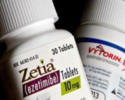
In a press release to Reuters on Nov. 13, 2003 Merck & Co. Inc. and Schering-Plough Corp. announced that ezetimib (Zetia), a new cholesterol-lowering drug that is marketed by both companies, was found by their researchers to lower C-reactive protein (CRP) significantly. At the annual meeting of the American Heart Association in Orlando/Fla. these researchers presented a clinical trial showing that ezetimib when used in combination with small amounts of simvastatin (Zocor) lowered CRP by 33%. However, simvastatin alone lowered CRP only by 14.3%. Dr. Christie Ballantyne, a Baylor College cardiologist, pointed out that this new finding was very important. It was important, because it shows that these drugs do not only lower LDL cholesterol, which according to the Framingham study is a known risk factor for heart attacks and strokes. In addition it has now also been proven to lower CRP significantly at the same time, which is another known inflammatory component produced by the blood vessels also associated with heart attacks and strokes.

New Cholesterol-Lowering Drug Reduces Inflammatory Marker

New Cholesterol-Lowering Drug Reduces Inflammatory Marker

Merck and Schering-Plough are now developing a new formulation containing both of these medications as one pill (Vytorin). This has the advantage to lower the risk on liver cells of Zocor by being able to lower the dose in the pill. The Zocor component will mainly lower the LDL cholesterol in the blood (and the CRP somewhat as well) and the Zetia component will provide the beneficial effect of the CRP lowering (anti-inflammatory component and LDL lowering). There is another advantage of this combination: Zetia works by inhibiting absorption of cholesterol by the gut, Zocor works by inhibiting cholesterol synthesis in the liver cells. Whenever the mechanism of action is different two drugs in combination are usually better tolerated than if both would work through the same mechanism. However, the companies pointed out that more research and clinical trials are needed to check out side-effects of Zetia before it would be submitted to the FDA for approval for general prescription by physicians.

P.S. on Oct. 31, 2012: Read the following article about Zetia and Vytorin (the combination pill): http://www.nytimes.com/2008/01/14/business/14cnd-drug.html?_r=0

Links regarding further information about heart disease (Net Health Book).

Last edited December 9, 2012

Dec
01
2003

Help For Patients With Iron Overload

Patients who are born with an inborn enzyme defect that leads to iron overload (hemochromatosis) and others with secondary hemochromatosis due to sickle cell anemia will benefit from new research by Dr. Gavin Oudit, Dr. Peter Backx, Dr. Peter Liu and others. The researchers at the University of Toronto and Toronto General Hospital have published their findings in the Sept. 15 issue of Nature Medicine.

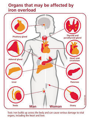
In animal experiments they found that the same calcium channels that transport calcium to vital organs are also the channels through which poisonous levels of iron are introduced with iron overload disease. In both animal experiments and in the clinical situation, human iron overload affects mainly the pancreas, the heart muscle and the pituitary gland. The authors of this study found that in hemochromatosis patients the calcium channel blockers, such as amlodipine (Norvasc), verapamil or diltiazem will stop the accumulation of toxic levels of iron in these organs.

Dr. Peter Backx, professor of physiology and medicine at U of T in the Heart & Stroke/Richard Lewar Centre of Excellence and senior author of the paper, explained that more detailed research determined that the L-type calcium channels that play a role in the normal calcium transport across the cell membrane are the same channels that allow the iron molecules into the heart muscle cells and into the cells of the other organs that get damaged with hemochromatosis. By using calcium channel blockers, heart drugs that are already on the market, it is possible to prevent accumulation of iron to the point of toxic levels. Up to now the only approach to therapy was to remove excessive iron from the body by expensive iron chelation medication that had to be given intravenously.

Help For Patients With Iron Overload

Further clinical trials on a larger patient population are necessary to determine who will benefit most from this approach of treating iron overload conditions with calcium channel blockers and what dosage to take. Dr. Peter Liu is another senior author regarding this study and is a cardiologist at the Toronto General Hospital and director of the Heart & Stroke/Richard Lewar Centre of Excellence and professor of medicine and physiology at U of T. He stated that this alternative therapy for heart failure from iron overload cardiomyopathy will likely open the doors for those patients worldwide who could not afford to have expensive chelation done, which is presently the only treatment method to remove the excessive iron. People of North American, European, Mediterranean or Asian descent are more prone to genetic hemochromatosis, thalassemia and sickle cell anemia that can all lead to iron overload requiring this type of therapy.

Last edited December 9, 2012

Nov
01
2003

Stroke Risk Increases With Carotid Artery Disease

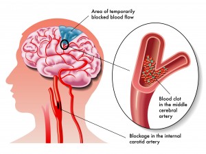
An important study about the risk of strokes and mini-strokes (called”transient ischemic attacks”) was published in the Oct. 27 edition of the Archives of Internal Medicine. Dr. Daniel J. Bertges and his group followed 1,004 patients between 1988 and 1997 with ultrasound studies of the carotid arteries (carotid artery duplex ultrasound scans). The studies took place at the Pittsburgh Veterans Affairs Medical Center/University of Pittsburgh School of Medicine. Patients were followed with regard to events such as strokes on the side of where the narrowed carotid artery was.

Reversible mini-strokes (medically correct term:”transient ischemic attack” or TIA) were also registered. A total of 1,701 narrowed arteries (called “stenotic arteries”) were found with this ultrasound method. All of the patients initially had no symptoms of the carotid artery stenosis (no dizziness, no fainting, no absence spells or symptoms of TIA or stroke). In 75% of the patients the carotid stenotic lesions were less than 50% meaning that the carotid artery blood flow was acceptable.

Here are some of the risks as the study went on over the years: both TIA and CVA risk in a given patient occurred at a rate of 3.3% per year. Regarding a specific involved artery the risk of developing a TIA as a result of this was 2% per year and the risk to develop a stroke was 2.1% per year. The investigators found that two main factors determined the ultimate progression into a TIA or a stroke and they were as follows. First, if the artery was severely stenosed at the outset, the probability was high that this would progress and be the cause of a stroke. Secondly, the degree of progression when checked with a follow-up duplex ultrasound was another important factor in terms of leading to a subsequent TIA or stroke.

Stroke Risk Increases With Carotid Artery Disease

Carotid artery clot can cause stroke

The composite risk of developing either a TIA or a stroke with a worsening stenotic carotid artery lesion was 1.68-fold. To develop a stroke alone in this scenario the risk was 1.78-fold. Clinical risk factors were of no help in predicting which cases would go on to develop TIA’s or strokes. However, the finding of further progression of a stenotic carotid artery lesion documented on serial duplex ultrasound studies was highly significant.

The authors concluded that there is value in doing serial carotid artery duplex scan studies in the same patient to screen for progressing stenotic lesions in the carotid arteries. When a stenotic lesion is significant enough or progressing fast, intervention by a cardiovascular surgeon with carotid endarterectomy can be done to prevent a stroke or TIA.

Here is a link to a chapter on strokes from the Net Health Book.

Last edited October 26, 2014

Oct
02
2003

Heart Scan Saves Lives In Diabetics

A simple new nuclear perfusion study of the heart when applied to healthy appearing diabetics (adult onset or “type 2 diabetics”) showed silent hardening of the coronary arteries in 21.6%. This large study of an American medical team was recently presented at the 18th Congress of the International Diabetes Federation in Paris/France. Dr. F. Wackers, professor of medicine from Yale University school of medicine and one of the lead investigators, explained that 1,124 patients with diabetes in the age range of 55 to 75 years who were all thought to not have any heart blood vessel disease, either had nuclear perfusion studies performed and a control group did not.

As indicated above to the surprise of the investigators 113 patients of 522 (=21.6%) had positive heart scans showing perfusion difficulties of the heart muscle. Further testing with other methods revealed that 73% indeed had perfusion defects and 27% had other heart disease, electrocardiogram abnormalities and other heart dysfunctions. Conventional assessment tools such as a smoking history, determination of degree of obesity, blood pressure,kidney disease , high blood lipid levels, high C-reactive protein levels, the diabetes test hemoglobin A1C or homocysteine levels in the blood were also assessed. However, these conventional tests did not help in predicting that these patients would have developed perfusion defects in their heart muscle. This was due to hidden narrowing of the heart blood vessels (=coronary arteries) and this affected the supply of nutrients and oxygen to the heart even though these patients were completely symptom free at the beginning of the trial.

Heart Scan Saves Lives In Diabetics

Heart vessels and nuclear scan

Dr. Vivian Fonsega, a professor of medicine and pharmacology at Tulane University in New Orleans and co-researcher of the team, added that after a follow-up of 1 year those who had normal initial nuclear perfusion studies of the heart only 1% developed serious heart disease. These control patients who have now been followed for 3 years overall remained very healthy. In other words a normal (called “negative”) nuclear perfusion test in diabetics predicts a better longterm outcome than a positive perfusion test.

With this heart scan the cardiologist can identfy the high risk group among diabetics and can subsequently concetrate on doing something actively about the identified diseased heart blood vessel(=”coronary artery”) disease. Identified narrowing in the coronary arteries (“stenotic arterial lesions”) can be overcome by prying them open and placing heart stents across the affected section utilizing catheters (angiography). In other cases heart bypass surgery can be done by the heart surgeon to improve the perfusion of the heart muscle. The researchers stressed that those diabetics at risk can be identified with this test and the life expectancy of this high risk group of patients can be significantly prolonged. The study will continue for several more years so that the longterm results of any intervention can be measured when compared to controls.

Based on The Medical Post (Sept. 23, 2003 ): p. 55.

Here is a link to a chapter on diabetes and here is a link to heart attacks.

Last edited December 9, 2012

Sep
01
2003

Poor Lungs And Heart Attacks Related To Leptin Levels

It is known from the medical literature that poor lung function can often lead to heart attacks making it one of the important causes of premature death for patients with poor lungs (due to emphysema, chronic bronchitis, COPD etc.).

A research team led by Dr. Don Sin from the University of Alberta, Edmonton, Canada, asked the question recently whether there may be a circulating factor that would be responsible for this association of poor lung function and increased cardiovascular disease.

They studied serum leptin and a variety of other inflammatory markers such as C reactive protein, leukocytes, and fibrinogen in 2808 participants in the Third National Health, Nutrition, and Examination Survey. Apart from blood tests they also measured lung function by spirometry (forced expiratory volume in 1 second, called FEV1). The leptin levels found in these patients were then divided into 5 groups from low to high levels. They also carefully adjusted the data for body mass index, sex, age and other factors. They compared the group with the lowest leptin concentration (lowest quintile) with the highest group of leptin concentration (highest quintile) and looked for any significant differences in any of the markers.

Results: The highest quintile group (high leptin in blood samples) had also the highest other inflammatory markers in their blood (C-reactive protein, leukocytes and fibrinogen). This group was the one that was associated with advanced lung diseases as well as heart disease. The authors of this study, which was recently published in a medical journal (Thorax 2003;58:695-698), concluded that leptin plays an important role, if not the major role, in the development of both chronic lung disease and cardiovascular complications.

Poor Lungs And Heart Attacks Related To Leptin Levels

Poor Lungs And Heart Attacks Related To Leptin Levels

Links to lung disease: http://www.nethealthbook.com/articles/chronicobstructivepulmonarydisease.php
Links to heart attacks:

http://www.nethealthbook.com/articles/cardiovasculardisease_heartdisease.php

Last edited December 9, 2012

Jul
01
2003

Obesity And Metabolic Syndrome

In the June 10, 2003 edition, following page24, of The Medical Post there was a minisymposium on obesity and the metabolic syndrome (also known as the “syndrome of hyperinsulinism”).

Four specialists had a discussion about this topic: Dr. Ehud Ur (endocrinologist, Dalhousie University, Halifax, N.S., Canada), Dr. Robert Dent (Director of the Weight Management Clinic, Ottawa Hospital, Ont.), Dr. Dominique Garrel (Director of Department of Nutrition and endocrinologist, University of Montreal, Quebec), and Dr. Arya Sharma (Prof. of Medicine, McMaster University, Hamilton, Ont.).

Introduction:

Obesity is now a health threat that about 25% of the North American population is suffering from. There is still a lot of discussion what the exact criteria should be, but the Third Report of the National Cholesterol Education Program Expert Panel on Detection, Evaluation, and Treatment of High Blood Cholesterol in Adults (ATP III) has simplified the detection of the metabolic syndrome.

Obesity And Metabolic Syndrome

Obesity And Metabolic Syndrome

The experts agree that when three or more of the criteria mentioned in this table are positive the person would be considered to have metabolic syndrome.

There is a wide age-related variety: in one study only 7% had metabolic syndrome in the age group of 20 to 29. The same study found 40% of study participants had the metabolic syndrome in the age group of 70 years and older. It is thought that too many calories coupled with too little activity over a longer period of time, perhaps coupled in some people with a genetic tendency to develop metabolic syndrome, leads to an accumulation of abdominal (so-called”visceral”) fat.

Because fat cells have their own hormone systems (leptins etc.) there is a change of metabolism including an elevation of the insulin level with associated loss of “insulin sensitivity”. So, the more obese a person becomes, the less effective insulin becomes in transporting blood sugar through cell walls. At the same time the liver metabolism is changing with the good cholesterol (HDL) being less produced and the bad cholesterol (LDL) being overproduced. The liver will produce a different mix of coagulation factors, which leads to a tendency to form clots in the veins of the legs and in the lungs. As the pancreatic capacity for insulin production gets exhausted over a period of time, the patient eventually develops type 2 diabetes mellitus. Due to the risk of the coronary arteries clogging up with the cholesterol changes and the accelerated hardening of arteries from diabetes, the risk for getting severe heart attacks in obese people with the metabolic syndrome when compared to a normal weight population is about 4-fold.

Elements leading to the diagnosis of “metabolic syndrome”
Finding: Comments:
abdominal obesity waist circumference more than 102 cm in men or more than 88 cm in women
elevated triglyceride level level of 150 mg/dl or higher
low HDL cholesterol level under 40 mg/dl in men or under 50 mg/dl in women
elevated blood pressure systolic or diastolic blood pressure exceeding 130/85 mm Hg
high fasting blood glucose level fasting glucose higher than 110 mg/dl

Treatment of metabolic syndrome:

The experts agreed that a reduction of only 5% to 10% of the body weight through a sensible combination of a mild exercise program (e.g. walking 30 to 45 minutes every day) and a calorie reduced food intake will make a significant difference in terms of normalization of the body chemistry. It is my estimate that perhaps 70% to 90% of all cases of obesity and metabolic syndrome can be treated this way.

However, the remaining cases should continue to see their physician and be followed like the doctor would follow someone who has high blood pressure. There are two types of medications available and they have nothing to do with the Phen-Fen diet pills from not too long ago that were found to cause pulmonary hypertension. These new diet pills are fairly safe and show weight loss results provided the patient co-operates with regard to a modified to low fat diet and some degree of regular exercise.

1. Sibutramine (brand name: Meridia) is a specific brain hormone inhibitor in the area where the appetite zone is located (serotonine and norepinephrine reuptake inhibitor). This medication helps the patient by experiencing satiety sooner so that the patient does not feel deprived despite less calorie consumption.

It is the medication of choice for those who tend to eat a lot. Like with other anti-depressants side-effects are a dry mouth, heart rate increases and sleep loss (insomnia).

2. Orlistat (brand name: Xenical) inhibits fat uptake at the level of the gastrointestinal wall (gastrointestinal lipase inhibitor). This leads to an inhibition of fat absorption by about 30%. The patient needs to keep the fat intake down to about 2 oz. (=60 gm) per day. If the patient consumes more fat, the side-effect of orlistat will be flatulence, abdominal cramps and diarrhea. If the patient is on a strict low fat diet, there would not be enough fat in the gut for the medication to be effective.

At this point it is not known how long the patient should be on such weight loss medication, if this was the chosen route. The experts felt that 1 year would be reasonable, but that the patient should be observed by the treating physician and it may be necessary after some intermission to go for another year of therapy all the way attempting to permanently change eating and exercise habits as an ongoing maintenance program.

Here is a link to another reference about the metabolic syndrome (syndrome of insulin resistance).

Last edited December 9, 2012


Jul
01
2003

High Blood Pressure On The Increase In The US

High blood pressure on the rise

Dr. Hajjar, of the University of South Carolina in Columbia and Dr. Kotchen, of the Medical College of Wisconsin in Milwaukee have compared a large group of people in the US in the years 1999-2000 with prior studies in 1988-1991and 1991-1994 where peoples’ blood pressures were measured.

The latest study involved more than 5000 people, the prior studies more than 9000 people each. Almost 29% of the population has a blood pressure of 140/90 or higher in the latest study.

Compared to the 1988-1991study this is a significant increase of 3.7%. More than half of this was explained on the basis that the population’s weight (measured by body mass index calculation) had increased. The sub groups who had the largest increase in blood pressure were as follows:

1. diabetics with high blood pressure. In this high risk group only about 25% had a blood pressure readings of less than 130/85, which is the target value.

2. Mexican Americans had a significantly poorer awareness and control of their elevated blood pressures than the non-Hispanic whites and non-Hispanic blacks.

3. Women as a group were poorer in blood pressure controlling than men.

4. People above the age of 60 had a much higher rate of uncontrolled blood pressure as well.

The study concluded that by concentrating campaigns and efforts on these four target groups significant gains could be made in terms of control of blood pressure, reduction of strokes, heart attacks and kidney damage.

Here are some links regarding high blood pressure, prevention of strokes and heart attacks to various chapters of my nethealthbook.com:

High blood pressure link

Heart attack link

Stroke link

Last edited October 26, 2014

Jun
01
2003

Vitamin B-6, Vitamin B-12, Folic Acid To Control Hardening Of Arteries

The May 2003 issue of the medical journal “Clinical Practice” contains an article by the dietician Andrea Miller (pages 46 to 49) about supplementation with these vitamins in relation to hardening of the arteries from too much of the amino acid homocysteine.

Homocysteine is an amino acid that contains sulfur, which is formed from the metabolism of methionine, an essential amino acid that we depend on in our food for maintaining a normal metabolism. There are complex metabolic pathways that involve several enzymes to function normally, which involve the three vitamins B-6, B-12 and folic acid (folate) to balance everything.

If one of these vitamins or any of the enzymes involved malfunction, a surplus of homocysteine results with an accelerated hardening of the arteries throughout the body. A certain percentage of the population does not have the full concentration of one or more of the metabolic enzymes in their system. If the food we eat is also relatively deficient in the above named vitamins, the body runs out of alternative metabolic options and produces dangerous levels of homocysteine. A normal blood level of homocysteine is between about 5 and 10 micro-mol per liter. This is not routinely measured by screening tests unless the doctor suspects a genetic trait in the family where family members tend to get heart attacks at an early age (mid 30’s to early 40’s). When the level of homocysteine is elevated to 12 micro-mols per liter, a supplement with folic acid of 1 mg per day would reduce this by 25% to safe levels of 9 micro-mols per liter. This results in a 15% decrease of the death rate from heart attacks.

Vitamin B-6, Vitamin B-12, Folic Acid To Control Hardening Of Arteries

Vitamin B-6, Vitamin B-12, Folic Acid To Control Hardening Of Arteries

Vitamin B-12 is found in animal products such as meat, poultry and dairy products. Vegetarians may not get enough Vit. B-12 and should supplement as should persons above the age of 50. A lack of B-12 vitamin leads to pernicious anemia.

Vitamin B-6 is found widely distributed in meat, fortified grains and poultry and usually is in our food supply to a satisfactory degree.

Folate (folic acid) is contained in orange fruits, beans, lentils and green vegetables. However, not everybody eats well banced meals and the food supply could be marginally deficient in folate.

Recommendation: The take home message is that a good multivitamin tablet containing about 400 to 600 micrograms (=0.4 to 0.6 mg) of folate (folic acid), 5 mg of vitamin B-6 and 20 micrograms of vitamin B-12 would be providing an adequate amount of these vitamins and reduce homocysteine levels in most people to safe levels. It is not recommended to take single vitamin supplements of these vitamins as this could lead to toxicity. Also, there is no point in taking higher doses of the vitamins as this also would lead to toxic reactions.

Link to information about balanced nutrition: http://www.nethealthbook.com/articles/nutrition.php

Last edited December 9, 2012