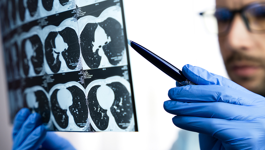
In 2013 the US Preventive Services Task Force recommended a yearly lung cancer screening program. The target population was age 55 to 80. Specifically, this program was to screen people who currently smoke, or had quit within the last 15 years and had a smoking history of 30 or more pack-years. Screening occurs with a special CT scan using low-dose radiation for lung screening. In the US Medicare and Medicaid reimburse residents for the cost of this procedure. The BCMA Journal describes the introduction of a similar lung cancer screening program in BC since May 2022. In the US the lung cancer mortality experienced a 20% drop since the introduction of the lung cancer screening program. This is because physicians now find lung cancer at stage 1 where treatment with surgery, radiotherapy or chemotherapy is much more effective.
Feasibility of a lung cancer screening program
Typically, with the conventional plain X-ray screening of symptomatic patients 40% of them, which radiologists diagnosed had lung cancer at a late stage, namely stage 4. At that stage the 5-year survival is less than 10%. However, now they diagnose patients with early lung cancer at stage 1 using a low-dose CT scanner with the lung cancer screening program. At this stage the 5-year survival rate is 73% to 90%. We know that the main lung cancer cause is cigarette smoking, the second cause is the aging process.
Lung cancer screening program free for patients
Similarly to the US the government sponsors the BC Lung Cancer Screening Program with no cost to the patient. With yearly checks the low-dose CT scanner detects early lung lesions that are highly suspicious of lung cancer. The screening program includes the higher age group and the ones who were heavier smokers. This is the highest lung cancer risk group, which benefits most from the lung cancer screening program.
What happens when the lung cancer screening program identifies early lung cancer?
With all the nodules that the CT scan screening finds, some have the features of suspicious nodules that require biopsy to check histologically whether or not there is lung cancer present.
Various methods to do lung biopsies
The simplest way for the physician to do this is by way of a bronchoscopy, where he inserts a needle into the nodule and retrieves a tissue sample. The pathologist analyzes this biopsy under the microscope. Not all suspicious nodules are within easy reach by bronchoscopy. If a lesion is located close to the lung surface the physician can do a needle biopsy through the skin (transcutaneous biopsy or transthoracic biopsy). Some patients require a biopsy using video-assisted thoracic surgery, which is performed under general anesthesia. Other patients require an open biopsy, which the chest surgeon performs under general anesthesia. In this case the chest surgeon opens the chest cavity and removes a piece of lung tissue, which the pathologist later analyzes for cancer.
Test to determine the extent of the lung cancer
The lung cancer stages are: stage I, II, IIIA, IIIB and IV. Following the initial X-ray, the physician will order an MRI or CT scan in order to determine whether the lung lesion was the only finding or whether there were metastases nearby. The MRI/CT scan can show whether or not there is involvement of the lymph glands in the chest or not. If there are lymph glands in the chest, a thoracic surgeon may be called in to do a mediastinoscopy, where the surgeon can look into the space between the lungs and the rib cage and assess the extent of the metastases in this otherwise difficult to assess space.
Distant metastases
The oncologist will want to continue to do the staging tests by doing CT scans of the liver, the adrenal glands and the brain to determine whether distant metastases are present. Blood tests and bone scans will rule out bone metastases. Finally, when all this information is gathered, the oncologist can do what is called an” extent of disease evaluation”. The following could be found for the various stages.
Extent of disease evaluation: Staging of lung cancer
Stage:
I : solitary lung tumor of less than 3 cm (=1 1/4″) in diameter
II : tumor more than 3cm(= 1 1/4″) in diameter, local lymph gland metastases on the same side as the tumor
IIIA : peripheral lung tumor: invaded the chest wall; central lung tumor: invaded distal mediastinal nodes on the same side
IIIB : same as stage IIIA, but more extensive lymph gland invasion involving mediastinal organs and pleural cavity
IV : Any of the above stages, but in addition distal metastases
Is it wasted time to do the staging procedure?
Why are oncologists “wasting time” to do the staging procedures? Studies over several decades have taught us that treatment of cancer without staging often gives everyone a false sense of security, where they learn later that the real extent of the cancer was much worse than originally thought. While everyone was thinking no further therapy was necessary, the cancer quietly multiplied and spread until it was too late to do anything about it. With the progress in the treatment of childhood leukemia oncologists learnt that long-term survival and cure rates could show significant improvement with adequate staging in the beginning and by following appropriate treatment protocols. In the last few years this has paid off for lung cancer as well.
Treatment of lung cancer
When the oncologist does an “extent of disease evaluation” he can discuss with the patient and the family what stage the lung cancer is in and what the chances of survival for the lung cancer are based on a vast amount of knowledge. There is a discussion of treatment options in detail and the oncologist can tailor the therapy to the needs of the patient. In principle, the approach to treat stage I and II is mainly by surgery to remove all cancer within the healthy surrounding tissue.
Surgical risk and treatment of stage III and IV
With an oncological or thoracic surgeon this kind of surgery has only a mortality of 1% to 8%. In younger patients this risk is lower, in patients above 75 years of age the risk is higher. With surgery higher survival rates are achievable (up to 80 % in stage I, up to 50% in stage II). Stage IIIA can be managed surgically, but stage IIIB needs another approach. Usually with this stage as well as with stage IV radiotherapy and combination chemotherapy is needed.
Lung Cancer Screening Program
Conclusion
In the US a lung cancer screening program is in place since 2014. Since then, lung cancer mortality has dropped 20%. Also, in 80% of cases lung cancer is in stage I, the earliest form of lung cancer. In the past the majority of diagnosed lung cancer was in stage IV with a 5-year survival of only 5-10%. Now with the CT scan lung cancer screening program the 5-year survival is 73% to 90%. Treatment is mostly surgical for earlier-stage lung cancer (stage I, II and IIIA). For stage IIIB and stage IV a combination of surgery, radiation therapy and possibly chemotherapy is in use. The emphasis is on smoking cessation and yearly screening with a low-dose CT scanner.