Mar
19
2016

Book Review: “Healing Gone Wrong – Healing Done Right”, By Ray Schilling, MD

This book entitled “Healing Gone Wrong – Healing Done Right” (Amazon, March 18, 2016) is dealing with the practice of medicine then and now. Medical errors, false diagnoses and wrong treatments are nothing new in the history of medicine. It happened in the past, and it is happening now. My first book was about anti-aging. The title was “A Survivor’s Guide to Successful Aging” (Amazon 2014).

Book overview

Chapter 1

Here I describe describe that famous people like President Kennedy, Elvis Presley, Churchill, Beethoven or more recently Michael Jackson have something in common: all of them suffered the consequences of blatant medical mistakes. In Beethoven’s time lead containing salves to plug the drainage holes from removing fluid from his abdomen caused lead poisoning. In this chapter I review also how doctors treated the illnesses of the above-mentioned celebrities, but then ask the question: “What better treatments have offered to prevent some of the disastrous treatment outcomes?”

Chapter 2

Modern drugs seem to come and go. We learn that twenty-first century medications that are supposed to be the latest therapeutic agents are having their potentially deadly consequences too: COX-2 inhibitors, the second generation arthritis drugs cause strokes and heart attacks! Your doctor may still prescribe some of these dangerous drugs for arthritis now.

Chapter 3

This chapter deals with the fact that medical treatments for people’s diseases may be inappropriate when the doctor treats only symptoms, but the doctor does nothing about the causes of their illnesses. This is a scary thought.

Chapter 4

What does it take to prevent these poor health outcomes, so that we will be able to prevent any disastrous outcomes pertaining to our own health care in the present and future? As we will see, the problem today is still the same as it was in the past, namely that many physicians still like to treat symptoms instead of the underlying cause of an illness. Big Pharma has the seducing concept of a pill for every ill, but it is not always in your best interest, when these medications have a slew of side effects. “Gastric reflux” means a mouthful of stomach acid. Big Pharma simply offers the patient with the symptom of gastric reflux a multitude of medications to suppress this symptom. But it is more important to dig deeper to find the reason for the illness and treat the underlying cause.

Chapter 5

We all need our brain to function. This chapter concentrates on the brain and how we can keep our brains functioning optimally until a ripe old age. This review spans from prevention of head concussions to avoiding type 3 diabetes (insulin sensitivity from overconsumption of sugar). It manifests itself in Alzheimer’s disease. It is a form of diabetes of the brain that leads to deposits of a gooey substance. Prevention of this condition is also reviewed .

Chapter 6

This chapter reviews what we now know about how to keep a healthy heart. Certain ingredients are necessary such as regular exercise, a healthy Mediterranean diet, supplements etc. The good part is that what is good for the heart is also good for the brain. You are preventing two problems (brain and heart disease) at the same time.

Chapter 7

What should we eat? And why does healthy food intake matter? Without the right ingredients of our body fuel, the body machinery will not work properly. The Mediterranean diet is an anti-inflammatory diet that is particularly useful.

Chapter 8

We need healthy limbs, bones and joints. We are meant to stay active in our eighties and nineties and beyond. No osteoporosis, no joint replacements, no balance problems that result in falls! Learn about how to deal with problems like these in this chapter.

Chapter 9

This chapter deals with detoxification. What do we do as we are confronted with pollution, with radiation in the environment and poisons in our daily food? A combination of organic foods, intravenous chelation treatments and taking supplements can help us in that regard.

Chapter 10

I am dealing here about reducing the impact of cancer in our lives. A lot of facts have come out in the past 10 years telling us that reduction of sugar and starchy food intake reduces cancer. Curcumin, resveratrol and vitamin D3 supplements also reduce cancer rates as does exercise and stress management. All of this is reviewed here.

Chapter 11

This chapter tells you all you need to know about your hormone status. Women need to avoid estrogen dominance; both sexes need to replace the hormones that are missing. By paying attention to your hormonal status and replacing the missing natural hormones with bioidentical ones, most people can add 10 to 15 years of useful, active life!

Chapter 12

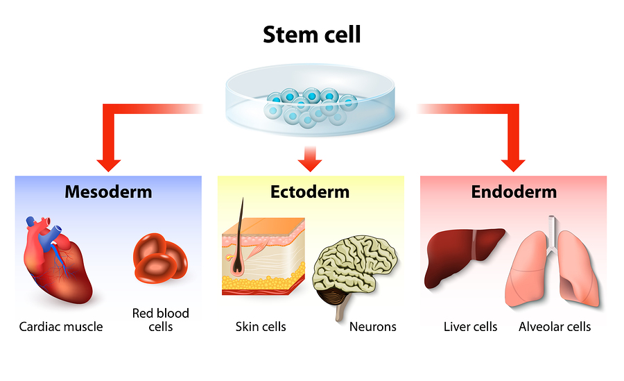
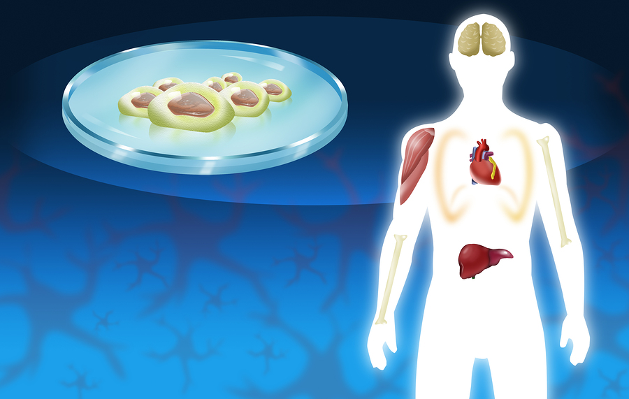
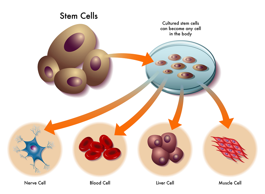
Here you will learn more about anti-aging. You will learn about the importance to keep your mitochondrial DNA healthy. Apart from that there are ways how to keep your telomeres longer; certain supplements that are reviewed will help. Also your lifestyle does make a big difference in how old you can turn.

Chapter 13

This chapter investigates the limits of supplements. Many supplements are useful, but you do not want to overdo it and get into toxic levels. More is not necessarily better!

Chapter 14

Here is a review of an alternative approach to treating ADHD. Attention deficit and hyperactivity disorder has been over diagnosed, has been neglected and has been over treated with dangerous drugs. An alternative treatment plan is discussed, which includes a combination of therapeutic steps.

Chapter 15

This gives you a brief summary of the book.

Kirkus Review

Kirkus Reviews reviewed the book on March 17, 2016: “A retired physician details how various preventative measures can fend off disease and disability in this consumer health guide. Schilling (A Survivor’s Guide to Successful Aging, 2014) had a family medicine practice in Canada for many years before retiring. Although Schilling ventures into some controversial territory in his latest book, it’s generally an engaging, helpful synthesis of ideas that draws on reputable research from the Mayo Clinic and other sources. Overall, it serves as an intensely detailed wake-up call to the importance of preventative health. He largely brings an accessible and even-tempered tone to his narrative, warning readers, for example, that preventative health measures can only aid in “a delay of aging, not ‘eternal living.’ ” A thought-provoking, impassioned plea to be proactive about one’s health.”

Healing Gone Wrong – Healing Done Right

Healing Gone Wrong – Healing Done Right

Conclusion

In this book it becomes evident that it is better to prevent an illness whenever possible rather than to wait for illness to set in and cause disabilities or death. You heard this before: “Prevention is better than a cure” or “an ounce of prevention is better than a pound of cure”. I will give an explanation, based on scientific data that there is indeed evidence to support these notions on a cellular level.

Mitochondria, the energy packages within our cells

The mitochondria, the energy packages within our cells, are the driving force that keep people vibrantly healthy well into their nineties. All this can only happen when the mitochondria function properly. If toxins poison the mitochondria and as a result they malfunction, we are not looking at a person with vibrant health. Instead sixty or seventy year-olds may use a wheelchair. If you want a life without disabilities, a life without major illnesses and enjoy good health to a ripe old age, you are reading the right book.

The book is written in American English.

Available in the US: http://www.amazon.com/gp/product/1523700904

In Canada: https://www.amazon.ca/Healing-Gone-Wrong-Done-Right/dp/1523700904/  

In other countries the book is available through the local Amazon websites.

Feb
27
2016

Orthopedics Without A Knife

Dr. Fields gave a talk in Las Vegas about orthopedics without a knife. His talk took place at the 23rd Annual World Congress on Anti-Aging Medicine on Dec. 12, 2015 in Las Vegas. Dr. Fields gave a talk entitled “Regenerative orthopedics – non-surgical repair with stem cells/PRP/prolotherapy”. In essence the talk was about alternative treatments to surgeries in orthopedic medicine.

Dr. Peter Fields, MD, DC is a board certified medical physician and chiropractor. He is also the director of the Pacific Prolotherapy & Medical Wellness Center in Santa Monica, CA.

Introduction

Joints, muscles, tendons, ligaments and joint capsules control the movements in joints. Due to injuries and wear and tear these body parts can have a lack of function, which will lead to pain and disorders. The result can be weak, torn or damaged ligaments and tendons, arthritic changes, excessive joint motion, increased pressure, and a decrease in range of motion.

This is the common treatment cycle in medicine

Joint pain prompts you to see the doctor. You are told it is arthritis, and you get non-steroidal anti-inflammatories (NSAID’s). You come back with more pain, and you’ll get a stronger NSAID prescription. Eventually a cortisone injection is given, which helps for a few months, but then the pain reoccurs. The doctor arranges for an MRI scan. A referral to an orthopedic surgeon is likely to be the next step, and an arthroscopy (pinhole surgery) is arranged. In that case, if this does not resolve the pain, surgery like a knee replacement or hip replacement is suggested.

Common sayings when traditional medicine has nothing to offer

You may have heard some of these common sayings before. “Nothing more we can do about it!” -“I suggest you learn to live with it”- “You should never play that sport again!”- “Take these pain medications” and “The only alternative is surgery!”

The problem is, that none of these pieces of advice are really helpful. This type of approach does not treat the cause; it is directed against symptoms.

How to treat the cause?

Prolotherapy

Prolotherapy is a natural, non-surgical method to assist the body to heal torn soft tissues. It works in cases like torn ligaments, damaged tendons, cartilage, menisci or a torn labrum in the shoulder. Hyperosmolar dextrose solution is injected into the injured area. This stimulates the body’s healing forces and the body repairs what is damaged. More information is found here. In essence, prolotherapy fixes the cause, not just the effect; it heals, and it is permanent. Prolotherapy strengthens tissues, relieves pain and increases the range of motion in joints. There is 80 to 85% full pain relief and more than 80% improvement in range of motion. Prolotherapy promotes the healing of torn or damaged ligaments and tendons.

Conditions suitable for treatment with prolotherapy

Suitable conditions for treatment with prolotherapy are sports injuries, muscle tears, arthritis, tendinitis, bursitis, sciatica, TMJ problems, and fibromyalgia. Common areas treated with prolotherapy are the hip, knee, shoulder, ankle, neck, lower back and elbow. Dr. Fields showed MRI scans before and after prolotherapy treatments of ligament injuries within the knee and of shoulder ligament tears before and after treatment. Normally the physician expected these injuries to require surgery. But all that was done was one or two injections (prolotherapy treatments) with reactivation of the affected joint. There were astonishing results shown with MRI’s before and after herniated disc injuries and how they healed in a relatively short time following prolotherapy.

PRP prolotherapy

Platelet rich plasma (PRP) is a tool from regenerative medicine to amplify the healing response in connection with stem cell therapies .  The lab technician takes blood from the patient and subsequently spins it down in a centrifuge. The platelet rich fraction (PRP) contains all of the growth factors, which have the healing power of the blood. The physicians combines this with prolotherapy to make healing even more successful. This is particularly useful for labral tears in shoulders, meniscus tears in knees and other localized injuries.

Stem cell prolotherapy

Stem cell therapy has been the gold standard for repairing more serious problems. Dr. Fields combines stem cell therapy with prolotherapy to treat more serious injuries like end stage arthritis.  This is the case when bone rubs on bone, where conventional orthopedic medicine would offer a joint replacement in the hip or knee. Stem cell prolotherapy can repair any joint that has cartilage damage. A severe meniscus tear in a knee or a severe labrum tear in a shoulder would also be situations where stem cell prolotherapy is superior to surgery or to just using prolotherapy alone.

Here is a description of the procedure

Before the patient’s procedure the physician first harvests bone marrow stem cells by way of a pelvic bone aspirate; secondly the physician obtains mesenchymal stem cells from fatty tissue by aspiration of abdominal fat. A cell separator provides the stem cell fractions. The physician combines both types of stem cells, the bone marrow stem cells and the mesenchymal stem cells from fat as each one has its own strengths. These two stem cell types are more effective in combination to repair whatever tissue needs repair. Thirdly, the lab technician will draw blood from the patient to obtain PRP, which contains the growth factors needed to activate the stem cells to do their job of healing. The last step is that the physician now combines hyperosmolar dextrose (the prolotherapy part) with the stem cell preparation and mixed in PRP and injects this mixture into the injured area.

Conditions that respond to stem cell prolotherapy

This procedure has superior healing power. Before and after MRI scans of all of the major body regions showed impressive results. Several video recorded testimonials  complemented the MRI scans. It is surprising how quickly and completely fairly severe injuries can heal using stem cell prolotherapy. One particularly nasty condition is osteonecrosis of the hip, which can occur as a side effect of chronic cortisone treatment for arthritis, asthma or chronic obstructive lung disease. One or two stem cell prolotherapy treatments will heal this condition because the stem cells build up brand new bone and get rid of the old necrotic bone from the osteonecrosis. Conventional medicine has no answer for this condition. Regenerative orthopedics is successful by using stem cell prolotherapy.

What are the advantages of regenerative orthopedics?

Regenerative orthopedics reduces pain very quickly and it improves function rapidly. Healing occurs naturally, and it strengthens the tissues involved. Particularly complicated lower back pains or lower neck pains (due to degenerative disc disease, facet joint osteoarthritis, spondylolisthesis and significant foraminal stenosis) respond really well to stem cell prolotherapy, getting rid of chronic pain. The speaker showed before and after MRI scans. He also shared testimonials from patients about the various procedures.

End result following stem cell prolotherapy versus conventional surgery

This is quite in contrast to what conventional orthopedics has to offer: discectomy with fusion surgery, where the patient often has scar pain later. With a laminectomy to treat a foraminal stenosis the patient may have limited improvement of the chronic back pain for a couple of months, only to experience new back pain from a subsequent spinal stenosis as a late complication from the prior surgery. The end result with conventional orthopedics is disability, pain and suffering; the end result with regenerative orthopedics is a patient that is well, active, pain free and thankful.

Orthopedics Without A Knife

Orthopedics Without A Knife

Conclusion

There is a form of orthopedics without a knife: regenerative orthopedics. The tools are prolotherapy for minor musculoskeletal problems. Some very conservatively minded physicians still scoff at this, but wrongly so. PRP prolotherapy is suitable for more severe injuries that require more healing power. Stem cell prolotherapy is what the physician uses for the severe cases. All of the healing power (minus the knife) is put to use. Two types of stem cells initiate healing where there is a need for it. The stem cells transform into the cell types that do the repair.

Two types of stem cells needed sometimes

Research has shown in the past that the mesenchymal stem cells alone will not heal cartilage of joints very well, but in combination with bone marrow derived stem cells this heals quite well and efficiently. Healing osteonecrosis and complicated lower neck and lower back problems borders to miraculous healing. Regenerative orthopedics is definitely something to remember should you get into trouble down the road. There are alternatives to the knife!

Apr
25
2015

Rejuvenate With Stem Cells

We all age; but can we rejuvenate with stem cells? There is a limit to detoxification, to eating organic food, to exercising, to the effects of vitamins and supplements and even to the effect of bioidentical hormone replacements. The limit comes from our telomeres and from stem cells that get depleted in our body as we age. Some researchers report that in regions where we suffer from a disease stem cells are even more depleted than in the rest of the body.

We do not have all the answers yet. We would like to know why our stem cells in the fatty tissue or in the bone marrow do not migrate on their own into an aching back or a sore shoulder. There are all the aches and pains associated with old age. So, why do our own stem cells not help us? They seem to be locked away in fatty tissue and in bone marrow.

At the 22nd Annual World Congress on Anti-Aging Medicine in Las Vegas (Dec. 10-14, 2014) I learnt that there is a group of stem cell experts in California with affiliates all over the US. They simply take stem cells from the fatty tissue and sometimes also from the bone marrow, isolate the stem cells through a stem cell separator and infuse the stem cell rich fraction (minus fatty and connective tissue) in a bit of saline solution back into the vein of the patient. When the stem cells are in the blood stream, they get activated by the growth factors that are present in blood and can now find where they are needed and start the healing process.

Studies have shown that when stem cells are in circulation in the blood, they are very sensitive to signals from tissues that indicate that there is an inflammatory process. This is why stem cells will repair arthritic changes. The can repair a torn meniscus, a rotator cuff tear in the shoulder or repair a weak immune system. The interesting observation is that stem cells from fatty tissue, also termed mesenchymal stem cells, are pluripotent. This means they can develop into cartilage building cells (chondrocytes) and build up cartilage; this is badly needed in a person with severe osteoarthritis. But stem cells are flexible: they can turn into meniscus cells in a knee with a torn meniscus. They also can repair the damage and relief the patient of the chronic pain. In a shoulder with a rotator cuff tear they can turn into a tough ligamentous material mending the tear.

Some data even indicates that circulating stem cells can repair vital organs like the brain, heart, liver, kidneys and bone marrow; these latter observations were mostly done in animal experiments, but human data is starting to be published in the medical literature.

So, let’s examine what has been found useful with regard to stem cells that are taken from your fatty tissue or your bone marrow and injected into one of your veins.

Here is a website from Arizona that I am only showing as a typical example (I have no conflict of interest and no commercial connections to this group) of what I described above.

With websites like this it is also important to read the disclaimer: “Even though our treatments are done using autologous cells, our Stem Cell Therapies are not approved by the FDA. Stem Cell Treatments are not a cure for any condition, disease or injury, nor a substitute for proper medical diagnosis and care…” Another website from La Quinta, CA describes the use of mesenchymal stem cells for regenerative therapies.

Stem cell treatments are in flux. There is a large body of knowledge that has accumulated showing that with proper technique and aseptic conditions it is a safe procedure. The FBA has been watching this. There are publications regarding the safety of procedures with adipose mesenchymal stem cells; here is one example.

The next step is to show in clinical trials that a certain procedure with stem cells is effective in treating a certain condition.

Below I did a literature review, which are only a few examples, but does not claim to be complete; it highlights some of the problems with stem cell treatments.

Stroke treatment with intravenous administration of bone marrow mononuclear stem cells

This study from India showed no statistical difference of stroke patients treated intravenously with bone marrow derived mononuclear stem cells (the experimental group) and the control group that did not receive such treatment. The investigators examined both groups with functional brain tests and performed PET scans to look at the healing of the brain lesions. Unfortunately the tests showed no statistical difference, but did show that the stem cell procedures were safe. It may be that the wrong stem cells were used (mononuclear bone marrow stem cells) when adipose derived mesenchymal stem cells may have done better. In stark contrast to the study from India is the stem cell treatment for a severe stroke in the former hockey player, Gordie Howe that has gone through the media recently. His procedure was done in Mexico. The stem cells were administered via a lumbar puncture approach as well as intravenously. As you can see from this case, stem cell treatment is even possible in patients who are in their mid 80’s with impressive results.

Parkinson’s disease

Here is a feasibility study from March 2014. A 71-year-old Asian man with progressive supranuclear palsy, an aggressive form of Parkinson’s disease was treated with adipose tissue-derived mesenchymal stem cells that were administered intravenously and intrathecally (to get stem cells into the cerebrospinal fluid that bathes the brain). A remarkable functional recovery took place.

Possible side-effects

This is a report of pulmonary embolism after administering intravenous adipose tissue-derived stem cell therapy. The blood clots in the lungs were treated with anticoagulant therapy. Repeat CT scans of his lungs showed later that the emboli were dissolved spontaneously. It is not clear whether this was a case where familial clotting problems pre-existed as a relative of this patient experienced a similar occurrence after stem cell therapy as well.

A case of chronic autoimmune thrombocytopenic purpura

A rare form of autoimmune disease exists where the body forms antibodies against platelets that help your blood to clot. Here is a paper from June 2009 that describes how a man with this disease was cured using adipose tissue-derived mesenchymal stem cells that were injected intravenously.

Renal transplant survival in type 1 diabetes patient

This case report from India shows that adipose tissue derived mesenchymal stem cells that were given at the time of a kidney transplant to treat end stage kidney disease. The treatment stabilized the condition of this patient after a kidney transplant. At the same time some of the mesenchymal stem cells differentiated into insulin producing cells, which made it much easier to control this patient’s diabetes. In this case stem cells were providing stability following an organ transplant (kidney) and some stem cells turned into insulin producing pancreatic cells.

Osteonecrosis of hip treated with adipose tissue derived MSC

In this study from South Korea dated January 2012 two cases of osteonecrosis of the hip, where the hipbone died (osteonecrosis) are described. The following stem cell protocol helped: The fraction that contained the stem cells (called stromal vascular fraction) was mixed with platelet rich plasma and hyaluronic acid. Using a long needle this mixture was injected into the affected hip joint. Conventional medicine has nothing to offer except a total hip replacement. But here are two cases that showed complete resolution of their pain, regained hip function completely, and healing could be documented with the help of MRI scans.

Treating heart attack patients with stem cells

Here is a paper from The Netherlands, published in June 2014 that describes the problems with stem cell treatment in humans. It points out that much has been learnt from animal experiments. The problem following a heart attack is that there is a massive inflammatory response in the infarcted heart muscle, which makes it difficult for stem cells to establish themselves in the injured heart muscle. However, stem cells have been shown to prevent the development of cardiomyopathy that follows a massive heart attack and often is the cause of death. More refinements are needed for successful treatments, such as the ideal timing of stem cell injections in relationship to the time of the heart attack, the best treatment approach and what number of stem cells to inject are all questions that still need to be answered.

MS model in mice shows promise with adipose mesenchymal stem cells

Experimental encephalitis in mice is used as a model for MS in humans. It helps to preselect potentially effective treatments for MS in humans. In this 2013 paper from Australia researchers used mesenchymal stem cells from adipose tissue and injected them intravenously. To their surprise the mesenchymal stem cells were able to penetrate the blood/brain barrier and end up in the myelin lesions inside the brain. In contrast, bone marrow derived stem cells were unable to do that. The researchers stated that adipose mesenchymal stem cells should be considered “as a cell therapeutic that may be used to treat MS patients”.

A group from Iran published this paper in February 2015 further emphasizes that mesenchymal stem cells would be a logical way to treat MS in humans.

Immunosenescence

As we get older the immune systems weakens because of a process called immunosenescence.

A research group from Austria published a paper in December 2011 that is typical for the thinking that mesenchymal stem cells from fatty tissue have properties that help the immune system to get stimulated. Based on this human data it should be possible to stimulate the immune system by giving stem cells from the fatty tissue to the same person intravenously. This publication shows that this process, which would benefit people above the age of 50 or 60 when the immune system gets weaker, will indeed stimulate the immune system. However, at this point we do not have the data of large clinical trials where this would have been done with measurements of the immune function before and on several occasions after stem cell injection to get a feeling for how long the effect would last. We also do not know whether this procedure is associated with longevity.

Rejuvenate With Stem Cells

Rejuvenate With Stem Cells

Conclusion

Stem cell therapy is definitely coming and many applications are already established as I discussed in a prior blog. It is only recently that physicians are no longer worried about creating tumors with stem cell transfer. Now we are in a phase where various stem cell transfer methods (intravenous, intrathecal, interstitial) are being tested as a treatment for various illnesses. It looks like stem cells from fatty tissue may soon be used intravenously, but I have not seen any such trials when checked on PubMed. The activation of stem cells by laser light has only been mentioned sparingly in the literature. This combination (laser activated, intravenous mesenchymal injection) has the potential for being useful for a multitude of chronic illnesses like fibromyalgia, MS, generalized arthritis, just to mention a few. Mesenchymal stem cells are anti-inflammatory, and they can mend defects without leaving scars.

Jan
30
2015

Prolotherapy And Stem Cell Therapy

This article describes prolotherapy and stem cell therapy. It is the 5th blog and the last one of a series that dealt with telomeres, lifestyle and stem cells, topics that were on the agenda of the 22nd Annual Anti-Aging Conference in Las Vegas (Dec.10 to 14, 2014). Here are summaries of two talks from this conference that dealt with methods of repairing damage to your joints or bones without surgery. Treatments consist of stimulating local stem cells through a treatment called “prolotherapy” where needles are used to inject concentrated dextrose. I will explain below why this method is effective.

Modification of original prolotherapy

A modification of this original prolotherapy is when the effect of it is amplified by growth factors from so-called platelet rich plasma (PRP), which is mixed with the dextrose injection. The ultimate healing jerk occurs when you mix in stem cells with the PRP into the injured tissues. In both lectures the speakers showed images before the procedures and images some time after with impressive results.

1. Dr. Fields’ talk had the title: “Repairing joints and spine without surgery: prolotherapy/PRP/stem cell therapy”

This talk concentrated on the use of prolotherapy with concentrated dextrose and prolotherapy with platelet rich plasma (PRP) with or without the addition of stem cells in the treatment of various musculoskeletal injuries.

When the doctor decides to do prolotherapy by itself, the therapeutic modality is 12.5% Dextrose. The therapist injects it into the area of injury. Dr. Fields said that the reason it works is that local stem cells in the area of injury undergo activation from the Dextrose. Since Dextrose activates stem cells, they now can do the healing. The physician can improve the results further by injecting a small amount of PRP very focally to an area of ligament rupture. Centrifuging blood from the patient’s vein separates the red blood cells from plasma, which is where the platelet rich plasma (PRP) originates from.

Stem cells and PRP

The physician discards the red blood cells, but the platelet fraction and some of the plasma becomes the PRP preparation. The PRP fraction amplifies the effects of the stem cells from bone marrow and from fatty tissue. The physician mixes all of them into the injection. Dr Fields explained that bone marrow is aspirated from the pelvic bone and in the same patient a liposuction is also done to receive adipose tissue. A cell separator is the next stage in obtaining pure stem cell preparations. This way stem cells that had their origin in bone marrow and mesenchymal stem cells that had their origin in adipose tissue are obtained.

Superior stem cell mix

The physician combines both fractions as they make a superior stem cell mix. Finally, the addition of platelet rich plasma activates the stem cell mix. This mix was used for bone fractures that were slow to heal, for ruptured tendons, ligaments, Achilles’ tendons and rotator cuff tears.

Healing without a residual scar

Dr. Fields showed before-slides and several weeks to months after-slides with MRI scans of the original injuries and the final healed tendons and ligaments. I have never seen such beautiful healing with no residual scar. Stem cells are the specialists of healing such defects. They change into whatever cell type is missing and they fill in the defects. This explains the perfect function after healing of the injury following stem cell and PRP injection. It also explains why many athletes who had this done went on to winning more medals after the repair. You do not hear about success stories that often after conventional surgery, because the range of motion and strength suffer from scarring following conventional surgical repairs.

Case histories

Several patients with knee injuries received prolotherapy. Dr. Fields recorded their video testimonials. They explained that their procedures only involved needles in the injured area, that they experienced almost complete pain relief on the day of the procedure and that they could rehabilitate right away.

Slides were also shown of specific knee ligament injuries involving the medial collateral ligament (MCL), the posterior cruciate ligament (PCL) and the anterior cruciate ligament (ACL). These are very important support ligaments within the knee.

Lower back injury with a ruptured disc

But this was not all: there were lower back injuries with ruptured discs. With conventional medicine the solution is a discectomy, but here these patients received prolotherapy. They experienced a stabilization of the weak areas, spontaneous resorption of the prolapsed disc and stabilization and strengthening of the weak spine. MRI scans of the spinal injury before treatment and several months after the treatment showed a complete normalization of the spine. In my work as a Medical Advisor for Workers’ Compensation cases that I was doing for 16 years I have never seen a single case like that.

Dr. Fields showed a similar spinal injury in the neck as well with a testimonial from that person. Again, following stem cell treatment there was minimal pain, immediate rehabilitation and a full range of motion several weeks after the injury.

What is treated with prolotherapy?

Basically all of the major joints lend themselves for treatment with prolotherapy: the shoulder, knee, back, the neck, ankle, elbow and hip. Typically, the types of injuries for prolotherapy treatment are sports injuries, fibromyalgia, sciatica, muscle tears, tendonitis, arthritis, bursitis and temporomandibular joint problems (TMJ).

Dr. Fields also stressed (and so did Dr. Purita, see below) that platelet rich plasma with its growth factors activates stem cells.

There was a presentation of two special cases, namely patellar tendinitis and Achilles tendinitis, which both respond very well to prolotherapy and PRP plus stem cell therapy. This provides complete healing of these otherwise very difficult clinical entities.

Testimony of a former Surgeon General of the US

An image was shown from the late C. Everett Koop, MD, the former Surgeon General of the Untied States who had this to say about prolotherapy: “I have been a patient who has benefited from prolotherapy. Having been so remarkably relieved of my chronic disabling pain, I began to use it on some of my patients.” This may yet be the strongest argument to at least consider prolotherapy in otherwise hopeless cases.

2. Dr. Joseph Purita gave a lecture on the “Effects of PRP And Stem Cell Injections”

As explained above PRP stands for platelet rich plasma, which is a “soup” of various growth factors and exosomes =cell-to-cell mediators). He discussed the importance of the proper harvesting of PRP. He explained that apart from white blood cells (WBC) and platelets an important component of PRP are very small embryonic like stem cells (VSELs). With a microscope you can visualize VSELs. The missing link has been the observation that white blood cells produce inflammatory substances, which have been detrimental when stem cell injections with PRP were done in the past (poor survival rate of stem cells). Research showed that photo-activation of the PRP before injection leads to anti-inflammatory behavior of the WBC in PRP.

Light activated PRP

Dr. Purita calls this “light activated PRP”, which leads to the best results with stem cell/PRP injections. Soft laser stimulation with red, green and blue soft lasers have been shown to improve tissue healing significantly when stem cells and light activated PRP are used. As already described in Dr. Field’s talk the main sources for good stem cells are the fat tissue (from the “love handles”) and the bone marrow (obtained from pelvic bone). Dr. Purita also explained that nitric oxide and electrical stimulation also help to improve stem cell survival and reduce inflammation. All of these methods revolutionized orthopedics where injured tissues can heal with the help of injecting stem cells and activation of PRP by laser treatments. Dr. Purita showed very detailed technical aspects of these procedures with various applications.

Treatment of osteoarthritis

For instance, he showed a slide regarding treatment for osteoarthritis of the knee. All this is contained in the plasma that is used to inject PRP into a joint with degenerative arthritis. You can mix this with stem cells from bone and from adipose tissue and inject it into arthritic knees. This is the ideal remedy for a person with degenerative arthritis. The stem cell/PRP mix calms down the degenerative process with instant pain relief. The stem cells are transforming into cartilage cells (chondrocytes) building up hyaline cartilage. The end result is a new knee surface;  the old and the new knee surfaces form one seamless unit.

The doctor can use PRP by itself successfully for repairing bursitis of shoulders and rotator cuff tears, muscle tears and sprains, meniscus tears of the knee, mild to moderate osteoarthritis of various joints and spine disorders, particularly facet joint problems.

Prolotherapy And Stem Cell Therapy

Prolotherapy And Stem Cell Therapy

Types of stem cells for joint injections

Dr. Purita gave a thorough overview of stem cells. He pointed out that stem cells fulfill two criteria:

  1. They are undifferentiated and they are capable of self-renewal by replication
  2. They can undergo differentiation into specific cell lineages.

From a practical point of view as already mentioned in Dr. Field’s talk there are two sources for stem cells that are important: stem cells from adipose tissue (also with the name MCS or mesenchymal stem cells) and stem cells from bone marrow, obtained usually from the pelvic bone. When the physician mixes them and stimulates them with PRP they are the miracle mix that helps to heal all these injuries.

Stem cells and other factors for healing

What does the FDA say to stem cell therapy? “The FDA states it is ok to use these cells as long as they are put back into the same patient and they are minimally manipulated.”

Dr. Purita listed a host of other factors besides platelet rich plasma. It supports stem cells, increases their survival on transplantation and stimulates them to differentiate and heal the defect. This happens at the recipient site as quickly as possible. Photoactivation of platelet rich plasma with low level lasers (soft lasers) will release exosomes. They are tiny particles, which platelets and white blood cells release. They contain proteins and genetic material required for wound healing and stimulation of stem cells.

Patient with avascular necrosis

Towards the end of the talk Dr. Purita showed an MRI scan of a knee with avascular necrosis (dead bone) before and after treatment with stem cells, PRP and low level laser therapy. There was a complete resolution of the avascular necrosis without any surgery. With a second case the initial MRI scan showed a complete tear of the MCL. MCL stands for medium collateral ligament (MCL tear) of the knee. The follow-up scan showed the same ligament intact. The physician achieved this without surgery, just by treating the patient with an injection of stem cells; PRP and using low-level laser therapy to activate PRP.

Conclusion

Prolotherapy and stem cell therapy are the hottest new treatment modalities. They can treat ruptured tendons, ligamentous injuries, and disc herniations in the neck and in the lower back. You will not get this from your primary care physician or your regular orthopedic surgeon. At the present time they tend to not have the insight. Besides, they profit from the conventional procedures. But you owe it to your health to try these alternatives first as they are much less invasive. They involve your own cells that will heal the defects with a very high probability. You still have the option to seek the advice of an orthopedic surgeon, should these alternative procedures fail. Unfortunately most insurance carriers will not pay for this service at this time.

Disclaimer

Dr. Schilling has no conflict of interest with regard to Regenexx . He also has no conflict of interest with any of the other companies of which images were shown. They simply displayed the best images with regard to the many illustrations in this blog.

Incoming search terms:

Oct
14
2014

New Material To Fill Bone Defects

Disfiguring head injuries from car accidents, work injuries or personal injuries left plastic and cranial surgeons with the problem of how to fill the gaps. This problem may soon be medical history as a new polymer foam material has been invented that serves as a scaffold. With its interconnected pores it allows bone to fill the gaps and subsequently gets absorbed leaving new bone in the place where the scaffolding foam had been placed by the surgeon. Texas A&M and Rensselaer Polytechnic Institute researcher Melissa Grunlan who is an associate professor in the university’s Department of Biomedical Engineering develop this material together with her co-workers.

It has interesting properties. The shape of memory polymer foam (SMP foam) is malleable when treated with warm saline water and can be fitted into the bone gaps from the injury. After a short time it hardens in place and provides the scaffolding needed for new bone growth.

This new material lends itself to fill gaps from birth defects (like cleft palates), from facial injuries or from the removal of bone tumors in the head, jaw or face.

Traditionally, bone grafts were used to fill gaps. They had to be taken from an area like the hip, but there were problems attaching these grafts to the underlying bone to which the graft would hopefully attach to in the future. Many complications could occur like graft absorption leaving the original defect or infection of the grafted material.

With the new polymer foam material there is instant attachment, instant shaping of the surface of the material and very quick population of the foam with bone cells from the surrounding bone. In addition, the polymer foam has all its surfaces coated with a bioactive substance that attracts bone-forming cells, called osteoblasts. Experiments have shown that after only three days the coated sponge channels attracted five fold more osteoblasts than that of uncoated control foam material.

New Material To Fill Bone Defects

New Material To Fill Bone Defects

Traditional bone grafting

Traditionally, bone grafts are used to help with complicated fractures in the healing process. In spinal surgery bone grafts may be required to over bridge facet joints with end stage arthritis, as following successful healing after fusion surgery the back pain will improve significantly. Bone grafts have also been used when bone defects were present from severe infection, injury or congenital defects. In these cases the bone graft is used to fill in the defect. Finally, bone grafts are sometimes necessary to fill in bone following surgical hip joint replacements or after repairs of fractures with plates and screws. More about these traditional bone graft techniques can be found through this link.

Complications of traditional bone grafting

Complications depend very much on the location of the surgery and what type of procedure is being used. For instance when it comes to fusion surgery in the lower neck or lower back region, the traditional success rate of fusion surgery is about 70%, which means that in 30% of cases the procedure did not work. On the one hand the combination of mechanical stabilization through instrumentation with bone grafting has increased the success rate of fusion surgery above 70%. But smoking, older age, osteoporosis, obesity, diabetes, and prior spinal surgery have decreased the success rate as indicated in this review.

This overview about autografts (bone graft in the same patient), allografts (bone grafts with bone from a bone donor center), xenografts (bone from an animal) as well as synthetic bone grafting explains more details about these topics.

Another website also contains useful information about this topic.

Other applications for memory polymer foam

In the same facility Duncan Maitland, associate professor in the university’s Department of Biomedical Engineering, is researching the use of SMP foam for the treatment of brain aneurysms in humans. A flat piece of SMP foam is introduced into the aneurysm, a sac-like pouch of a brain blood vessel and activated through heat from a laser beam. This actives the foam to become round and fill the pouch of the aneurysm, closing off the area that could have ruptured and led to bleeding. This application has to be proven in clinical trials as well as does the use of the SMP foam for bone defects.

Conclusion

Often in medicine new ideas were necessary to lead to progress in treatments. For instance when hemoglobin A1C was detected by an Iranian team of doctors, all of a sudden a powerful  tool to monitor treatment of diabetes had become available. Prostate specific antigen (the PSA test) is another example of a test that allows for a much earlier diagnosis of prostate cancer. Earlier detection of prostate cancer leads to a more successful treatment of this cancer.

With memory polymer foam (SMP foam) physicians will soon be given a new tool of closing big bone gaps to help their patients overcome unsightly appearances or unstable situations in the spine or in other body locations. The brain aneurysm application will help prevent brain hemorrhages. More research is needed for these clinical applications, but the initial research shows a lot of promise.

More information on bone cancer where following surgery large bone defects can remain: http://nethealthbook.com/cancer-overview/bone-cancer/

Last edited Nov. 8, 2014

Feb
12
2013

Stem Cell Treatments That Are Currently Available

In the first place I am discussing stem cell treatments that are currently available. By the same token stem cells got a bad reputation in the beginning of stem cell research. As a matter of fact, researchers at that time thought that embryonic stem cells were necessary to treat degenerative conditions. Be that s it may, immediately this raised an ethical flag, as physicians would harvest embryonic stem cells from a dying fetus. Similarly, this created a lot of unnecessary division among the public and scientists. To put it another way, it turns out that for most treatments there are enough stem cells in the body. As I will explain below,  this was not general knowledge in the beginning.

Dolly, the sheep

With this in mind, the next bad rep came from Dolly, the sheep that was born on July 5, 1996. She only lived for 6 years, because the telomeres were short like that of an old animal and she died of a progressive lung disease. It is important to realize that researchers used a complicated stem cell technique to create Dolly. The method went under the name of “somatic cell nuclear transfer”. Three animals were necessary to achieve this. Researchers used an unfertilized egg from one animal from which they extracted the nucleus. The second animal donated a cell nucleus of a breast gland cell. A third animal served as a surrogate mother. After some cell divisions the researchers introduced the blastocyst into the uterus of the surrogate mother who carried baby Dolly until birth.

Mesenchymal stem cells and bone marrow stem cells

Fast forward to 2013. We now know that stem cells are fragile cells that do not like too much manipulation. Stem cells have long telomeres so they are younger than the typical cells of the body. Stem cells are present in the bone marrow, in fatty tissue and many other organs. In the last few years research has shown that this has already translated into practical applications. Originally researchers thought that stem cells were cells that could develop into any variety of body cell. Physicians at that time thought that stem cells would derive from bone marrow or from fetal tissue. Scientists called these cells “pluripotent cells”.

Induced pluripotent stem cells and mesenchymal stem cells

In the last years researchers could show that it was possible to turn off the SP100 gene could also prompt a body cell to become such a versatile, pluripotent stem cell. They are called “induced pluripotent stem cells” to distinguish them from embryonic stem cells, which have been highly contentious with religious groups. In recent years matters have been simplified with the observation that mesenchymal stem cells are distributed freely throughout the body and can be harvested fairly easily from fatty tissue by liposuction. Physicians prefer this latter technique, as it does not involve any manipulation of the stem cells themselves.


Stem Cell Treatments That Are Currently Available

Non-Surgical Space Lift, Before and After

Here are a few examples of what is happening now and what developments are probable in the future:

Minimal invasive face-lift, called “space lift”

Mesenchymal stem cells are freely available in fatty tissue. Plastic surgeons in the US and elsewhere are using mesenchymal stem cells for a minimal invasive plastic surgery. In this “space lift” procedure the surgeon removes fat by liposuction. A cell separator separates the fat graft into a fat cell fraction and into a mesenchymal stem cell rich fraction. The physician injects fat cells and stem cell rich fraction together into various areas of the face where subcutaneous tissue is needed.

Facial rejuvenation

The mesenchymal stem cells enable the fat cells that were transplanted to latch on to the small vessels in the transplanted area so that they survive permanently. The result is a more youthful appearance of the face (see image). This is the secret of film stars in Hollywood. Nowadays the plastic surgeons in Beverly Hills do not do the conventional facelifts so much as they lead to artificially looking faces. They rather do the minimal invasion space lift for the natural look.

Knee problems, hip problems and lower back problems

Why wait with degenerative diseases of the knees, hip or of the lower back until there is permanent scarring and disabilities? Physicians can inject the mesenchymal stem cell rich fraction (without the fat cells this time) into diseased joints. This is a useful tool for degenerative changes in knee joints, hips and facet joints along the spine. The result is that the transferred mesenchymal stem cells recreate cartilage and the patient becomes pain free and regains mobility! But as with other diseases it is important to intervene early enough before permanent damage has set in.

Organ failures

Another exciting development is intravenous injection or injection via an arterial catheter into failing organs. Heart failure is an end-stage heart disease, where conventional medicine has nothing to offer other than symptomatic supportive medication. In contrast, physicians can inject mesenchymal stem cells into the blood. The pluripotent stem cells will find the weak areas in the heart muscle.  It is there that they transform into heart cells and give the existing heart cells a boost.

The result is that the heart pumps more forcibly and the symptoms of heart failure disappear.

Parkinson’s disease and liver failure

Similarly, there are trials that show that Parkinson’s disease can be positively influenced with injections of the mesenchymal stem cell rich fraction.

Treatment of patients with liver failure using mesenchymal stem cells is being investigated, but is still in its infancy.

Chronic kidney failure

With regard to mesenchymal stem cell treatment of chronic kidney failure early human experiments on 30 patients showed very encouraging results.

Although this field is very promising, more caution is in order with regard to laboratory-manufactured stem cells. Researchers need to do more trials to show that they are ready for use in regenerative medicine. They have to show similar or even better successes as the present results achieved with mesenchymal stem cells. Physicians harvested mesenchymal stem cells directly and did not change them as described above.

More information on:

1. stem cells for stroke victims: http://nethealthbook.com/news/stem-cells-help-stroke-victims/

2. stem cells for knee osteoarthritis.

Apr
01
2007

Disc Transplantation New For Herniated Disc

Back problems can be a source of chronic pain and it is a significant reason for disability in the general population. Often the patient lives with the problem, and treatment with pain killers or over the counter remedies proves to be useless. Once the symptoms are becoming more severe and there is a feeling of numbness that extends into the leg or foot, even the most stoic back pain sufferer will seek out medical advice. Tests such as a CAT scan or MRI scan will reveal that the reason for the chronic pain is a herniated disk.

The only treatment that could be offered has been a discectomy which is performed by an orthopedic surgeon. So far fusion surgery has been the modality of choice, which has the unfortunate consequence of predisposing patients to degeneration in other levels of the spine as the wear and tear following fusion surgery is higher at the adjacent levels.

Dr. Dike Ruan from the Navy General Hospital in Beijing reports about a new treatment in the form of disc transplantation instead of fusion. Artificial materials have been tested and the results have been very variable. Contrary to organ transplantation it is possible to work with donor material from non-related persons as the inert disk material does not cause transplantation rejection. The herniated disc is removed including the adjacent end plate and a fresh-frozen composite disk with end plate is introduced. Five patients who underwent the procedure had improved neurological status after three months. Also the bony union at the end plate was nearly complete after three months. Long term follow up, which continued for at least five years, continued to show improvement.

Disc Transplantation New For Herniated Disc

Disc Transplantation New For Herniated Disc

Contrary to organ transplants in which anti-rejection drugs have to be used, the disk transplants caused no immunological reaction. Motion and stability remained excellent, and only minor degenerative changes were observed after five years.

With further refinements, such disc transplants will likely become an effective alternative treatment for degenerative disc disease.

More information about disc herniation treatment suggestions: http://nethealthbook.com/arthritis/lower-back-pain/treatment-low-back-pain-2/disc-herniation-treatment-suggestions/
Reference: The Lancet, 2007; 369-993 and p.999

Last edited November 2, 2014

Feb
01
2007

King Tut’s Death Demystified By CT Scan

CT scans are important diagnostic tools in medicine. They are generally performed in radiology departments or departments for diagnostic imaging in hospitals. They are extremely useful to get detailed information that surpasses the simple findings an x-ray can provide.
Recently CT scanning has been used as a tool by archeologists to examine a patient that has passed away 3,300 years ago. Tutankhamun, the Egyptian king, died very young. After an x-ray examination in 1968 which seemed to detect bone fragments in the boy king’s skull, it was speculated that he had been a victim of foul play. Dr. Ashraf Selim, a radiologist at Cairo University and leader of the CT examination of King Tut, did not find any evidence of this. During the discovery of the mummy by the Englishman Howard Carter in 1922 Carter and his cronies were quite rough, when they tried to remove the pharaoh’s golden mask, and as a result some bone fractured, which also matched a defect within the first cervical vertebra. This being an injury long time after death excluded foul play. What was obvious in the CT finding was a fracture to the femoral bone, which occurred before the death of the young king. While researchers cannot assess how this injury happened, the findings suggest that the injury was likely an open wound that became infected and led to the untimely death of the king (no antibiotics there at that time).

King Tut's Death Demystified By CT Scan

King Tut’s Death Demystified By CT Scan

It is rare that archeologists will draw on CT scans to uncover a mysterious death, but CT scans are not only tools for specialists like orthopedic surgeons or neurologists. They can be a helpful tool to assist in other areas of medicine such as forensic medicine to find valuable insights.

Reference: The Medical Post, January 16,2007, page 16

Last edited December 5, 2012

Jan
01
2007

Less Invasive Surgery Through Body Orifices

Natural orifice transluminal endoscopic surgery (NOTES) has become increasingly popular during the last few years. This is not surprising, as a surgical procedure which does not need an incision is less threatening for the patient. Unsightly skin scars are not a concern and recovery is usually less complicated. Procedures have become more varied. Transvaginal procedures have been performed for a number of years, such as vaginal hysterectomies.
In 2004 Anthony Kalloo, a gastroenterologist from John Hopkins University reported on the feasability and safety of gastrointestinal surgery and research has proceeded at a stunning rate. The idea behind NOTES is to use a body orifice (the oral cavity in the case of gastro-intestinal surgery) as an entry point to the surgical area. An incision into the abdominal wall is avoided.

The concept of minimally invasive surgery is not new, as laparoscopic procedures for gallbladder surgery and hernia repair are currently in use. Joint surgery through arthroscopic surgery is common and surgery to the pineal gland in the brain has been performed by using the nostrils as an entry to the area.

Experience in gastrointestinal surgery through natural orifice transluminal endoscopic surgery at this point is limited. Seven transgastric appendectomies have been performed by the surgeons Dr. N.Reddy and Dr.V.G Rao in Hyderabad, India.

Less Invasive Surgery Through Body Orifices

Removal of gallbladder through colorectal approach

The research group from John Hopkins is aware that this new technique will revolutionize surgeries, but there is the sentiment that things should not be rushed. The procedures need to be thoroughly researched to ensure that they are safe and beneficial to surgical patients. To this effect the Natural Orifice Surgery Consortium for Assessment and Research (NOSCAR) has been formed in the US, which promotes research and monitors safety of these new approaches of less invasive surgeries.

Reference: MD Consult news, January 3, 2007: “New procedure aims to avoid surgery”

Last edited December 5, 2012

Incoming search terms:

Feb
01
2003

A Slipped Disc…Do Surgery

In the Dec.31, 2002 edition of the Medical Post a report of the recent North American Spine Society’s annual meeting in Montreal was entitled: “Aggressive approach to slipped discs needed”. A ground breaking study from England was presented that will change the attitude of many physicians about “slipped discs”.

Discs in the lower back do not really “slip”. The disc can bulge, protrude or herniate. In the center of the disc is a more liquid part (nucleus pulposus), which can leak out when the fibrotic shell of the disc tears.

This occurs commonly with aging and also with obesity. Dr. Keith Greenfield from the University of Bristol presented data together with the Walton Neurosurgery Centre in Liverpool showing that the borderline cases (“bulging discs”)that previously were treated without surgery do much better when early surgery is done (discectomy). This is a shift for many European countries and Commonwealth countries including Canada. In the US back surgeons always had a higher surgical rate as MRI scans have been used much earlier as the standard and the hidden minor disc protrusions and bulging discs are visualized earlier. Dr. Greenfield’s study involves 88 patients who belong into the group of patients who are borderline cases between those who definitely need surgery because of a sciatic nerve compression and those who do not need surgery (normal MRI scan). Clinically the “slipped disc” group (with disc bulges) has moderate back pain and some pain that radiates into the leg.

A Slipped Disc...Do Surgery

A Slipped Disc…Do Surgery

They have moderate disabilities with problems of walking, sitting, travelling and standing. The investigators treated half of the patients with microdiscectomy and the other half with conservative treatment (physiotherapy etc.). One year after the surgery a large percentage of them had returned to work and are feeling fine. The control group is quite the opposite: a large percentage of them has slipped into total disability that makes it impossible for them to return to work. Many have entered into chronic pain syndromes that might keep them disabled for a long time. The study is ongoing and the group will report about the two year follow-up point in Vancouver/B.C./Canada in May 2003 at a conference of the International Society for the Study of the Lumbar Spine. For now it seems that microdiscectomy is the treatment of choice in the bordrline cases of disc bulges and mild disc protrusions.

Comments: The rate of back surgery in the US has been 10 times that in England. Perhaps it is time that back surgery is being standardized and MRI scans are done routinely in every more significant back case to find out what’s going on earlier in the course of back pain. The direction medicine is going is that back pain that persists for more than 1 month likely should be MRI scanned. In case of a positive finding (bulge, protrusion or disc herniation), this study suggests that doing a microdiscectomy would be the new standard of therapy. Early mobilisation is the other key, which sports medicine physicians have been aware of and used for the last decade. If it’s good for athletes, it is likely good for the public at large.

Other link: Low back pain.

Last edited December 10, 2012