Oct
26
2019

Steroid Injections Are Doing Harm, But Stem Cells Help

When a person has osteoarthritis, steroid injections are doing harm, but stem cells help. A new study published in the journal “Radiology” came to the conclusion that steroid injections are doing harm.

The article was published on October 15, 2019. 459 patients with osteoarthritis of knees or hips received injections with intraarticular corticosteroids or placebo to conduct this study. As a matter of fact steroid injections are a standard treatment for moderate to severe osteoarthritis. The injections consisted of triamcinolone 40 mg mixed with a local anesthetic. 36 of these patients (8%) had adverse events in their joints.

Here is the break down of these negative events following intraarticular corticosteroid injections.

  • Accelerated osteoarthritis progression (6%)
  • Subchondral insufficiency fracture (0.9%)
  • Osteonecrosis (0.7%)
  • Rapid joint destruction with bone loss (0.7%)

Critical analysis of effects of intraarticular corticosteroid injections

The authors reviewed the literature and came across an evidence-based review by the Cochrane Musculoskeletal Group involving 1767 participants. The review showed an overall low evidence for all outcomes. Intraarticular corticosteroid injections were inconsistent and variations between trials were high. Certainly, there was a moderate improvement of joint pain, and physical joint function improved as well.

But then again more severe osteoarthritis in knee or hip joints showed a lack of improvement and in many cases even joint deterioration. With this in mind, these cases often ended up with total hip or total knee replacements. Overall the Cochrane review showed a lack of objective evidence of symptom improvement after intraarticular corticosteroid injections in people with osteoarthritis.

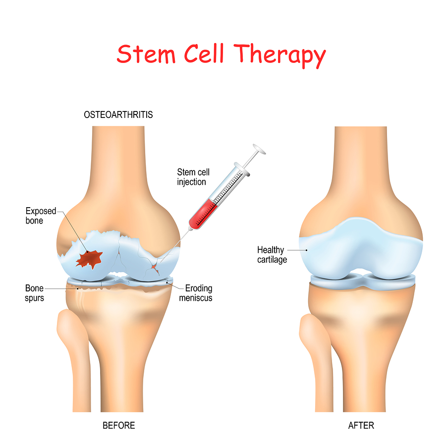
Mesenchymal stem cell injections for osteoarthritis

A fairly recent alternative approach to treating osteoarthritis is by mesenchymal stem cell injection with platelet-rich plasma activation. Researchers conducted a meta-analysis of 35 studies regarding knee osteoarthritis and mesenchymal stem cell injections in 2018. There were minor adverse events like knee pain and swelling. No serious side effects of the stem cell treatments were noted. The outcome of the treatment was classified as “very low” to “low”. The reason for this were poor study designs, large heterogeneity among the studies and wide variation among the 95% confidence intervals regarding study outcomes.

Australian study of mesenchymal stem cell injections into symptomatic knees

A short-term study from Australia examined 30 patients with mesenchymal stem cell therapy for knee osteoarthritis. There were two treatment groups. One group received 100 million adipose tissue derived mesenchymal stem cells into symptomatic osteoarthritis knee joints. Another treatment group received 100 million mesenchymal stem cells twice, namely at baseline and at 6 months after the first injection. The third group served as a control with continued conservative management. All of the groups had baseline MRI scans (magnetic resonance imaging) to assess the knee conditions. Both treatment groups had pain and inflammation at baseline. After 1 year following the initial treatment the patients had repeat MRI scans. They showed modification of the disease process in the treatment arm, while the controls had deteriorating osteoarthritis. The researchers concluded that adipose derived mesenchymal stem cell treatment of knee osteoarthritis has the potential of preventing disease progression.

Why stem cells help with osteoarthritis of the knees and the hips

Here is a research project between researchers from Palm Harbor, Florida and researchers from Serbia. The Center for Molecular Medicine and Stem Cell Research, Faculty of Medical Sciences University of Kragujevac, Serbia reviewed the literature on treatment of osteoarthritis.

The researchers explain that the physician can easily harvest mesenchymal stem cells by doing a liposuction of fat cells. These stem cells maintain their differentiation potential and there is only minor immunological rejection because of low histocompatibility antigen expression. As a result mesenchymal stem cells stay in the joint where they are injected.

Two processes that occur with injected stem cells

Two main advantages of mesenchymal stem cells stand out. Mesenchymal stem cells are capable of building up hyaline cartilage and in addition they have immunosuppressive characteristics. In comparison with controls patients who receive intraarticular injections of mesenteric stem cells have less pain and less swelling.

This review paper cites all the major animal and human studies regarding mesenchymal stem cell therapy for osteoarthritis. It concludes that stem cell therapy is most effective in mild to moderate osteoarthritis cases. It stresses the point that stem cells can regenerate joint tissues and that the inflammatory process of osteoarthritis is interrupted by the stem cells. This stabilized the osteoarthritic condition.

Steroid Injections Are Doing Harm, But Stem Cells Help

Steroid Injections Are Doing Harm, But Stem Cells Help

Conclusion

Mesenchymal stem cells derived from fatty tissue will repair tissue defects in joints afflicted by osteoarthritis. The stem cells also take care of the inflammation, which takes care of pain and swelling. Range of motion of the joint increases following stem cell treatment. Other studies have shown that intraarticular steroid injections do not interrupt the degenerative process. MRI scan studies have shown deterioration of the joint tissues following steroid injections. All in all, stem cell treatments of joints with osteoarthritis are superior to a treatment with steroid injections.

Dec
09
2017

Stem Cells Cure Back Pain

A person with chronic back pain has several treatment options, but only stem cells cure back pain. Stem cell treatment has been available in the US and Canada and many other countries for approximately 10 years.

I come from a family with a strong history of back pain (mother, maternal grandmother and maternal grandfather). They all got their back pain in their mid to late 40’s. From my growing up years I remember that they complained about chronic back pain on and off. Sometimes they had to cancel events they wanted to attend because they could not tolerate sitting. In those times there were no CAT scans or MRI scans. If you had back pain, you just had to put up with it.

My personal experience

Given my family history of back pain I was surprised that my back pain was only a more persistent problem in the last 1.5 years, but not earlier. Normally a monthly chiropractic adjustment would keep my back symptoms under control. But in the last 1.5 years I needed to see a chiropractor more often than that. I took omega-3 fatty acid supplements for the past several years (two capsules twice per day) thinking that this should halt the development of degenerative arthritis in the lower back joints. When I turned 71, it was clear to me that I was now at the point where my immediate relatives were when they were in their late 40’s. Therefore, diet, exercise, weight loss, good nutrition and supplements can only do that much for you. If there is a familiar disposition, it will eventually catch up with you.

Conventional medicine’s approach to lower back pain

I have practiced as a general practitioner for 16 years in the past. In addition I joined Workers’ Compensation for another 16 years as a medical advisor. From this clinical activity I knew of hundreds of cases first hand what the steps were in the treatment of chronic back pain. First of all, physiotherapy treatments or chiropractic treatments were the treatment protocol. In minor back pain cases this would often help the pain symptoms. Furthermore, if residual pain persisted, the patients received anti-inflammatory medication (non-steroidal anti-inflammatory drugs or NSAID’s). Finally, if symptoms continued to persist, a CT scan or MRI scan was necessary for assessment. If it showed moderate changes like my findings, the patient received intermittent physical therapy, chiropractic therapy or acupuncture therapy. 

Surgical procedures for chronic lower back pain

If there were more severe degenerative changes or spinal stenosis with severe degenerative changes, a referral to an orthopedic surgeon or neurosurgeon would be necessary. But this was often the point of no return. If the surgeon felt that the condition was severe enough to do back surgery, various procedures could follow. For disc herniations irritating one of the nerve roots, laparoscopic discectomy was the treatment of choice. For severe spinal stenosis or intractable pain from end stage facet joint disease instrumentation was an option.

Fusion surgery

Under a general anesthetic the surgeon makes an incision in the patient’s back over the lumbar spine. The surgeon identifies the diseased disc level and places stabilizing stainless steel plates over the affected facet joints or the narrowed disc space. Many people think that fusion surgery would be the end of their trouble. In many cases this can actually be the beginning of chronic back trouble. The problem is that the body is designed to move. If the surgeon takes movement away in one area of the spine, the levels above and below have to work harder. It often takes only a few months or a couple of years, and the patient is back with excruciating pain from degenerative changes in the levels above and below the previous surgery. What does the surgeon usually do? He does more fusion surgery above and/or below the previous area of surgery.

Alternatives to back surgeries

New treatment options have opened up new possibilities. On the one hand there is prolotherapy treatment that I have described under this link. On the other hand stem cell therapy is another popular regenerative technique. Prolotherapy strengthens tissues, relieves pain and increases the range of motion in joints. There is 80 to 85% full pain relief and more than 80% improvement in range of motion. Prolotherapy promotes the healing of torn ligaments and tendons. There are many suitable conditions that lend themselves to the treatment with prolotherapy like the hip, knee, shoulder, ankle, neck, lower back and elbow. With prolotherapy the physician uses hyperosmolar dextrose injections into the affected area. Current thinking is that this irritates the tissues, which mobilizes local stem cells to heal the area.

In my case I had two prolotherapy treatments of my lower back, but it did not change my lower back pain.

MRI scans of my lumbar spine

We needed to find out what was happening in my lower back. My general practitioner ordered MRI scans of my lower back in summer of 2017. There are 5 levels of the lumbar spine from L1 to S1. In my case one level of 5 was normal. The other levels showed bulging of the discs. The scans also showed signs of arthritis in the small joints adjacent to the spine. Lucky for me, there was no sign of spinal stenosis. It was not good news: overall 4 levels of my lumbar spine showed signs of  degenerative disc changes. At the same levels I also had arthritic changes in the facet joints. This was enough to consider some intervention, or I would be headed for trouble in the future.

Stem cell treatment for chronic back pain

Following the failed prolotherapy for my lower back pain I needed to figure out what to do next. The MRI scans had shown degenerative changes in the discs of the lower 4 levels of the lumbar spine. There also was arthritis in eight facet joints (two on each side of each of the four L2 to S1 levels). Conventional medicine would have offered corticosteroid injections into the facet joint areas. My experience with many patients who had this procedure was that the effect of the corticosteroid injections wore off after 3 to 6 months. If a patient had more than 3  injections, there usually was a point of no return, and fusion surgery would be next.

Best therapy for my own chronic lower back condition

For me there was no question that stem cell therapy would be the best fit for treating my back condition. In addition platelet -rich plasma and low-level laser therapy could activate the stem cells. This would be the ideal non-invasive treatment option to treat my chronic lower back pain. I had met Dr. H. Michael Weber before. He is a well-known laser expert from Germany who has a double certification as an engineer and as an internist treating various clinical conditions with laser and stem cell therapy. In addition he is an expert of regenerative medicine methods. Also, he invented and designed the laser machines himself. I set up an appointment in the fall of 2017 at his clinic in Lauenförde, Germany.

First day of stem cell treatment

On the first day fat tissue was removed under a local anesthetic from my lower left buttock area. Next a cell separator divides the tissue into connective tissue, fat cells and mesenchymal stem cells. Two blood samples were also taken from me for processing platelet rich plasma (PRP). PRP is a natural stem cell activator. Growth factors and anti-inflammatory cytokines were also part of the mix together with the stem cells.

The very same afternoon I received the stem cell mix by injection. Eight needles, four on each side, were necessary to administer the stem cell combination. I also had a treatment on a light therapy bed with red light to activate stem cells in general. The stem cell injection was a pain free procedure, as I received a shot of a  local anesthetic in the area before. After that the physician inserted laser applicators through the interstitial needles.

Laser activation of injected stem cells

The next step was to use laser treatments with 5 different colors (infrared, blue, red, yellow and green) for 10 minutes for each of the 8 interstitial needles. The laser activation and the PRP mixed with the mesenchymal stem cells were the two main stem cell activators. They are crucial for activating the stem cells. But growth factors and anti-inflammatory cytokines also aided stem cell activation.

Second day of stem cell treatment

On the second day I received an infrared light treatment over my back for 20 minutes. Following that I received light therapy bed treatment for 20 minutes. The physician told me  that all of this was to activate the stem cells further. The next step was a bone marrow low-dose laser therapy.

Bone marrow stem cell activation by low-dose laser therapy

Often stem cell therapists mix mesenchymal stem cells from fat tissue with bone marrow stem cells which they harvest before from pelvic bone marrow. Dr. Weber told me that he would do a direct bone marrow laser activation of the pelvic bone marrow instead. He anesthetized the tissue above the pelvic bone. Following this he made a small hole into the pelvic bone through which he inserted a laser applicator into the bone marrow cavity. 5 different colored lasers were again applied for 10 minutes each to activate the bone marrow stem cells. Studies have shown, as Dr. Weber stated, that low-dose laser activates bone marrow stem cells. They can be found in the blood circulation within 1 hour. This is similar to mixing stem cells in a Petri dish and then injecting it as a mix, except it is a less invasive approach.

Further activation of stem cells

Following these procedures Dr. Weber felt that another light bed therapy was necessary for 20 minutes. He also gave me a Weber medical laser watch called “Regenerate+”. This device fits on the wrist. It is programmed to generate a number of different lasers to shine against the underside of the wrist. This is the area where the ulnar and radial arteries run close to the surface. This device will shine the laser lights for 30 minutes, and the laser light reaches the arterial blood. The circulating stem cells from the stem cell therapy are receiving a further boost this way. Dr. Weber told me to use this device twice a day on an ongoing basis. The Weber medical laser watch stimulates the immune system.  Jet lag also responds to, and it can stimulate stem cells as they circulate in the blood.

Stem Cells Cure Back Pain

Stem Cells Cure Back Pain

Conclusion

Medical tourism is flourishing. I have become a medical tourist myself because I did not want to get crippled by conventional medicine regarding my lower back pain. Two days after my stem cell treatment my back pain was significantly improved. There was mild pain in the area of the fat liposuction site. Four days after the treatment the lumbar spine pain was gone. Innumerable chiropractic treatments and two prolotherapy treatments had not given me relief. Now stem cell therapy in Germany has taken my chronic back pain away in only a few days. I realize that the healing process will take 3 to 6 months to complete, but as a patient what counts most is pain relief.

What, if someone criticizes me for choosing stem cell treatment?

It is difficult to argue with success. Whether somebody criticizes me for having followed a non-conventional treatment protocol does not matter to me. My question back would be: what do you do when conventional methods fail? Are you willing to suffer chronic pain and swallow pain pills that could either get you addicted or have serious side effects? I would try stem cell therapy again, if I had a problem that does not respond to conventional therapy.

Jul
08
2017

Stem Cells For Osteoarthritis

Many clinicians have used stem cells for osteoarthritis of the knee or other joints for some time. However, objective publications about the effectiveness of stem cells are only coming out now. Both stem cell types, derived from fat or stem cells from the bone marrow, are effective. Most doctors are using stem cells from fat (mesenchymal stem cells), because they are so much easier to harvest.

CNN reported about a man, Bill Marlette who had lost one of his arms in the past. He ended up overusing the other arm and as a result developed end-stage osteoarthritis in his wrist. He could not find relief with conventional methods of anti-inflammatories and pain pills. Next he went to a stem cell expert in Munich, Germany who treated him with mesenchymal stem cells from his fatty tissue. Only one treatment took away his chronic pain and helped him regain his wrist mobility.

Approval of stem cell therapy in Germany

Prof. Dr. Eckhard Alt, an expert in regenerative medicine has previously treated patients with end stage osteoarthritis and had good clinical outcomes with it. As a result the German regulatory agency has approved his treatment protocol.

Dr. David Pearce, executive vice president for research at Sanford Health in South Dakota said that Prof. Dr. Eckhard Alt was the first one to use fat cells as a source of mesenchymal stem cells to treat osteoarthritis. He went on to say: ”Those stem cells don’t have to be programmed in any way, but if you put them in the right environment, they will naturally turn into what the cell type around them is.” The physician harvests the stem cells through liposuction. An enzyme mixture is necessary to separate the stem cells from fat cells, oil and connective tissue. A cell separator can also help separating the stem cells from the rest of the cells and tissue.

A case of wrist osteoarthritis

As I mentioned before only one injection was necessary to relieve the chronic pain of Bill Marlette’s wrist. Since his return the doctors in the US have followed Bill closely. They took MRI scans and noted that the bony cysts associated with the severe arthritis have disappeared. His wrist and hand strength have returned to normal. The pain almost disappeared. There were no side effects whatsoever. Because the stem cells are of the same tissue type as all his other cells of his body, one would not expect any tissue rejection by the immune system. Bill Marlette did not need any pain pills following the procedure in August 2016. And he says: “I have more range of motion with my wrist, shaking hands didn’t hurt anymore,” he said. “My wrist seems to continue to improve, and there’s less and less pain all the time.”

Past experiences treating osteoarthritis with mesenchymal stem cells

A 2014 clinical trial from Korea involved 18 patients with osteoarthritis of the knee where adipose mesenchymal stem cells were injected. The high dose group did best. After 6 months there was significant improvement, also confirmed by arthroscopy. The previous cartilage defect in the femoral and tibial condyles had decreased in size. Range of motion in the knee joints and pain had also improved. There were no adverse effects from the treatment.

Chinese study

Mesenchymal progenitor cells have the propensity to develop into cartilage. At the Shanghai Medical College, Fudan University Shanghai, China the following experiment took place in 2015. The researchers grew human adipose mesenchymal cells in vitro. Later they injected these mesenchymal progenitor cells into the knees of rabbits with experimentally produced osteoarthritis. Despite doing xenotransplants (human cartilage to rabbits) with known HLA differences the cartilage grew and cured the osteoarthritis of the rabbits. The new cartilage had human HLA markers while the rabbit cartilage underneath had rabbit HLA markers. At 16 weeks the researchers examined the tissues under the microscope and another exam involved the HLA marker testing.

Tehran study

A study from Tehran, Iran was carried out on 18 patients with ankle, knee and hip osteoarthritis in 2015. Physicians injected stem cells from the bone marrow into the osteoarthritic joint. The doctors followed the patients and ordered occasional MRI scans for 30 months. All of the patients had improved significantly with regard to their joint function and pain. The MRI scans also showed thickening of the joint surfaces from new cartilage production.

French/German study

In a 2016 joint French/German study 18 patients with end stage knee osteoarthritis were treated with stem cells. The stem cells came from adipose tissue that went through a cell separator. Physicians injected the mesenchymal stem cell fraction into the osteoarthritic knees. This was a phase I study to rule out any adverse reactions, but none were evident. It also established that there were significant positive improvements in pain and mobility with regard to the affected knees.

General remarks about how stem cells heal osteoarthritis

The example above with end stage osteoarthritis of the wrist was just one example of where osteoarthritis can strike. Perhaps the more common other locations are hips, knees and the facet joints of the lower lumbar spine (causing chronic lower back pain).

The same treatment procedure, which Bill Marlette’s wrist benefitted from is useful for all these other locations. The common factor in osteoarthritis is that the cartilage is getting thinner and thinner until bone rubs on bone causing excruciating pain. It is here where mesenchymal cells can come to the rescue. The stem cells will assess what requires a repair after injection into an affected joint. They recognize that there is a lack of cartilage. Then they transform themselves into chondrocytes, which are cartilage-forming cells. How can stem cells do that? They come with a program to replace missing cells, particularly cartilage and bone cells. But if they are within fatty tissue, they cannot act within a joint that has osteoarthritis. The doctor has to transport the mesenchymal cells into the joint where they can then begin their healing function.

Other methods to treat osteoarthritis

Stem cells are only one of several regenerative treatment modalities for osteoarthritis. Another method are platelet-rich plasma (PRP) injections. Platelets have a lot of anti-inflammatory substances in them and also growth factors that can stimulate stem cells contained in the synovial membrane, the lining of any joint. To get PRP plasma, it is necessary to spin down blood and harvest the PRP fraction with a syringe. After three PRP injections were given into the knees of 90 patients with end stage osteoarthritis these patients were followed for two years.

In the beginning before treatment 100% of the patients had symptoms. After one year following the treatment with PRP their knee functions were normal in 67% of them. After two years only 59% had normal knee function. The investigators pointed out that this treatment modality initially helped to a certain point, but then the effects were slowly fading away.

Stem cell treatment of osteoarthritis of the knee

The literature on either bone stem cells or fat stem cell use for osteoarthritis of the knee in man is still sparse. Nobody has done larger clinical trials. Part of the reasons could be that total knee and total hip replacement in orthopedics is very lucrative. We are still in a symptomatic treatment mode. Physicians treat osteoarthritis conservatively with anti-inflammatories and pain pills. When bone rubs on bone, there can be excruciating pain. The physician refers the patient to an orthopedic surgeon who likely will do invasive surgical procedures. My own impression in general practice in the past is that these procedures do not always turn out the way they are supposed to work. Following total hip or knee replacement joint swelling often remains; pain issues are still there. There can be unequal height issues, balancing problems and so on.

Here is a review of mesenchymal stem cell therapy for osteoarthritis.  This publication is very conventional medicine. An attitude change by conventional medicine would be useful to catch up with what is happening in real life. Some patients will travel abroad to Munich as Bill Marlette did. But others may travel to other places like India, Mexico or wherever medical tourism takes you. Regenerative medicine is there to stay.

Stem Cells For Osteoarthritis

Stem Cells For Osteoarthritis

Conclusion

We have learnt about a case of severe osteoarthritis of the wrist that has been cured in Germany with one injection of mesenchymal stem cells. More common than wrist osteoarthritis is osteoarthritis of the hips, knees and the facet joints of the lower lumbar spine. The same stem cell therapy can be given for osteoarthritis in these locations. I find it very strange that progress in stem cell treatments is so slow in the US. The FDA has decided to be open to clinical trials with stem cell treatments, but progress seems to be much slower than in other countries. Why? We may never know. In the meantime, patients may seek treatments in other countries where such treatments are offered. In real estate sales there is a saying: “Buyer beware”.

Be cautious, if you get treated abroad

The same goes for stem cell treatments in another country. Should you contemplate doing this, do your homework; ask about the qualification of the treating physician, about safety records and whether the local authorities have approved this procedure. In the case of Bill Marlette’s osteoarthritis of the wrist the procedure in Munich, Germany had been accepted by the European equivalent of the FDA, the European Medicines Agency. Safety is top priority, effectiveness is next.

Nov
02
2013

MS Is A Multifaceted Disease

A new study was recently released that showed that MS is a multifaceted disease. A significant number of people without multiple sclerosis have narrowing of their neck veins. There is a new theory that chronic cerebrospinal venous insufficiency due to narrowing of veins outside the skull may be responsible in causing MS in a significant percentage of patients. Using venograms in MS patients and in controls without MS a recent study from the University of British Columbia, Vancouver, BC showed that normal controls also had narrowing of neck veins and the authors felt that this invalidated the vascular theory of MS. This story is based on this Lancet publication.

Brain oxygenation is what counts

What was not mentioned in this publication was that venous blood flow on the surface of the brain can get obstructed inside the skull. It’s all about brain tissue oxygenation; if the brain gets enough oxygen, all is well. If there were a lack of perfusion due to venous congestion inside or outside the skull, the patient would be in trouble.  I will discuss this further below.

In this blog I will discuss first how to diagnose MS, then mention some newer studies about neck vein circulation with SPECT scanning. I will then review various causes of MS and return to a discussion of the Vancouver study from above.

How MS is diagnosed

The physician can combine a number of tests to diagnose MS. This includes the patient’s symptoms such as balancing problems, double vision, memory problems, fatigue etc. Neurological examination, imaging studies like MRI scanning, lumbar puncture to examine the cerebrospinal fluid and evoked potential studies is what a neurologist orders. The physician requires all of these findings to decide whether the criteria for making a diagnosis are in keeping with all the symptoms of MS. In 2001 an international neurological panel developed the McDonald criteria for diagnosing MS, which were revised in 2010.

MS Is A Multifaceted Disease

MS Is A Multifaceted Disease

Newer ways diagnosing perfusion problems in MS patients (SPECT scan)

One of the newer functional scans is a SPECT scan. It shows areas where there is a lack of blood supply to the brain, but can also identify areas where too much blood circulates. Here is a site where the technique of the SPECT scan is reviewed in more detail.

SPECT scan results in MS patients

SPECT scans in MS patients showed a significant reduction in blood flow to the frontal lobes and to the left temporal lobe. Reduced activity of the left temporal lobe on SPECT scans correlated with MS patients having a deficit in verbal fluency and having a problem with verbal memory. This indicates that a reduction in blood flow to these areas of the brain associates with developing MS.

Perhaps a SPECT scan of the brain (which is where the action of MS is) may be a better indicator for MS than looking for veins in the neck by ultrasound or venograms, as SPECT scans look directly at brain perfusion. The question is whether these blood circulation problems in MS patients may cause deficiencies in the brain of oxygen, nutrients and possibly of other internal mediators.

Known causes of MS

There are a number of known causes of MS, which I will review below.

Autoimmune disease

As this link shows, MS is a disease due to inflammation of the brain. The area where there is inflammation leads to demyelination from loss of the myelin sheaths, which causes the white lesions visible on MRI scans of the brain. One such cause is an allergy to wheat and wheat products. Gliadin antibodies and anti-tissue transglutaminase antibodies were positive in a significant number of MS patients, but not in controls. This suggests at least in part that immunological causes are at play. I agree with this blog that describes that there is significant evidence that gluten intolerance can lead to MS and the positive tests that were found by researchers are likely just the tip of the iceberg.

Overlap of celiac disease and MS

Dr. William Davis describes in Ref. 1 and 2 that you can have celiac disease with no gut symptoms, in other words a person can develop autoimmune symptoms from gliadin and gluten sensitivity without diarrhea or bowel cramps. Dietary lectins, particularly the ones found in wheat lead to leaky gut syndrome and subsequently to autoimmune diseases. One of these autoantibodies can cause MS by destroying the myelin sheath. In a mouse model Mayo Clinic researchers have shown that an antibody injection can be used to block autoantibodies against the myelin sheath. Investigations are ongoing with regard to whether this type of treatment would work in humans as well.

Genetic factors

It is known that a human leukocyte antigen (HLA -DRB1) shows an association with the risk for developing MS. Caucasians have a higher risk of developing MS and they also carry the HLA-DRB1 antigen more often. Another genetic factor is a variation of the IL7R gene. You can read about it under the HLA-DRB1 link. On average the risk of getting MS in the general population is about 10 to 20%.

Nutrition and dietary factors

It has been described that vitamin D3 levels when obtained from in MS patients are low. Vitamin D3 can prevent against MS to a certain degree, so does sun exposure. In countries where malnutrition is common, MS occurs more often.

According to Dr. Terry Wahls who is a clinical professor of medicine at the University of Iowa Carver College of Medicine, a diet of vegetables, fruit, meat, no grain, no dairy, no sugar, no corn and no potatoes can cure MS. Dr. Wahls herself had severe MS in the past and cured it with the help of this diet!

Infections

Certain infections can cause MS. Probably the best correlation was found between the mononucleosis virus (Epstein Barr virus) and the later development of MS.

Chronic cerebrospinal venous insufficiency

According to Dr. Zamboni from Italy clogged veins in the neck can cause MS. Dr. Zamboni placed stents into neck veins that showed narrowing.  He found that about 50% of MS patients had improvement with the placement of stents. This allows the blood from veins around the brain to drain normally. This could improve brain circulation in the areas described above where SPECT scans detected a lack of blood supply to certain parts of the brain.

Discussion of the Vancouver publication

It is important to note that certain areas of the brain were not circulating blood as well as others. SPECT scans depicted the blood circulation of the brain. 50% of patients with chronic cerebrospinal venous insufficiency experienced a cure from MS with simple venous stent procedures. This is remarkable. Sure, the Vancouver researchers found that normal controls also have a significant amount of venous abnormalities in their necks. But this does not explain the successes in those MS patients who got better with a simple venous stunt procedure. We also need to be cognizant that Big Pharma sponsored the Vancouver study researchers.

Measuring brain circulation in MS patients

SPECT scans on both the control groups and the experimental MS groups before and after stent procedures need to be done. This way we know whether the brain circulation following stent procedures improved or not. However, this is what I would have expected to see. In other words more research is necessary by other investigators. The question they need to answer is whether or not the surgical stents provided help. Did the surgical procedure help to normalize their brain circulation or not?

Conclusion

Our knowledge regarding MS is getting more multifaceted as new research is emerging. Diet appears to be a major contributing factor, as vitamin D3 is essential for normal brain function and for a normal immune system. At the same time researchers identified grains and wheat as a cause of MS in a subgroup of patients. Leaky gut syndrome can cause autoimmune antibodies, which subsequently can bring on MS. Avoid the foods Dr. Wahls described as being causative in developing MS and you can improve MS remarkably or get cured. The same is true for avoidance of wheat and wheat products as Dr. Davis described.

Multifaceted causes of MS

In my opinion not every MS patient benefits from a stent, but vitamin D3 deficiency or a history of mononucleosis infection in the past does not explain the causation of every MS case. We simply do not have all of the answers yet. But we do have enough information to thoroughly investigate MS patients;  the treating physician will then use clinical judgment to decide which treatment would be the most suitable one for an individual MS patient.

More information on multiple sclerosis: http://nethealthbook.com/neurology-neurological-disease/multiple-sclerosis/

References

1. Stern: Massachusetts General Hospital Comprehensive Clinical Psychiatry, 1st ed. “DSM-IV SUBTYPES OF MDD”. Copyright © 2008 Mosby

2.  William Davis, MD: “Wheat Belly. Lose the Wheat, Lose the Weight, and Find Your Path Back to Health”. HarperCollins Publishers LTD., Toronto, Canada, 2011.

3.  William Davis, MD: “Wheat Belly Cookbook. 150 Recipes to Help You Lose the Wheat, Lose the Weight, and Find Your Path Back to Health”. HarperCollins Publishers LTD., Toronto, Canada, 2012.

Last edited Nov. 7, 2014

Feb
01
2007

Avoiding Back Pain By Relaxed Sitting

This article is about “avoiding back pain by relaxed sitting”. Notably, parents and educators often reprimand children and students to “sit up straight”. The results however are not that favorable. Truly, a 90 degree angle in this position is causing problems. Specifically, it causes the largest movement of the spinal disc and a lot of strain. To put it another way, conventional wisdom has it that sitting up straight avoids back pain. It must be remembered, this has important implications for a large population group that has work situations with mostly sitting jobs. In addition, consequences of bad posture are also quite visible: lower back pain is the leading cause of disability in the U.S. and Canada and a major cause for missed work days.

Improper posture and back pain are related

Investigators have presented evidence, that a 135 degree body/thigh sitting position, leaning back, is the optimal position to avoid back pain. The author of the study, Dr.W. Bashir from the department of radiology and diagnostic imaging at the University of Alberta Hospital in Edmonton emphasizes, that the angle of posture has been under question for some time, and improper posture and back pain are related.

Positional MRI scans 

Positional MRI scans have shed more evidence on the spine. There are special MRI machines that take images when the patient is not in a supine position, but in a position that allows the patient free motion during the test. Spinal angles can be measured and spinal disk movement can be observed in various positions and angles.
The 90 degree angle showed most spinal disk movement, creating stress for the spine. Another unfavorable position is a slouching posture.

Avoiding Back Pain By Relaxed Sitting

Avoiding Back Pain By Relaxed Sitting

Scan results showed a reduction in spinal disk height, signifying more wear and tear of the lumbar spine. Spinal disk movement was least pronounced with the 135 degree angle. This more relaxed position poses less strain on spinal disks and the associated muscles and tendons. Employees sitting comfortably versus sitting up straight can make the job place friendlier to people’s backs.

More about back pain: http://nethealthbook.com/arthritis/lower-back-pain/

Reference: The Medical Post, January 16, 2007, page17

Last edited November 2, 2014

Mar
01
2005

Magnetic Therapy For Depression

Depression is a psychiatric condition, which is experienced by a lot of people. It is more than just a transient feeling of the “blues”. Depression can be a chronic and disabling disease, and in severe cases there is the risk of suicide. Great advances have been made from removing the social stigma from psychiatric illness, and cognitive therapy and effective medications can help most the patients to lead full and productive lives.
For a few patients however, depression can be drug-resistant and as a result, effective therapy will be much more difficult.
According to research by Dr. Gary Hasey at Mc Master University in Hamilton, Canada, magnets may have a future role in the treatment of mood disorders. TMS (standing for transcranial magnetic stimulation) has shown promising results in the treatment of some types of depression. A so far unpublished study found that 27% of 50 patients with drug -resistant depression achieved full response with TMS.

Simulated treatment in a control group did not produce this result in any of the patients. MRI scans have shown that depressed people have below-average brain activity in the frontal cortex of the brain. A magnetic field, which is created by passing an electric current through a hand-held magnetic coil, is aimed at the patient’s pre-frontal cortex, which stimulates the brain activity in this area. This treatment is vastly different from the well-known electro-shock treatment (ECT), where the patient needs sedation and close observation in a hospital setting. Contrary to this, TMS can be done without sedation.

Magnetic Therapy For Depression

Magnetic Therapy For Depression

The patient is conscious and can resume his normal activities after the treatment. The therapy was first discovered in the 1980’s, but a lot of research had been necessary before treatment could be made available. Other trials are also running in Great Britain, and promising results have been published in the medical paper “The Lancet”. Dr. Hasey cautions that there are still some details that have to be worked out. In the meantime Health Canada has approved TMS as treatment for drug-resistant depression.

More information about depression: http://nethealthbook.com/mental-illness-mental-disorders/mood-disorders/depression/

Reference: The Medical Post, February 1,2005, page 28

Last edited October 27, 2014

Incoming search terms: