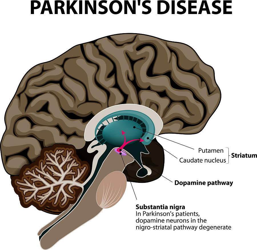
Dec
17
2022

Mast Cell Activation Syndrome

Mast cell activation syndrome (MCAS) also goes by the name of systemic macrocytosis. That is to say it is a syndrome where mast cells are multiplying abundantly and secreting the inflammatory substances histamine, leukotrienes and cytokines. Certainly, people who suffer from MCAS can get severe anaphylactic reactions, but an epinephrine injection can often stop this.  Indeed, the physician must look for potential triggering factors like alcohol, spicy foods, exercise, insect stings, possible heavy metal accumulation or certain medications. In some cases heavy metal accumulation could also be a factor that triggers mast cells to release histamine.  In these cases a series of 20 chelation therapy sessions would be stabilizing.

Symptoms of mast cell activation syndrome

  • When there is skin involvement in patients with mast cell activation syndrome, they get flushing, itching and skin rashes.
  • With gastrointestinal involvement patients experience nausea and vomiting, bloating, abdominal pain, diarrhea and reflux (GERD).
  • Patients with neurological symptoms develop brain fog, headaches, cognitive problems, tremors and anxiety/depression.
  • When MCAS affects your endocrine glands, you may develop bone pain, bone lesions or weak bones.
  • Patients where the heart is affected may be fainting, their blood pressure may fluctuate between with high or low readings and they may experience heart palpitations.
  • When your respiratory system is affected, your lungs may be wheezing and you may develop nasal congestion.
  • More symptoms
  • The most dangerous symptom is anaphylaxis. This is a life-threatening allergic reaction where your air way entry could close off.
  • Any of these symptoms can get triggered by heat, cold or temperature changes. Stress, friction, insect bites or stings can also trigger a reaction. Additional factors can be environmental odors or perfumes, certain foods or medicine, alcohol and contrast dyes.

Diagnosis of mast cell activation syndrome

The most appropriate specialist to see is an allergist or immunologist. Other specialists could be a dermatologist, gastroenterologist, hematologist or endocrinologist. You will need blood tests like a serum tryptase level, which is a marker for mast cell burden. It is best to get a baseline tryptase level and also get a tryptase level after a mast cell reaction. In addition, you need a 24-hour urine collection for a number of mast cell activators. Depending on where your mast cell activation syndrome is located you need a skin or bone marrow biopsy.

More possible tests

The physician may decide to do an endoscopy or colonoscopy of the gastrointestinal tract. The pathologist can do several staining procedures with biopsy material to specifically look at mast cells. If there is a strong family history of mast cell activation syndrome the physician may decide to do genetic tests. In order to assess mast cell damage, your doctor may order a bone density test and bone scans including CT scans of the abdomen and chest.

Treatment of mast cell activation syndrome

The treatment of mast cell activation syndrome consists of a combination of multiple steps. First, if there is a life-threatening anaphylactic reaction, the patient applies an epinephrine injection. The physician taught the patient earlier how to do an epinephrine injection. This stops the sudden, rapid release of mediators from mast cells. After the epinephrine injection the patient needs transport to the nearest ER of a hospital for follow-up care. It is important that any patient with this syndrome should carry injectable epinephrine( an Epi-Pen) at all times.The purpose of treatment against mast cell activation syndrome is to block reactivity of mast cells or to stop the effects of mast cell mediators.

A number of medications are available to do this.

You can lower your risk of getting mast cell activation syndrome by watching your diet. Here is a list of the foods that will protect you:

Mast Cell Activation Syndrome

Mast Cell Activation Syndrome

Conclusion

Mast cell activation syndrome is a complex disease entity. Often there are several factors that contribute to this. Conventional medicine still cannot offer a treatment modality that will cure this condition, the only possibility is to control it. The physician must therefore use a combination of treatment modalities in order to help the patient with this condition. In cases of heavy metal accumulation several treatments with chelation therapy are beneficial. With an acute anaphylactic reaction, the

Mast Cell Activation Syndrome applies an epinephrine injection, which will stabilize the condition. But the patient should now follow this up with a series of blood tests in the emergency department of a hospital.

Treating mast cell hyperactivity

The purpose of treating mast cell activation syndrome is to block reactivity of mast cells or to stop the effects of mast cell mediators. H1 and H2 antihistamines help for gastrointestinal hyperactivity. Cromolyn sodium and ketotifen are mast cell stabilizers. Leukotriene inhibitors such as montelukast help to stabilize the mast cells to not secrete cytokines, which cause inflammation. Aggressive mast cell disease may require chemotherapy treatment, similar to what is needed to treat cancer.

The purpose of treatment is to help the patient control the mast cell hyperactivity. At this time medical science does not have all the answers. Unfortunately, at this point conventional medicine has no cure for this syndrome, but it can be managed with a lot of attention to the symptoms.

Incoming search terms:

Nov
19
2022

Lack of Sleep Harms the Immune System and Causes Inflammation

A research group from Boston, MA and New York, NY found that a lack of sleep harms the immune system and causes inflammation. This was summarized in this CNN article.

Specifically, they first conducted experiments with a mouse model. They studied the effects of sleep disruption and sleep deprivation and could later confirm identical changes in man. The observation was that a lack of sleep caused the hematopoietic cells in the bone marrow to proliferate, but the cell diversity was less than in people with normal sleep patterns. The same pattern of bone marrow proliferation was present in mice. This research was published Sept. 21, 2022 in the Journal of Experimental Medicine.

Chronic sleep deficit

A chronic sleep deficit caused chronic inflammation and eventually autoimmune diseases. Again, this was a pattern present in both the mouse model and in humans. Next the researchers observed what happened with sleep recovery. In the past it was assumed that with sleep recovery all of the physical changes from sleep deprivation would disappear. However, the opposite was true: both in mice and in humans the bone marrow stimulation and the lack of cell diversity persisted.

In the mouse model the researchers could show that there were permanent epigenetic changes, which were caused by sleep deprivation. The same is true with humans, but this is more difficult to show than in the mouse model. The researchers came to the conclusion that sleep deprivation stimulates bone marrow maturation, but restricts the clonal differentiation. In doing so the body initiates inflammation, which becomes chronic even with sleep restoration.

Human sleep studies

There were 14 volunteers that were the test subjects. One group was the normal sleep control. The other group underwent chronic sleep deprivation. Each group did this for 6 weeks. There was a 4-to-6-week washout period. Following this the previous normal sleep group started a 6-week sleep deprivation program. On the other hand, the prior sleep-deprived group switched to 6 weeks of normal sleep. All of the participants had daily late afternoon blood tests.

There are many sleep disruptions, which cause a sleep deficit

In modern life sleep gets disrupted in many ways. There can be sleep fragmentation, sleep restriction, jet lag, obstructive sleep apnea (OSA), and insomnia.

People with these conditions often oscillate between these various types. They may have a few days of normal sleep, but then have sleep deprivation again for a few days. Every time they have sleep deprivation the bone marrow enhances hematopoietic activity. Normally there is a high leukocyte number in the blood at the end of the day and in the morning a lower leukocyte count. But with sleep deprivation there is a high monocyte count in the blood that stays high even when subjects switch back to a normal sleep pattern.

Epigenetic effect of sleep deprivation on bone marrow cells

The authors found that sleep deprivation affects the genetic control of hematopoietic cells in the bone marrow. They called this the epigenetic effect of sleep deprivation. This is responsible for the evening leukocyte response, the monocytosis and the tendency for autoimmune diseases. They summed this up by saying: “Our findings support the hypothesis that periods of poor sleep, even if followed by sleep recovery, have sustained consequences on immunological health.”

Lack of sleep harms the immune system and causes inflammation says the literature

There is ample evidence that a lack of sleep causes cardiovascular disease, diabetes, depression and more frequent infections. Healthy sleep is important when you want to age well without complications. But enough sleep is also necessary to prevent obesity, diabetes and cardiovascular disease. Experts consider getting enough high-quality sleep as essential as a balanced diet and regular exercise.

Lack of Sleep Harms Immune System and Causes Inflammation

Lack of Sleep Harms Immune System and Causes Inflammation

Conclusion

So far, most researchers believed that when you miss some sleep for a few nights that a afternoon nap or a few nights of longer sleep would compensate for the sleep deficit with no sequelae. Think again, because new research from a group in Boston, MA and New York, NY found that lack of sleep harms the immune system and causes inflammation permanently. Sleep deprivation stimulates the bone marrow cells to multiply and causing proliferation of monocytes, called monocytosis as well. Despite afternoon naps and recovery sleep this condition remains  and can lead to autoimmune diseases. All this was unknown up to now. Our bone marrow cells need regular sleep hours to stay diversified and to optimally fight infections in the body. This prevents autoimmune diseases and keeps our defenses against viral diseases strong.

Feb
13
2022

How healthy are Carbohydrates?

A recent review article asked: how healthy are carbohydrates? The three food components that occur in natural food are carbohydrates, fats and protein. Among the carbohydrates it is important to distinguish between simple carbohydrates (such as sugar) and complex carbohydrates. Simple carbohydrates are readily absorbed into the blood, which causes an insulin peak. After a few hours the peak is gone, and there is a “crash”. You will know the feeling of feeling hungry just a few hours after eating doughnuts! Complex carbs like peas, beans, fruit and vegetables take longer to get digested. The final breakdown product of the digestive process is sugar as well. But this process takes longer meaning that the concentration of sugar in the blood is much lower. There is also no “crash”. The result is that complex carbs cause less insulin secretion into the blood.

Long-term effect of eating too much sugar

Integrated over several decades of life, this means that a person who constantly consumes beverages with sugar and snacks containing sugar is at a higher risk of developing type 2 diabetes. In contrast, a person who eats well balanced meals where the insulin secretion is low, will not develop diabetes and have much less hardening of the arteries. This translates into a lower risk to develop heart attacks and strokes.

The types of carbohydrates

Before I discuss the health effects of various carbohydrates, we need to look at the types of carbohydrates.

Simple carbohydrates

Table sugar is a disaccharide, which consists of one molecule of glucose and one molecule of fructose.

The enzyme amylase very quickly breaks down the chemical bond between fructose and glucose and creates these monosaccharides in the small intestine from which they are rapidly absorbed. Milk sugar is a disaccharide, which consists of a molecule of glucose bound to a molecule of galactose. Milk contains 2 to 8% of milk sugar. We have to watch these simple carbs, because they trigger insulin production and lead to accelerated hardening of the arteries, heart attacks and strokes.

Complex carbohydrates

In contrast, complex carbs are healthy, because they take some time to be digested in the digestive tract. They consist of polysaccharides, long chains of sugar molecules. Both starches and dietary fiber consist of complex carbohydrates. They often are present in vegetables and many fruit. Complex carbs slow down the absorption of their breakdown products and minimize the insulin response. Dietary fiber is the indigestible part of fruit and vegetables. It contributes to good gut health as the beneficial gut bacteria can multiply on the fibre particles.

Healthy carbs

When simple carbohydrates dominate in our food intake, we are in trouble because they are loaded with calories. Overconsumption of them leads to weight gain and obesity, to diabetes, heart attacks, strokes and even cancer. On the other hand, consumption of complex carbohydrates is healthy. We get them from eating apples, bananas, berries, vegetables like spinach, tomatoes and carrots. Other healthy complex carbs are whole grain flour, quinoa and brown rice. Black beans, lentils, peas and garbanzo beans are also healthy complex carbs. Dairy products like low fat milk, yogurt and ricotta cheese are healthy as well.

Mediterranean diet as an example of a healthy, balanced diet

In 2019 a study was published where women with polycystic ovary syndrome (PCOS) were either put on a Mediterranean diet or not. This study showed that a Mediterranean diet was anti-inflammatory, reduced insulin resistance and reduced testosterone levels in PCOS patients.  But the same is true in a general population. The Mediterranean diet is one example of a healthy, balanced diet with complex carbohydrates. It prevents insulin resistance, inflammation and hormone disbalance. Other diets have similar effects like the DASH diet, the Zone diet and the Pritikin diet.

Quantity and quality of your food intake matters

A 2018 study from India showed that it matters how many carbohydrates we consume.  On average Indians eat a diet with 65-75 percent of calories coming from carbohydrates. Many of these carbs are the unhealthy simple carbohydrates. How healthy are  carbohydrates? The authors recommended to reduce complex carbohydrates to 50-55% and to add 20-25% protein, mostly from vegetable sources and add 20-30% from fat. The fat consumption needs to include monounsaturated fats (e.g., olive oil, nuts and seeds). Among the carbs a lot of green leafy vegetables help to balance the diet. This prevents the development of type 2 diabetes, heart attacks and strokes.

The above addresses the issue of quality of food. But it is also important what quantity of food we are eating. This is where counting or estimating calories comes in. If we overeat, we will very quickly gain weight and eventually can develop obesity.

The glycemic index and glycemic load

In order to help you with the choice of right carbohydrates the glycemic index was developed.

Here is another reference about the glycemic index/glycemic load.

In table 1 towards the end of the last link you find a column designated “GI” for glycemic index. All the foods that have a value less than 55 are foods that you can eat freely.

Problematical carbohydrate foods

Baked russet potatoes and boiled potatoes are very high on the GI index list. Puffed rice cakes, doughnuts, jelly beans and corn flakes measure high on the glycemic index list. But water melons, dried dates, white bread and white rice are also items to be avoided.

You best avoid anything with a glycemic index above 55. The column to the right of GI shows you a serving size and the last column on the right the glycemic load. The lower the glycemic load per serving, the better it is for your health. The glycemic index and the glycemic load are useful concepts of helping you to sort out your diet items.

My wife and I used this in 2001 to shed weight. We both lost 50 pounds (=22.72 kilograms) each in a period of 3 months.

Fasting mimicking diet (FMD)

According to Dr. Longo intermittent fasting stimulates the stem cells of the bone marrow. This leads to new clones of lymphocytes (B cells and T cells), which are part of the immune system. Your immune system becomes stronger from this.

Dr. Longo has done detailed mouse experiments, which inspired him to develop a new diet plan. Patients would receive a fasting mimicking diet (FMD) on 5 consecutive days per month. The rest of the month consists of a normal, balanced diet. 5 days of the month the person consumes a low 600-800-calorie diet. This reduced calorie intake is enough to ensure adherence to the diet, but low enough to lead to enormous positive metabolic changes including youth-preserving stem cell stimulation.

I am following the FMD

I have followed a FMD since December 2017. It helps me to keep my weight (BMI) in the 21 to 22 range. I feel more energetic and have managed to stay in good health.

The above chapter on the FMD was previously published here.

How healthy are Carbohydrates?

How healthy are Carbohydrates?

Conclusion

Healthy eating consists of 50-55% calories from complex carbohydrates; add to this 20-25% protein, mostly from vegetable sources and add 20-30% of total calories from fat. The fat consumption needs to include monounsaturated fats (e.g., olive oil, nuts and seeds). Unfortunately, most “convenience foods” (=processed foods) are incompatible with a healthy lifestyle. They contain too much simple carbs (sugar). Many people live on 65-75 percent of calories coming from simple carbohydrates, which are too many carbs. It should be complex carbs that digest slower and that do not induce insulin resistance. The glycemic index and glycemic load are useful concepts to help you chose the right foods that keep you healthy. The fasting mimicking diet can help you to take the last few pounds off that may be difficult to shed. Weight loss and weight maintenance are possible when you choose the right foods.

Apr
03
2021

Pollen Allergies Make Covid-19 Infection Rates Worse

A recent study showed that pollen allergies make Covid-19 infection rates worse. This was published in the Proceedings of the National Academy of Sciences (PNAS) in March 2021.

The study determined that airborne pollen exposure enhances susceptibility to respiratory viral infections. Specifically, this includes SARS-CoV-2 infections as well. There were 130 test sites in 31 countries across 5 continents where measurements were made. Pollen concentration, air humidity and temperature, population density and lockdown effects on Covid-19 figures were measured. In countries with high pollen counts, high humidity and higher temperatures the Covid-19 rates were up to 44% higher than in countries with low pollen counts and colder climates.

PNAS study in more detail

In the following I am discussing the PNAS study in more detail. The SARS-CoV virus from the SARS epidemic in 2002 and the present SARS-CoV-2 virus are both capable to suppress the body’s interferon response to either virus. Additionally, there are intracellular proteins with the name “inflammasomes”, which the SARS-CoV-2 virus activates. With excessive activation this causes a cytokine storm, where inflammation spreads through the whole body. In the blood this leads to disseminated coagulopathy with multi organ failures. In the lungs severe acute respiratory syndrome occurs with severe viral pneumonia. On average mortality is 3.4%.

Tree and weed pollen can weaken the immune response

A study from South Korea examined what happens with exposure in asthmatic and allergic school-aged children to tree and weed pollen. https://www.sciencedirect.com/science/article/pii/S0091674919311856.

Allergic reactions make allergic children more prone to rhinovirus infections by reducing interferon in the blood. In addition, allergic reactions stimulate inflammasomes. When the SARS-CoV-2 virus affects an allergic child, both interferon depletion and excessive inflammasome activation make Covid-19 much more severe than in a child without allergies.

Warm spell in the Northern Hemisphere

On March 12, 2020 the WHO announced the Covid-19 pandemic when over 33% of the world’s countries were affected by the SARS-CoV-2 virus. However, at the same time there was a large-scale warm spell across the Northern Hemisphere with tree pollens being distributed across the same regions. This resulted in an exponential increase of Covid-19 cases. The researchers determined that the rates of Covid-19 infections were highest in areas where there was a high tree pollen count, crowding of people and high humidity/temperatures. The researchers used data from 248 airborne pollen monitoring sites in 31 countries. The highest exponential growth rates of Covid-19 occurred in the countries with the highest pollen counts. 6 out of 8 countries studied with regard to high pollen counts showed a significant correlation with regard to Covid-19 infections in excess to just person-to person virus transmission.

Population density and lockdown affecting daily SARS-CoV-2 virus rate

Some countries had a complete lockdown when rates of infection were high. This reduced transmission of the SARS-CoV-2 virus by 50%. Those countries with only a partial lockdown still experienced a significant reduction of infection rates. Rural areas had significantly less daily SARS-CoV-2 virus rates compared to very densely populated cities.

The researchers observed the following:

  • There was a lag effect of 4 days between the increase of pollen concentration in the air and infection increase with the SARS-CoV-2 virus
  • Pollens in the air caused infection rates of SARS-CoV-2 to rise by 10 to 30%, but in some high pollen areas even up to 44%.
  • Lockdowns reduced infection rates of SARS-CoV-2 by 50%
  • Higher environmental temperatures and higher humidity of the air also increased infection rates of SARS-CoV-2, although this may have occurred indirectly by increasing the pollen count in the air

Discussion

  1. The authors added a thorough discussion of the multiple factors regarding the increase of the infection rate of Covid-19 in 2020. They pointed out that climatic factors, air pollutants, or pollen, often exert their effects at the same time. They quantitated the contribution of the pollen count in the air easily. In contrast, pollution and climatic factors were not predictable in their effects.
  2. The infection rate of the SARS-CoV-2 virus always lagged behind the increase in pollen count by 4 days. The researchers observed this in all those countries where increasing pollen counts were a significant factor.
  3. The epithelial lining of the nasal cavity is the target of inhaled pollen. The researchers cited several publications regarding reduced interferon production as a result of exposure to pollens in the nasal mucous membranes. This leaves the immune system with a weakness, which the SARS-CoV-2 virus exploits. Recently specialists discussed the use of intravenous interferon to interrupt the cytokine storm caused by the SARS-CoV-2 virus.
Pollen Allergies Make Covid-19 Infection Rates Worse

Pollen Allergies Make Covid-19 Infection Rates Worse

Conclusion

In a recent publication researchers showed that pollen allergies make Covid-19 infection rates worse. The investigators had 130 test sites in 31 countries across 5 continents where they took measurements. They measured pollen concentration, air humidity, temperature, population density and lockdown effects on Covid-19 figures. In March of 2020 there was a warming trend in the Northern Hemisphere. This caused pollen counts to significantly rise in many countries. The result was that the mucous membranes in the nasal cavity weakened. This made it easier for the SARS-CoV-2 virus to multiply and invade. A lag period of 4 days occurred between the rise of the pollen count and the start of SARS-CoV-2 infection. The authors recommend that those who react to pollens in the air should wear pollen filtering masks in the spring season. This minimizes the danger of getting viral infections including SARS-CoV-2 infections following pollen exposure.

Jan
09
2021

Melatonin Is More Than a Sleeping Aid

Notably, the January 2021 issue of the Life Extension magazine informs you that melatonin is more than a sleeping aid. It contains an interview between Dr. Roman Rozencwaig and a Life Extension (LE) magazine reporter. It must be remembered that Dr. Rozencwaig dedicated much of his career to the healing effects of melatonin. Another keypoint is that in 1987 Dr. Rozencwaig published a paper together with two other researchers. Specifically, it showed that melatonin production by the pineal gland declines in older age. Markedly, they stated that this is the reason why people age and why diseases of aging develop. Another key point is that Dr. Rozencwaig also stated that taking oral melatonin can promote a healthier life.

Melatonin deficiency causing aging and various illnesses

With the aging process the pineal gland calcifies and melatonin production is steadily declining. Surely, along with this is a deterioration of the circadian hormone rhythm. Meanwhile, the neuroendocrine system in the brain gets disorganized. Accordingly, this causes various diseases to occur. To emphasize, Dr. Rozencwaig says that a proper balance between melatonin and neurotransmitters is what we need to maintain health and longevity. As a result, a daily intake of melatonin supports healthy aging and longevity.

The many clinical effects of melatonin

Oral melatonin tablets help you to fall asleep easier, particularly the population that is older than 60 years.

But besides that, melatonin has many other clinical effects.

  • Melatonin improves immunity, which improves resistance against infections. It helps also in cancer prevention
  • Melatonin maintains the circadian hormone rhythm by synchronizing pituitary and hypothalamic hormone production
  • It protects the brain and may prevent Parkinson’s disease, Alzheimer’s disease, multiple sclerosis, autism, and others
  • Melatonin modulates anti-inflammatory cytokinins in different diseases

Dr. Rozencwaig mentioned that melatonin slows down the aging process. There are multiple intertwining reasons for this. 

Melatonin’s actions against the aging process 

  • Melatonin regulates gene expression. This means that some signs and symptoms of aging can be reversed through genetic switches
  • Because melatonin regulates the immune response, the body is more protected against viral, bacterial and parasitic infections
  • Melatonin helps to overcome chronic inflammation that produces cytokines
  • Melatonin is also liver-protective through stimulation of an enzyme (AMPK). This enzyme regulates cellular metabolism.
  • There are other processes that melatonin is involved in: energy metabolism by protection and restoration of mitochondria.
  • Melatonin protects against osteoporosis by balancing and regulating bone formation versus bone loss.

More actions of melatonin

  • An important function of melatonin is the stimulation of antioxidant enzymes like glutathione peroxidase and superoxide dismutase (SOD)
  • Melatonin regulates sirtuins, which are proteins that maintain cellular health. They protect you from obesity, type 2 diabetes, cancer, heart attacks and strokes, dementia and more
  • As already mentioned, melatonin is a neuroprotective agent and may prevent Alzheimer’s and dementia
  • Melatonin stimulates apoptosis of cancer cells.
  • Oral health and melatonin are related. Melatonin suppresses herpes infections and periodontal disease. Melatonin prevents oral cancers to a certain degree. In addition, dental implants survive better when melatonin is present in saliva.

Prevention of cognitive decline

Dr. Rozencwaig mentioned that melatonin stops much of the cognitive decline of aging. To achieve this the following processes take place.

  1. Melatonin improves the sleeping pattern and increases the amount of REM sleep.
  2. During sleep melatonin removes toxic amyloid and tau proteins. We know that with Alzheimer’s disease these are the proteins that accumulate in the brain.
  3. Melatonin improves myelination of white matter in the brain. This prevents brain atrophy of old age.
  4. The brain is metabolically very active and produces toxic free radicals. But melatonin is a strong antioxidant dealing with free radicals. Melatonin can cross the blood brain barrier and stimulates enzyme production to eliminate toxic reactive oxygen species.
  5. Chronic inflammation also increases with age, but melatonin deals with this condition in the brain.
  6. Here are 3 subtypes of melatonin receptors. The body integrates the multitude of actions of melatonin with the help of these receptors.
Melatonin Is More Than a Sleeping Aid

Melatonin Is More Than a Sleeping Aid

Conclusion

Melatonin is a powerful antioxidant that has many other useful protective qualities as explained. The body integrates various functions like anti-aging, anti-free radical activity, neuroprotection in the brain and more. Melatonin even synchronizes pituitary and hypothalamic hormone production. This helps to integrate the effect of melatonin, which benefits the body in many ways. Melatonin prevents Parkinson’s and Alzheimer’s disease, multiple sclerosis, autism, obesity, type 2 diabetes, cancer, heart attacks, strokes and dementia. Melatonin production deteriorates from the age of about 60 onwards. It is important to supplement with melatonin at nighttime from that age on. Usually, you only need small amounts of melatonin, between 1mg and 3 mg at bedtime. This prevents most of the serious diseases of old age, stimulates your immune system and lets you age gracefully.

Incoming search terms:

Dec
19
2020

The Use of Biologics for Treatment of Autoimmune Diseases

Notably, the use of biologics for treatment of autoimmune diseases is one of the newer achievements of medicine. In particular, a recent review summarized the use of biologics. For instance, chronic inflammatory conditions like skin eczema and asthma are some of the diseases where physicians use biologics.

Dupilumab (Dupixent)

It is important to realize that biologics are newer medications. They are mostly monoclonal antibodies developed in the lab and directed against various receptors. In particular, one of these is an interleukin-4 receptor. Specifically, this blocks inflammatory mediators such as interleukin-4 and interleukin-13. Dupilumab (Dupixent) is a monoclonal antibody. It must be remembered that it is a useful tool to treat atopic dermatitis (eczema), asthma and nasal polyps from chronic allergic rhinitis. For one thing, the common denominator for all these conditions is chronic inflammation. Here is more background information about Dupilumab. Specifically, this drug blocks certain proteins from attacking your own body. Besides, side effects of the drug are pink eye like inflammation of the eyes. Another side effect were mild skin rashes at the injection site.

Omalizumab (Xolair)

This drug is a monoclonal antibody also. It is given by injection into the skin every 2 to 4 weeks by a doctor or nurse. Originally it was developed for control of moderate to severe asthma. However, subsequently physicians treated moderate to severe atopic dermatitis cases also. Biologics are very expensive. It depends on your insurance carrier whether or not it is affordable for you.

Rheumatoid arthritis

Another disease that is autoimmune is rheumatoid arthritis. This can lead to crippling deformities in the hands and feet. Two of the earlier biologics for RA were etanercept (Enbrel) and adalimumab (Humira). But there are a host of other biologics that are effective as well.  Generally speaking, the physician will start with conventional medicine, like Methotrexate. If Methotrexate does not sufficiently control the symptoms of rheumatoid arthritis, the physician usually adds biologics. Often patients need a combination of Methotrexate and biologics.

Different biologics affect different aspects of the autoimmune response. The first biologic for RA was a tumor necrosis factor (TNF)-antagonist, etanercept (Enbrel). Other TNF antagonists are infliximab (Remicade) and adalimumab (Humira). A different approach is an interleukin (IL)-1 inhibitor, called anakinra (Kineret). This biologic interrupts the inflammatory pathway of RA. Another biologic interrupts the T-cells or killer cells; it is called a T cell co-stimulation blocker, abatacept (Orencia). A different mechanism of action is the B-cell depleting agent, rituximab (Rituxan and Mabthera). This suppresses the formation of RA-autoantibodies from B cells.

The rheumatologist has a wide range of biologics from which to choose. The key is that the specialist individualizes the treatment protocol according to the response of each patient.

Crohn’s disease

Crohn’s disease and ulcerative colitis belong to the inflammatory bowel diseases (IBD). They are also autoimmune diseases where biologics can be useful.

There are three categories of treatment that are worth mentioning.

  • Anti-Tumor Necrosis Factor Agents

Adalimumab (Humira) was one of the first anti-tumor necrosis factor agents. The physician uses Humira in moderate to severe cases of Crohn’s disease and ulcerative colitis. It will calm down the symptoms of Crohn’s/ulcerative colitis and will maintain the disease in this symptom-free state. There are 8 other anti-tumor necrosis factor agents on the market.

  • Integrin Receptor Antagonists

These medications block a protein that coats the inflammatory cells. This arrests the cells, so they don’t move out into blood vessels and to tissues where they could cause tissue destruction. Examples are vedolizumab (Entyvio) and natalizumab (Tysabri). Unfortunately, natalizumab can have a serious side effect, a brain condition called progressive multifocal leukoencephalopathy (PML), This is caused by John Cunningham (JC) virus, which is a virus that 60% of the population carry. Natalizumab suppresses the immune system, which allows the JC virus to flare up and cause PML in the brain. Vedolizumab (Entyvio) is an alternative drug among the integrin receptor antagonists. Contrary to natalizumab it does not enter the brain. In a large clinical trial, it did not cause PML. This drug is infused over 30 minutes initially, then after 2 weeks, 6 weeks and every 8 weeks for maintenance.

  • Interleukin-12 and -23 Antagonist

Two inflammatory kinins, interleukin-12 and interleukin-23 are involved in causing inflammation in Crohn’s disease. They are proteins and the interleukin-12 and -23 antagonist helps to suppress the inflammation. The FDA approved ustekinumab (Stelara) for moderately or severe Crohn’s disease cases where conventional treatment did not show adequate responses. The physician administers the first treatment intravenously. The follow-up treatment occurs subcutaneously every 8 weeks by a nurse. Alternatively, the patient trains to self-inject the drug subcutaneously and administers the drug every 8 weeks.

The Use of Biologics for Treatment of Autoimmune Diseases

The Use of Biologics for Treatment of Autoimmune Diseases

Conclusion

Biologics have entered the treatment world of autoimmune diseases. Biologics can be monoclonal antibodies that inactivate part of an inflammatory cause, such as interleukins. Others may counter certain hyperactive immune cells. One of the side effects can be that the immune system is weakened. This allows latent viruses such as the John Cunningham (JC) virus to suddenly flare up. This is the case with progressive multifocal leukoencephalopathy (PML) following natalizumab (Tysabri) treatment for Crohn’s disease. Due to the development of new medications, this treatment is no longer the best option. Vedolizumab (Entyvio) is an alternative drug among the integrin receptor antagonists where PML does not develop.

Such varied conditions like rheumatoid arthritis, atopic dermatitis (eczema), Crohn’s disease and ulcerative colitis respond to biologics. In addition, nasal polyps from chronic allergic rhinitis and asthma also respond to these drugs. The physician has to carefully match the treatment option to the condition of the patient. The more specific the targets of biologics are the less immunosuppressive side effects they have.

Incoming search terms:

Nov
21
2020

Antibody Treatment for Rheumatoid Arthritis Was Superior

Researchers found that antibody treatment for rheumatoid arthritis was superior to conventional therapy. In particular, rheumatoid arthritis is an autoimmune disease where autoantibodies attack the synovial lining of joints. In this case, subsequently macrophages are activated, which attack joint surfaces. As an illustration, this process leads to crippling joint deformities.

The original study was published in June, 2019, but this is difficult to understand. The magazine Sciworthy published a review article on August 24, 2020 with more understandable language.

To emphasize, in mouse experiments the researchers found that only a small subfraction of activated macrophages caused symptoms of rheumatoid arthritis. They were folate receptor beta (FR-β) positive macrophages. It is important to realize that the researchers found them both in mice with rheumatoid arthritis and in man. The evidence in humans were the same findings in human tissue samples of people with autoimmune diseases.

Details of mouse experiments

Folate receptor beta (FR-β) positive macrophages are key in mouse model of RA

Explicitly, the researchers started experiments with a mouse model of rheumatoid arthritis, because it is easier to do than human research. They found that the key to developing rheumatoid arthritis was the presence of folate receptor beta (FR-β) positive macrophages. Chiefly, macrophages remove cell debris, bacteria or viruses from the blood. However, once they are activated and they carry FR-β receptors on their surface, they destroy joints. Certainly, in the mouse model monoclonal F3 antibodies were developed that kill activated macrophages. On the contrary, the human equivalent for the F3 antibodies is monoclonal antibodies with the name m909. They are directed at the FR-β receptors on the surface of activated macrophages. But first to the mouse experiments.

Inflammation from intraperitoneal injection of thioglycollate

In the first place, the researchers created an inflammatory condition by injecting thioglycollate into their peritoneal cavity. They could demonstrate that inflammation did occur. With this in mind they found macrophages in the peritoneal fluid. There were a lot of activated macrophages in it. Histological slides were analyzed with the help of F3 antibodies. In this case they visualized the activated macrophages. Subsequently the researchers treated mice with varying concentrations of monoclonal F3 antibodies. They found that the higher concentrations cured intraabdominal inflammation of the mice.

Researchers used monoclonal F3 antibodies to treat mouse model of RA

The researchers treated collagen-induced arthritis next in a number of experiments. Several concentrations of monoclonal F3 antibodies were given to these mice. Other experiments showed that monoclonal F3 antibodies specifically attacked only the activated macrophages and killed them. The killing of the activated macrophages in the mouse model of rheumatoid arthritis cured the arthritis. Fig. 5 shows this.

Mice treated with maximum concentrations of monoclonal F3 antibodies showed decrease in bone density

Next the researchers treated rheumatoid arthritis mice with maximum concentrations of monoclonal F3 antibodies to treat the arthritis. The swelling of their paws went down completely. CT scans of the bone in the paws showed decrease in bone density, while untreated controls showed significant loss of bone density. Monoclonal F3 antibodies were indeed a cure for RA in mice (Fig. 6).

Human experiments

Researchers confined human experiments to tissue samples from patients with various autoimmune diseases. Skin biopsies from patients with psoriasis, scleroderma, and sarcoidosis showed the distribution of FR-β-positive macrophages by special staining. This staining technique used human monoclonal antibodies (m909) against human FR-β receptors on activated macrophages. The publication depicts images that show abundant activity in all three autoimmune diseases (Fig. 1).

Researchers examined tissue samples from other autoimmune diseases with monoclonal antibodies (m909) against human FR-β receptors. The activated macrophages including rheumatoid arthritis, Crohn’s disease, ulcerative colitis and idiopathic pulmonary fibrosis lit up on fluorescence microscopy. In addition, nonspecific interstitial pneumonia, chronic obstructive pulmonary disease, systemic lupus erythematosus, psoriasis, and scleroderma tested positive as well.

Future therapy possibilities of rheumatoid arthritis with monoclonal antibodies

A series of experiments showed that two mechanisms can eliminate FR-β-positive macrophages: complement-dependent cytotoxicity and antibody-dependent cell cytotoxicity. It means that there is a strong possibility that autoimmune diseases may be treatable with monoclonal antibodies. Fig. 2 summarizes these experiments.

Conventional therapy for rheumatoid arthritis

To explain, the conventional treatment approach of rheumatoid arthritis is to induce a disease remission with drugs. To this effect doctors use anti-inflammatory drugs like ANSAIDs, disease modifying anti-rheumatic drugs (DMARDs). For example, drugs like methotrexate and sulfasalazine belong into this category. Unfortunately, the conventional drugs have many serious side effects that often make the rheumatoid arthritis patient’s condition worse.

In contrast, the integrative medicine approach to rheumatoid arthritis is to use dietary measures to reduce the inflammation. The fasting mimicking diet is able to reduce the severity of the inflammation in RA patients.

Other authors described the use of the Mediterranean diet to reduce inflammation. In addition, there are a number of regenerative methods that help improve the condition of RA patients. Research described here proved that antibody treatment for rheumatoid arthritis was superior to conventional therapy in a mouse model.

Discussion

Monoclonal antibodies (m909) against human FR-β receptors targeting activated macrophages opened up a new therapy method against rheumatoid arthritis. The equivalent F3 antibodies in mice were a useful tool to expedite research in this field. The publication that I reviewed here was able to combine mouse experiments and work on human tissue samples essentially showing the same results . Monoclonal antibodies (m909) against human FR-β receptors work potentially like a broad-spectrum anti-inflammatory drug. The monoclonal antibodies reduce the accumulation of inflammatory immune cells, which treats the cause of rheumatoid arthritis. This will likely be the future cure of rheumatoid arthritis and other autoimmune diseases. We urgently need clinical trials to prove in humans that the findings from a mouse model and human tissue samples are correct.

Antibody Treatment for Rheumatoid Arthritis Was Superior

Antibody Treatment for Rheumatoid Arthritis Was Superior

Conclusion

Researchers recently showed in a mouse model that a small portion of activated macrophages cause rheumatoid arthritis (RA). But they also examined many biopsies from patients with autoimmune diseases. The findings in human tissue samples were identical to the findings in a mouse model. Activated macrophages are sensitised to attack the linings of joints as is the case with rheumatoid arthritis. These macrophages develop special receptors, called folate receptor beta (FR-β), and the macrophages release cytokinins. The cytokinins (TNFα, IL-1, IL-6, IL-12 and others) cause inflammation and make the RA worse. They also recruit more neutral macrophages and convert them into activated macrophages. The research group found that monoclonal antibodies against human or mouse FR-β receptors killed the activated macrophages. This alleviated the arthritic symptoms and after enough antibody treatments cured the RA. There were no negative effects on the rest of the immune system.

Physicians will cure human autoimmune diseases with monoclonal antibodies in the future

Researchers demonstrated this mostly in a mouse model. But the authors have manufactured human monoclonal antibodies against the FR-β receptors of activated macrophages. This has set the stage for curing human autoimmune diseases with monoclonal antibodies as well. At this point there is a need for clinical trials with various autoimmune diseases including rheumatoid arthritis with monoclonal antibodies against activated macrophages.

Nov
07
2020

Removal of Senescent Cells Can Extend Life

Several animal and human studies by the Mayo Clinic showed that removal of senescent cells can extend life. Researchers Xu et al. showed in 2018 that senescent cells weaken the body. Senescent cells are damaged cells that are still living. They can cause the release of inflammatory cytokines. The researchers showed in mouse experiments that intermittent senolytics increased life expectancy by 36%. Senolytics are drugs that dissolve senescent cells; the senolytic cocktail consisted of dasatinib plus quercetin.

In these mouse experiments their risk of dying was reduced by 65% compared to control mice that did not take senolytics.

Senescent cells causing premature aging

In the past 5 years research on aging and on chronic diseases made a lot of progress. Researchers realized that the accumulation of senescent cells is what causes both. All this happens because the process of apoptosis, the removal of dead cells, is impaired in the aging person. It appears that in older age there is a problem with dying cells and their removal. Instead they linger on and start producing cytokines, which cause inflammation. This can damage other cells and lead to organ failures. All this explains why older people often get chronic diseases and do not reach their normal lifespan. The accumulation of senescent cells also blocks regenerative factors that improve one’s health.

Senolytics

Dasatinib is a kinase inhibitor that was developed to treat acute myelogenous leukemia in adults and children. Researchers did animal experiments with a combination of dasatinib and quercetin for several years. They also have started smaller pilot clinical trials in humans. It appears that the human findings are very similar to the animal findings. But more research is needed to answer questions about side-effects and effects of removal of senescent cells.

Details about animal experiments with senolytics

The Mayo Clinic research showed that old mice treated with senolytics (dasatinib and quercetin) live 36% longer than controls that did not receive senolytics. Another part of this series of experiments showed that senescent cells are indeed what kills prematurely. They took senescent cells from old mice and transplanted them into young mice. Soon the young animals showed deterioration health wise and they died prematurely. Another control group were older mice that received senescent cells from old mice. They too died prematurely. Treatment with senolytics (dasatinib and quercetin) improved physical functioning and also survival.

Details about human trials regarding senolytics

For three days 11 participants received senolytics (dasatinib and quercetin). The effect of the drugs was evident for 11 days. The subjects took 100 mg of dasatinib daily and 500 mg of quercetin twice per day for 3 consecutive days. This dose was repeated twice more on a weekly basis for a total of 3 weeks. These patients had idiopathic pulmonary fibrosis. This is an incurable disease where senescent cells accumulate. These patients showed significantly improved gait speed, walking endurance, chair rise test performance and scores of other physical performances. All this occurred on day 5 after the initial dose of senolytics.

Alternative senolytics, so removal of senescent cells can extend life

Dasatinib as a senolytic has significant side effects.

For this reason, researchers looked for alternatives. Theaflavins, isolated from black tea fits this bill. It is non-toxic, but it is also effective as a senolytic. Researchers from Life Extension have developed a senolytic product containing theaflavins and quercetin. Instead of regular quercetin they included quercetin phytosome, which has 50-times more potent bioavailability. One capsule contains 74 mg of quercetin phytosome (the equivalent of 1250 mg of regular quercetin) and 275 mg of theaflavins.

Discussion

Future research needs to show whether or not the Life Extension senolytic indeed does what it promises. It claims that only one capsule per week stimulates apoptosis, reduces cytokines and increases longevity. I would like to see a clinical study that examines all these parameters. One measure of longevity is to determine the length of leukocyte telomeres. All the other laboratory tests are readily available. Research in this field will certainly continue and scientists will likely develop other senolytics.

Removal of Senescent Cells Can Extend Life

Removal of Senescent Cells Can Extend Life

Conclusion

The accumulation of senescent cells causes both aging and chronic diseases. Research showed that in older age the process of apoptosis, the removal of dying cells, is incomplete. As a result dying cells accumulate. They produce inflammatory cytokines, can damage other healthy cells and lead to chronic organ failure. In addition, cancer cells can develop and the patients can die prematurely. Senolytics are substances that clear out senescent cells. In mouse experiments they have already led to improved survival and health. Clinicians performed a clinical trial on patients with idiopathic pulmonary fibrosis, which is an incurable disease where senescent cells accumulate. They showed significantly improved gait speed, walking endurance, chair rise test performance and scores of other physical performances. One pill once per week with dasatinib and quercetin can achieve this. More research in this area can clarify why senolytics work and what the side effects are.

Oct
31
2020

Blood Type Has Some Bearing on the Severity of Covid-19 Coronavirus

Two independent research publications concluded that blood type has some bearing on the severity of Covid-19 coronavirus infections. One was published in Denmark, the other one in Canada.

In the US the 4 common blood types occur with this frequency: group O: 45% (O positive 38%, O negative 7%); group A:  40% (A positive 34%, A negative 6%); group B: 11% (B positive 9%, B negative 2%) and group: AB 4% (AB positive 3%, AB negative 1%). Positive and negative stands for the Rh group (the rhesus factor, which is another type of blood group).

Two separate publications

Denmark study

Briefly, the Denmark study showed that when positive and negative tests for the SARS-CoV-2 virus were checked in relationship to blood groups, blood group O had 13% less coronavirus infections, group A had 9% more infections, group B had 6% more infections and group AB had 15% more infections than negative controls. This means that blood group O is relatively protected from the SARS-CoV-2 virus. The investigators were fast to add that this does not give people with a group O blood type a licence to go to the pub and celebrate.

Canadian study

The Canadian study looked at 125 critically ill people with positive SARS-CoV-2 virus tests. Of these 95 had ABO blood types available. All these patients were admitted to the ICU. Here are the significant findings: 32% of blood group A required intubation versus 84% of AB patients, 35% of O group patients and 61% of B patients required intubation. 12% of A patients and 32% of AB patients, but only 5% of blood group O patients and 9% of B patients required kidney support (continuous renal replacement therapy). In addition, group O and group B patients required a median ICU stay of only 9 days. In contrast, group A and AB had to stay in the ICU for 13.5 days.

Gene study to determine susceptibility for severe Covid-19 disease

In a European genetic study from Italy and Spain 835 patients and 1255 control participants had genetic studies done. It turned out that the genetic loci that determined the severity of Covid-19 followed the blood groups. Blood group A patients had a 45% higher risk of developing severe Covid-19 disease, while group O patients had a 35% lesser risk compared to other blood groups of developing severe Covid-19.

Discussion

Dr. Mypinder Sekhon, an intensive care physician at Vancouver General Hospital stated that people with a blood group O make less of a key clotting factor, which makes them less prone to clotting problems in the blood. Clotting is a major driver in complications of Covid-19. Other possible explanations are the blood group antigens and how they interact with antibodies from the infection with Covid-19 coronavirus. Finally, it could be related to the genes of the blood groups and how they interact with receptors of the immune system. It is interesting to also note that there are genetically different risks that go along the line of the blood groups with group A having much higher risk than group O to develop severe Covid-19 disease.

Blood Type Has Some Bearing on the Severity of Covid-19 Coronavirus

Blood Type Has Some Bearing on the Severity of Covid-19 Coronavirus

Conclusion

Both research in Denmark and in Canada confirmed that blood group O had less coronavirus infections. In the Denmark study there were 13% less severe Covid-19 cases in people with the blood group O than in negative controls. In the Canadian study of 95 patients with severe Covid-19 physicians had to admit them into the ICU. They noted that 84% of blood group AB patients in the ICU required intubation. In contrast, only 35% of O group patients required intubation. With regard to kidney support, 32% of AB patients, but only 5% of O patients required this during their ICU stay.

More research required to understand these findings

The researchers added that it is not clear why there are such differences among patients with different blood groups. They mentioned that more research is necessary. This will reveal why group O has a milder course. It will also show why group O patients require less intubation and shorter ICU stays. Separate genetic studies showed that severe Covid-19 disease develops with blood group A patients (45% higher risk). In contrast group O blood group patients have milder Covid-19 disease (35% less risk). It is with these investigations that we can now understand some of the peculiarities regarding the Covid-19 disease.  It explains why some people develop severe Covid-19 disease while others develop only mild symptoms.

Sep
05
2020

How to Manage Clot Formation with Covid-19

A publication in the Canadian Medical Association Journal describes how to manage clot formation with Covid-19. A significant amount of cases among Covid-19 patients come down with clotting problems. This means that an infection with SARS-CoV-2 (or Covid-19 coronavirus) may initially present with a fever and cough. But a few days later it can suddenly turn into a dangerous disease with severe clots, multiple organ failures and death.

Clot occurrence with Covid-19

It is important to realize that most patients with SARS-CoV-2 do not need hospitalization. But physicians admit 10 to 15% of patients to the hospital. Of these 20% end up with treatment in the Intensive Care Unit (ICU). Of all the hospitalized patients between 5% and 30% develop some form of thrombotic event. Notably, complications of clot formation can be a stroke, a heart attack, a pulmonary embolism or a deep vein thrombosis in the leg. In a recent study from the US 400 random hospitalized patients with Covid-19 144 patients were admitted to the ICU. 4.8% had radiologically confirmed deep vein thrombosis. Overall there were 9.5% with thrombotic events that developed during the hospital stay.

How does a coagulopathy develop with Covid-19?

Truly, SARS-CoV-2 enters the body cells through an interaction of its viral spike protein with the angiotensin-converting enzyme 2 (ACE2) receptor. To explain, numerous organs and tissues express this receptor. This includes lung alveolar type 2 epithelial cells, endothelium, the brain, heart and kidneys. To emphasize, ACE2 leads to angiotensin II degradation. With the SARS-CoV-2 stimulation of the ACE2 receptor there may be an accumulation of angiotensin II, which causes a procoagulant state. Injury of the endothelium explains inflammation in the lining of the blood vessels in multiple organs. Commonly affected organs are lungs, heart, kidneys and intestines. The inflammatory reaction is what can lead to clot formation. When part of an organ has died off because of mini clots that destroyed part of the organ, this process can eventually lead to organ failure. Lung failure, heart failure and kidney failure can develop in these sick patients.

Adequate vitamin D blood levels are important for the immune system

By all means, vitamin D is very important for the integrity of the immune system. With vitamin D blood levels below 15 to 20 ng/mL (37.5–50 nmol/L) the immune system is paralyzed, and any viral or bacterial infection tends to overwhelm the body. Of course, this is the reason why the mortality due to Covid-19 coronavirus is highest in patients with these low vitamin D blood levels. People with secondary illnesses (diabetes, arthritis, autoimmune diseases, cancer) and patients above the age of 60 have the lowest vitamin D blood levels and have the highest mortality rates. This publication describes this in more detail.

Best vitamin D blood level is in the upper normal range (50-80 ng/mL)

Above a vitamin D blood level of 30 ng/mL (=75 nmol/L) a patient’s immune system is functioning normally. However, the immune system is strongest at a vitamin D blood level of 50–80 ng/mL (125–200 nmol/L), which is the upper range of the normal level for vitamin D in the blood.

Keep in mind that vitamin D toxicity occurs only above 150 ng/mL (375 nmol/L).

Specific effects of vitamin D on Covid-19

There are three major effects that vitamin D has.

  1. A strengthening of the epithelial barrier not allowing the coronavirus to penetrate into the lung tissue as easily.
  2. Release of defensins and cathelicidin, two crucial antiviral polypeptides that eradicate any virus in the system.
  3. Interruption of the “cytokine storm”, an overwhelming inflammation which is responsible for viral pneumonia to develop. Without the cytokine storm there is no damage to the lungs and people do not need treatment in the ICU. This is particularly important for people above the age of 60 and for people with pre-existing diseases.

In like manner, with the stabilizing effect of vitamin D regarding the immune function more severe forms of Covid-19 can turn into less severe forms with a better outcome.

Treatment of patients with Covid-19 who have clotting problems

Patients need to be assessed with respect to their risk of developing clots. This publication describes that high risk patients have elevated D-dimer levels. When blood clots dissolve the body produces D-dimer, a protein fragment. Normally the D-dimer test is negative in a person that does not produce clots. But in sick patients with Covid-19 who form clots this blood test typically shows D-dimer >2500 ng/mL. In addition the tests show high platelet counts (more than 450 × 109/L), C-reactive protein (CRP) >100 mg/L and an erythrocyte sedimentation rate (ESR) >40 mm/h.

Indeed, with this constellation of blood tests in a severe Covid-19 case in the ICU setting, the physician uses heparin intravenously or subcutaneously to counter clot formation. However, this needs to be balanced against the risk of causing severe internal bleeding.

Separate from the anticoagulant effect, heparin seems to also suppress inflammatory cytokine levels. In addition, heparin suppresses neutrophil chemotaxis and migration. Physicians rescued many patients from death using heparin therapy.

Risk versus benefit clinical trials of heparin therapy are required

At this point there are only retrospective clinical trials available to describe risk versus benefit of heparin therapy. Some show no difference, others do. There are two international clinical trials on their way to shed more light on this situation. Until the results of these clinical trials are available, physicians need to treat patients to the best of their knowledge.

How to Manage Clot Formation with Covid-19

How to Manage Clot Formation with Covid-19

Conclusion

Clot formation in sick Covid-19 patients is responsible for many deaths in Covid-19 patients. The SARS-CoV-2 (or Covid-19 coronavirus) causes a cytokine storm with injury to the lining of the arteries. This can affect multiple vital organs and the condition may lead to organ failure. This activates the clotting system and causes clots all over the body. When this process occurs, patients get very sick and the death rate climbs. Physicians were able to rescue some patients with heparin therapy. Two international clinical trials are on the way. Hopefully  these trials answer questions about this newer treatment method. The downside of heparin therapy is the complication of massive bleeding, which causes deaths as well. When it comes to Covid-19, don’t rely on curative medicine. Strengthen your immune system by preventative therapy like vitamin D3 that can interrupt the cytokine storm.

And even with a “well-prepared” immune system it is extremely important to follow all the guidelines of distancing, disinfecting and wearing face masks. We need all the help we can get!