Apr
25
2020

Exosomes can Regenerate Your Stem Cells

Dr. Douglas J. Spiel gave a talk on how exosomes can regenerate your stem cells. In essence, this was at the 27th Annual World Congress on Anti-Aging Medicine in Las Vegas from Dec. 13 to 15th, 2019. His original topic was: “Placental MSC Exosomes for Longevity and Chronic Disease”. Notably, MSC stands for “mesenchymal stem cells”. Dr. Spiel recommended this website to look at applications of exosome therapy.

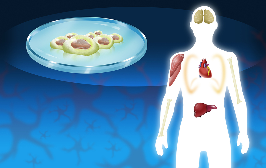
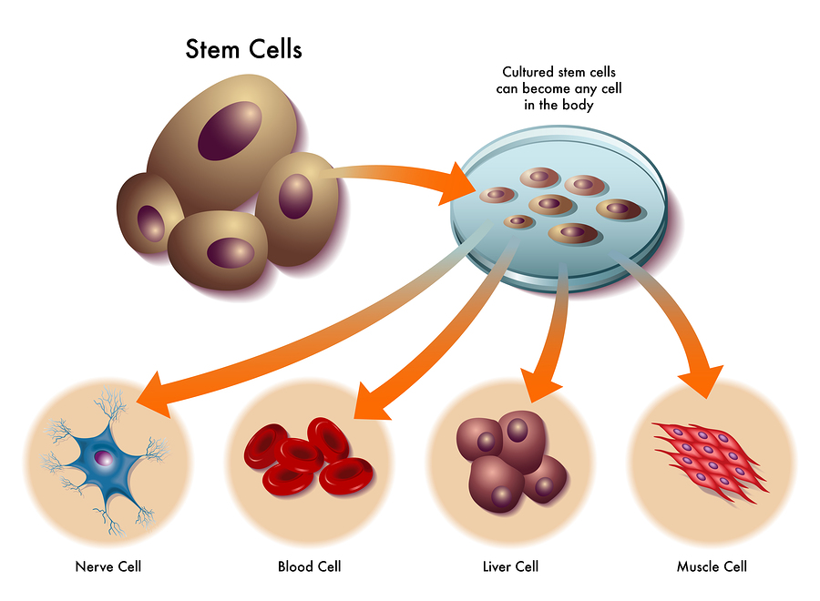
Essentially, what scientist found is that certain factors from stem cells can activate your own stem cells to regenerate tissues that grow old. These factors are messenger RNA (mRNA) and micro RNA (miRNA), which come as tiny particles of 40‐100 nm.

Advantages of administering exosomes

To emphasize, exosomes can be given systemically as infusion, and they can regenerate your stem cells, if they are in need of treatment. They cross the blood brain barrier, so it is possible to treat brain diseases. That is to say, there is no first-pass removal in the lungs as it is with mesenchymal stem cells (MSC). The potency is related to the age of the donor and his/her stem cells. Notably, exosomes are easy to store, freeze and administer.

Exosomes influence the growth of target cells and promote regeneration. In addition, exosomes stimulate immunomodulation and have anti-inflammatory and anti-fibrotic properties. To clarify, the only limitations are that the strength of the exosomes is related to the age of the blood donor. The exosome fraction comes from mesenchymal stem cells. That is to say, it circulates in the plasma portion of the blood, which is obtained by spinning blood cells down in a centrifuge. To emphasize, exosomes can regenerate your stem cells.

Applications of exosomes for various clinical conditions
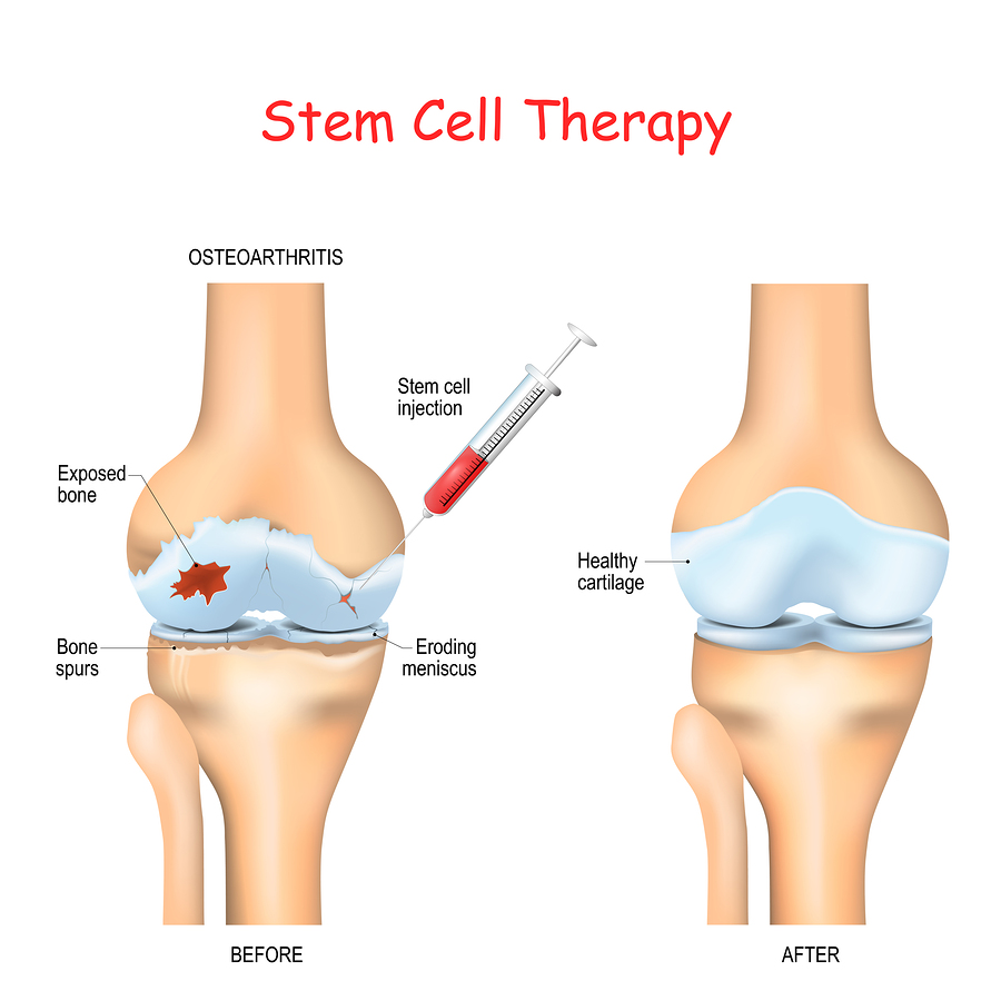
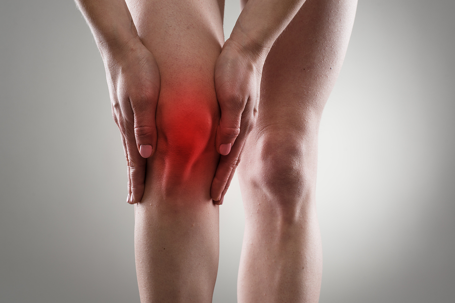
Joint inflammation

Mesenchymal stem cells are useful to treat arthritis. But it is important to realize that exosomes from mesenchymal stem cells are doing the same by stimulating the body’s own stem cells situated in the joints. In fact, several target cells have been identified that are stimulated by exosomes. These are chondrocytes, chondrocyte progenitor cells, cartilage-derived stem cells and synovium‐resident multipotent progenitor cells. In addition, other target cells are osteoblasts and osteoclasts in resident MSC within the subchondral bone and chondrogenic cells in the knee joint.

Disc degeneration  

Degenerative intervertebral discs respond to exosome treatments. The IL1 beta cytokine is involved in intervertebral disc degeneration. Exosomes inactivate these cytokines and have antioxidant and anti-inflammatory effects. Exosomes are not all the same. Different sub-fractions were isolated that have anti-inflammatory, immune-stimulating, antioxidant and other effects on the body.

Aging research

Researchers were able to pinpoint aging to various factors that contribute to premature aging. To clarify, when there is a decrease of catabolic processes and an increase of anabolic processes, an older person can combat premature senescence. Another key point, aging is also linked to redox homeostasis. Simply put, oxygenation processes in the body need to be balanced by reduction processes. This keeps the body in a healthy state. ADP/NADH production can be stimulated by exosomes.

Longevity comes from good lifestyles

With the use of exosomes, the aging process slows down, as oxidative stress is neutralized, damaged mitochondria are removed and cellular debris as well. That is to say, this improves inflammation and premature aging.

As has been noted, in the past 200 years life expectancy has doubled in most countries. 4 areas where longevity is particularly common are: Okinawa, Japan; Sardinia, Italy; Nicoya, Costa Rica and Loma Linda, USA. Only 7% of longevity stems from genetic factors, the rest is from lifestyles we adopt. In the final analysis, people who die prematurely followed a very poor lifestyle causing them to develop diseases, which ultimately killed them.

Clinical diseases from aging

Ultimately, advanced aging puts you at risk of getting cardiovascular disease (heart attacks and strokes), cancer and neurodegenerative diseases (Alzheimer’s disease, Parkinson’s disease). From the third decade onwards, there is the risk of bone loss, which causes osteoporosis. As has been noted, loss of cartilage causes osteoarthritis. Loss of muscle strength and muscle mass is called sarcopenia. With aging there is often an accumulation of abdominal fat. Hormones are disbalanced. Blood pressure is often elevated and blood lipids as well. Insulin resistance can develop and the blood vessels become stiffer. This causes heart attacks and strokes.

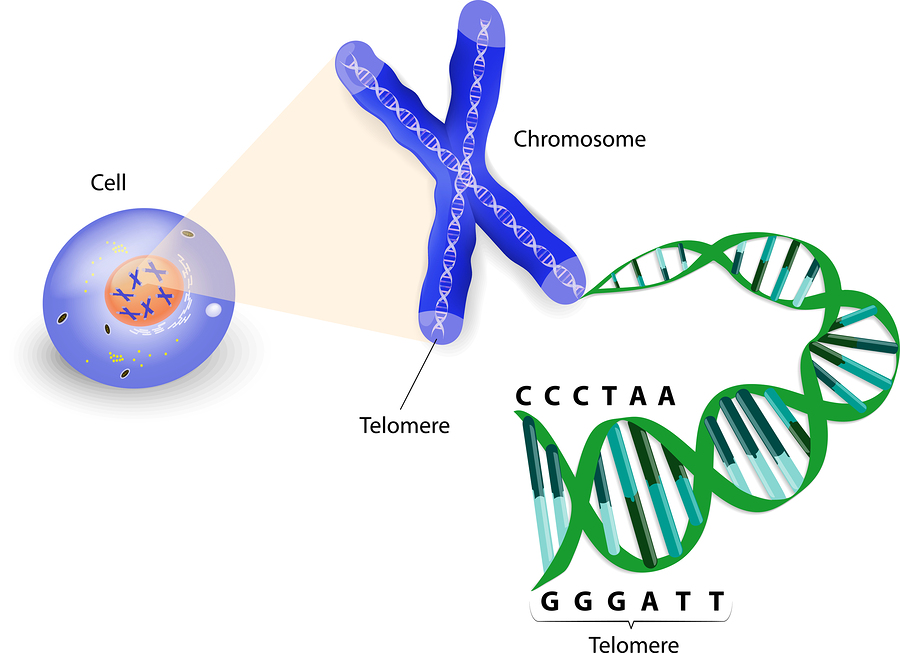
The details of the aging process are much more complicated than originally thought of. There is a combination of aging of the DNA, mitochondrial aging, stem cell exhaustion and a change of intercellular communication due to dysregulated endocrine signalling. In addition, there is a decline of the immune system and epigenetic factors that can turn off longevity genes.

Oxidative stress as a cause of premature aging

Dr. Spiel pointed out that reactive oxidative species (also known as free radicals) cause damage to mitochondria and mitochondrial DNA. But we need the energy from the mitochondria for a comfortable life. In essence, antioxidants can neutralize free radicals. Age-related conditions due to oxidative stress are: cardiovascular disease, chronic kidney disease and type 2 diabetes, chronic obstructive pulmonary disease, cancer, neurodegenerative disease, frailty and sarcopenia. Surely, both reactive oxygen and reactive nitrogen are free radicals. They have one or more unpaired electrons and all aerobic body cells produce them. Reactive oxygen and nitrogen species (RONS) cause oxidative damage to our cells and contribute to the development the diseases just mentioned.

Antioxidants help to prevent diseases

But antioxidants can contain these free radicals in various ways. The body has five built-in enzymatic ways to protect itself and five non-enzymatic ways (bilirubin, vitamin E, beta-carotene, albumin and uric acid). In addition, there are antioxidants that a person can take as supplements to inactivate RONS. These are: vitamin C and E; phenolic antioxidants like resveratrol, phenolic acids, flavonoids, oil lecithin, selenium, zinc and drugs like acetylcysteine.

Without control of the oxidative stress RONS can lead to cellular senescence and chronic inflammation. This leads to a vicious cycle where chronic oxidative stress and inflammation feed on each other leading to premature diseases.

Causation of several diseases

As we age, the body reduces the inborn antioxidant enzymes (superoxide dismutase and glutathione peroxidase). Before we can understand how to live longer, we need to be aware what happens in various health scenarios as follows.

  • The lack of inborn antioxidant enzymes leads to vascular endothelial dysfunction, high blood pressure and premature hardening of the arteries. This can become a precursor to heart attacks and strokes.
  • Elevated blood sugar in the case of type 2 diabetes leads to increased sugar concentration of body cells and formation of free radicals.
  • Oxidants from cigarette smoke activate macrophages and epithelial cells to produce inflammatory cytokines. Continued smoking releases proteases in the process that break down connective tissue and cause emphysema and COPD.

There are more diseases

  • Chronic kidney disease comes from oxidative stress affecting the filter units of the kidney, called glomeruli. With a lack of blood supply to the kidneys secondary high blood pressure develops and endothelial dysfunction. It also leads to chronic inflammation.
  • In the brain oxidative stress leads to cognitive impairment and dementia.
  • Oxidative stress and chronic inflammation are important ingredients for the development of cancer. RONS and cytokines release NF-kB, which activates cancer genes. RONS can also directly attack the DNA of cells and cause cancer through carcinogenesis.
  • Sarcopenia and frailty come from the action of RONS on the skeletal muscles. In old age there are less inborn antioxidants available. This leads to decreased muscle quantity or sarcopenia. Eventually frailty results with the risk of falls and fractures. 

Preventative measures for slowing the aging process

There is a number of steps that in combination help to slow the aging process.

  • A Mediterranean diet combined with a fasting mimicking diet or other calorie restricted diet
  • Regular physical activity
  • Cognitive training
  • Vitamin D3 supplementation
  • Reducing your risk to develop vascular disease
  • Certain drugs turn on the longevity gene (metformin, rifampin)
  • Spiel warned that due to limited compliance and variable response these steps alone may not be enough to prevent age-related problems

How to live longer

It is important to recognize the importance of antioxidants to counteract the development of these diseases. As already mentioned, the following counter the effect of free radicals: vitamin C and E; phenolic antioxidants like resveratrol, phenolic acids, flavonoids, oil lecithin, selenium, zinc and drugs like acetylcysteine. Mesenchymal stem cells can also stop the action of free radicals. In addition, exosomes, which  are products of mesenchymal stem cells can do the same. Mitochondria, the power houses within the cells, create energy, but also release free radicals. In his clinic Dr. Spiel administers intravenous exosomes to counter the oxidative stress. Numerous studies linked mitochondrial dysfunction to various age-related diseases. There are markers in blood tests that the physician can order to analyze malfunctions in the body. Dr. Spiel showed 4 slides that contained a lot of medical information that is too technical. I omitted it for this review.

Intravenous infusions of exosomes

The important thing to remember is that epigenetics can be changed by exosome infusion and lifestyle changes mentioned above. Dr. Spiel said that generally he uses 15 ml of exosomes by intravenous infusion every 12 weeks for longevity and performance enhancement. This treats conditions like infertility, osteoporosis, osteopenia, heart, liver and kidney weaknesses. Here is the dosing for intravenous exosomes by weight:

20-50 lb: 5 ml; 50-90 lb: 10ml; more than 90 lb: 15 ml; more than 220 lb: 20 ml. Unfortunately, one exosome treatment costs between 500.00 and 922.00 USD, an amount that most people cannot afford.

Contraindication to the use of stem cells or exosome therapy

It is important to realize that a person who has cancer should not receive either mesenchymal stem cells or exosomes. Indeed, exosomes do not differentiate between cancer cells and healthy cells, but stimulate cell division. For the same reason people with myeloproliferative disease (sickle cell anemia, bone marrow dysplasia) should also not receive exosomes. To clarify, other conditions where the physician will not order exosomes are primary pulmonary hypertension, acute bacterial infection or an immune-compromised state. In addition, macular degeneration with neovascularization is also a condition where the health professional does not administer exosomes.

Exosomes can Regenerate Your Stem Cells

Exosomes can Regenerate Your Stem Cells

Conclusion

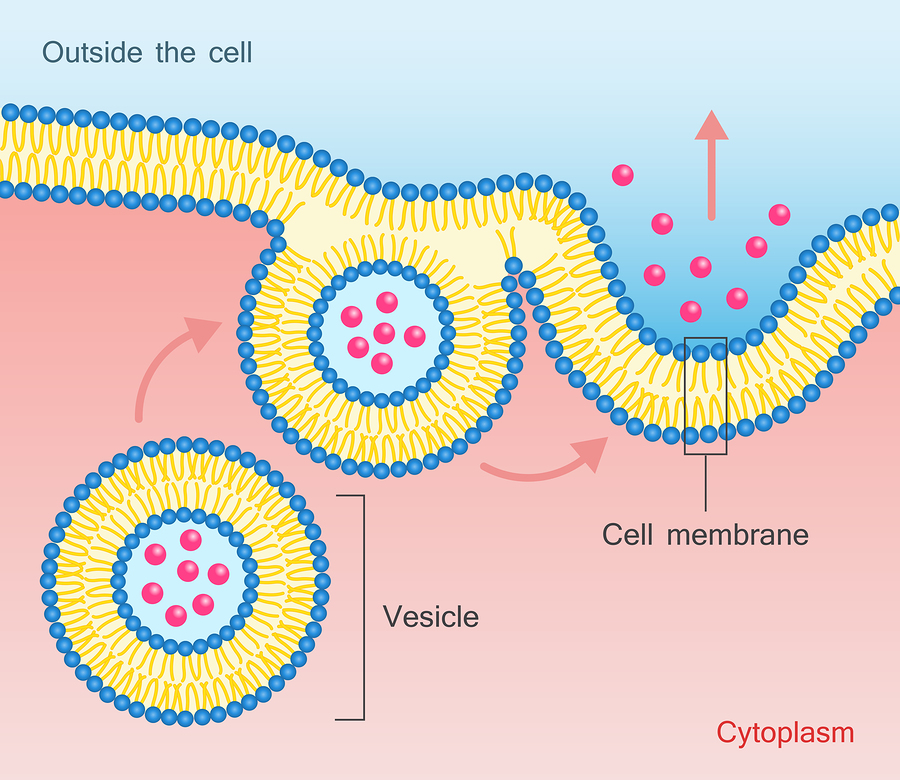
Dr. Douglas J. Spiel gave a talk on how exosomes can regenerate your stem cells. Specifically, this was at the 27th Annual World Congress on Anti-Aging Medicine in Las Vegas from Dec. 13 to 15th, 2019. Dr. Spiel explained how disease processes age our organs. Reactive oxygen and nitrogen species (RONS) cause oxidative damage to our cells and contribute to the development of diseases. This involves the mitochondria in the cells as well. The good news is that a healthy lifestyle can counter these damaging processes to a certain extent. But it takes another step to re-establish the balance of our cells, exosome infusions. Exosomes are tiny particles that are shed by stem cells and that circulate in the blood. They can reenergize stem cells that are ailing to become functional again.

Expensive exosome infusions

He recommended an infusion with exosomes every 12 weeks for maintenance of good health and as a “fountain of youth”. Obviously, there are some limitations. As mentioned, it is not suitable for all patients, like cancer patients, patients with sickle cell anemia, acute bacterial infections or pulmonary hypertension. In addition, it is also not a treatment which many patients will seek out as the cost is prohibitive. One exosome treatment cost between 500.00 and 922.00 USD, an amount that most people cannot afford.

Oct
26
2019

Steroid Injections Are Doing Harm, But Stem Cells Help

When a person has osteoarthritis, steroid injections are doing harm, but stem cells help. A new study published in the journal “Radiology” came to the conclusion that steroid injections are doing harm.

The article was published on October 15, 2019. 459 patients with osteoarthritis of knees or hips received injections with intraarticular corticosteroids or placebo to conduct this study. As a matter of fact steroid injections are a standard treatment for moderate to severe osteoarthritis. The injections consisted of triamcinolone 40 mg mixed with a local anesthetic. 36 of these patients (8%) had adverse events in their joints.

Here is the break down of these negative events following intraarticular corticosteroid injections.

  • Accelerated osteoarthritis progression (6%)
  • Subchondral insufficiency fracture (0.9%)
  • Osteonecrosis (0.7%)
  • Rapid joint destruction with bone loss (0.7%)

Critical analysis of effects of intraarticular corticosteroid injections

The authors reviewed the literature and came across an evidence-based review by the Cochrane Musculoskeletal Group involving 1767 participants. The review showed an overall low evidence for all outcomes. Intraarticular corticosteroid injections were inconsistent and variations between trials were high. Certainly, there was a moderate improvement of joint pain, and physical joint function improved as well.

But then again more severe osteoarthritis in knee or hip joints showed a lack of improvement and in many cases even joint deterioration. With this in mind, these cases often ended up with total hip or total knee replacements. Overall the Cochrane review showed a lack of objective evidence of symptom improvement after intraarticular corticosteroid injections in people with osteoarthritis.

Mesenchymal stem cell injections for osteoarthritis

A fairly recent alternative approach to treating osteoarthritis is by mesenchymal stem cell injection with platelet-rich plasma activation. Researchers conducted a meta-analysis of 35 studies regarding knee osteoarthritis and mesenchymal stem cell injections in 2018. There were minor adverse events like knee pain and swelling. No serious side effects of the stem cell treatments were noted. The outcome of the treatment was classified as “very low” to “low”. The reason for this were poor study designs, large heterogeneity among the studies and wide variation among the 95% confidence intervals regarding study outcomes.

Australian study of mesenchymal stem cell injections into symptomatic knees

A short-term study from Australia examined 30 patients with mesenchymal stem cell therapy for knee osteoarthritis. There were two treatment groups. One group received 100 million adipose tissue derived mesenchymal stem cells into symptomatic osteoarthritis knee joints. Another treatment group received 100 million mesenchymal stem cells twice, namely at baseline and at 6 months after the first injection. The third group served as a control with continued conservative management. All of the groups had baseline MRI scans (magnetic resonance imaging) to assess the knee conditions. Both treatment groups had pain and inflammation at baseline. After 1 year following the initial treatment the patients had repeat MRI scans. They showed modification of the disease process in the treatment arm, while the controls had deteriorating osteoarthritis. The researchers concluded that adipose derived mesenchymal stem cell treatment of knee osteoarthritis has the potential of preventing disease progression.

Why stem cells help with osteoarthritis of the knees and the hips

Here is a research project between researchers from Palm Harbor, Florida and researchers from Serbia. The Center for Molecular Medicine and Stem Cell Research, Faculty of Medical Sciences University of Kragujevac, Serbia reviewed the literature on treatment of osteoarthritis.

The researchers explain that the physician can easily harvest mesenchymal stem cells by doing a liposuction of fat cells. These stem cells maintain their differentiation potential and there is only minor immunological rejection because of low histocompatibility antigen expression. As a result mesenchymal stem cells stay in the joint where they are injected.

Two processes that occur with injected stem cells

Two main advantages of mesenchymal stem cells stand out. Mesenchymal stem cells are capable of building up hyaline cartilage and in addition they have immunosuppressive characteristics. In comparison with controls patients who receive intraarticular injections of mesenteric stem cells have less pain and less swelling.

This review paper cites all the major animal and human studies regarding mesenchymal stem cell therapy for osteoarthritis. It concludes that stem cell therapy is most effective in mild to moderate osteoarthritis cases. It stresses the point that stem cells can regenerate joint tissues and that the inflammatory process of osteoarthritis is interrupted by the stem cells. This stabilized the osteoarthritic condition.

Steroid Injections Are Doing Harm, But Stem Cells Help

Steroid Injections Are Doing Harm, But Stem Cells Help

Conclusion

Mesenchymal stem cells derived from fatty tissue will repair tissue defects in joints afflicted by osteoarthritis. The stem cells also take care of the inflammation, which takes care of pain and swelling. Range of motion of the joint increases following stem cell treatment. Other studies have shown that intraarticular steroid injections do not interrupt the degenerative process. MRI scan studies have shown deterioration of the joint tissues following steroid injections. All in all, stem cell treatments of joints with osteoarthritis are superior to a treatment with steroid injections.

Dec
30
2017

Fasting Mimicking Diet

The fasting mimicking diet (FMD) was at the center of this year’s anti-aging conference in Las Vegas. This was the 25th Annual World Congress on Anti-Aging Medicine in Las Vegas, Dec. 14-16, 2017. Dr. Valter Longo, PhD reviewed some of the research he had done on longevity in yeast cells, worms and mice.

Fasting mimicking diet relevant in humans

Dr. Longo pointed out that this type of research has relevance in humans. If there was a cure for cancer, heart disease, stroke and diabetes, we would live 13 years longer. But if we stimulated longevity with this pulsed calorie restricted diet, we would live on average 30 years longer. There is a rare genetic abnormality where people are deficient for IGF-1, a growth factor produced in the liver. These genetically IGF-1 deficient people live longer and do not develop cancer. Observations like these and detailed mouse experiments inspired Dr. Longo to develop a new diet plan. Patients would receive a fasting mimicking diet on 5 days per month. The rest of the month would consist of a normal, balanced diet. 5 days of the month the person would consume a low 800-calorie diet. This is enough to ensure adherence to the diet, but low enough to lead to enormous metabolic changes including youth-preserving stem cell stimulation.

Clinical Application of fasting mimicking diet in cardiovascular health

Dr. Joel Kahn, Prof. of Medicine at the Wayne State University School of Medicine lectured later that day. He is also the Director at the Kahn Center for Cardiac Longevity. His talk was entitled “The Fast Track to Slow Cardiac Aging: Fasting &Targeted Nutrition”. He mentioned that a fasting mimicking diet was a powerful tool in cardiology to prevent heart attacks and hardening of arteries. He explained in detail the complex aging pathways that involve three components, IGF-1, mTOR and PKA. When lifestyle choices stimulate these genetic markers, accelerated aging is a consequence. But with the inhibition of those markers longevity can happen. He added that researchers looked at heart cells, where the same principles apply. Dr. Kahn pointed out that the basic research of Dr. Longo enables clinicians to see positive results in patients who follow caloric restriction for 5 days in a month on a regular basis.

How does the fasting mimicking diet work?

It is best to let one of the users of this diet explain how it works. Once per month you eat calorie-restricted food with only 800 calories per day and you follow this regimen for 5 days. Some patients receive 1100 calories for the first of these 5 days, if they have difficulties switching from normal food to the boxed food. Dt. Longo has developed boxed food, called ProLon (from L-Nutra). ProLon stands for “pro longevity”. Dr. Longo and Dr. LaValle mentioned at the conference that these prepared meals make it a lot easier for patients to stick to the low calorie diet. Three hundred dollars for the boxed food for 5 days are a stiff price, and this may well be out of reach for you.

Alternative way to make your own 800 calorie food at home

Nevertheless, this should not stop you. You can look at the ingredients online and copy the boxed food by creating your own balanced 800 calories per day food at home. It is true: you have to do some research! But counting calories and finding information about the caloric content of food on the Internet is not difficult. And preparing these very, basic, small and simple meals does not require a degree in nutrition. Here is another testimony from a user of the fasting mimicking diet.

Effect of the fasting mimicking diet on the metabolism

In the past it was thought that only ketogenic diets or periods of fasting would trigger longevity genes. But the basic research of Dr. Longo and others has shown that a low calorie diet for only 5 days can achieve the same thing. Longevity genes are activated; the negative aging pathways including IGF-1, mTOR and PKA are suppressed. The immune system gets activated from this. It also  leads to lowering of LDL cholesterol, triglycerides, blood pressure, insulin resistance, and diabetes improves. With the fasting mimicking diet the stomach sees some food, but the cells are fasting. According to Dr. Kahn this combination down regulates the body’s key nutrient-sensing pathways, which activates cellular regeneration and rejuvenation.

Clinical observations

Dr. Khan observed a high compliance rate with 3 cycles of the fasting mimicking diet. 94% of a group of patients were compliant over 3 months. Mild fatigue, mild headaches and mild weakness were present, but improved with each cycle. In addition to the above findings Dr. Khan found that there was weight loss, abdominal fat loss and waist circumference loss. There was also a reduction in IGF-1 levels, a reduction of the C-reactive protein and stimulation of stem cells.

Inflammation reduced, autoimmune diseases improved

The reduction of the C-reactive protein proves that semi-fasting reduces inflammation. The finding of stimulation of stem cells explains that regenerative processes can take place. Pain disappears, people report more energy and are generally feeling better.

There are other clinical findings. The positive effects from following the fasting mimicking diet last for several months. Also, when patients are on chemotherapy for cancer, the FMD will protect the healthy cells from the side effects of chemotherapy.

Dr. Kahn and Dr. LaValle noted that autoimmune disease responded to FMD. This was shown in both animal experiments using mice and in clinical case reports. Dr. LaValle described a 46-year old former Olympic athlete swimmer who had multiple sclerosis. After FMD she lost all of her muscle aches and cured her optic neuritis. This was something conventional medicine could not do for her.

Clinical applications of fasting mimicking diet

Here are some of the conditions that will respond to it.

  • Obesity, because of the weight loss effect
  • Diabetes: insulin resistance becomes lower and blood sugar levels drop.
  • High blood pressure reduced: many patients were able to reduce their medications or discontinue them
  • Prevention of heart attacks and strokes
  • Pain conditions will improve as all kinds of pain disappears, an effect for which at this point is no explanation
  • Autoimmune diseases like MS and rheumatoid arthritis improve, likely because of the effect of increased stem cell circulation
  • Prevention of heart attacks because of reduction of LDL, triglycerides and CRP
  • Cancer cure rates improved by protecting normal cells and bone marrow
  • Longevity improved in mice with a 3-fold increase of their life span. Telomere length in humans was increased. Increased stem cells will find defective areas that need repair. This effect will open up a new chapter in medicine.

Maintaining the achievements of the fasting mimicking diet

At this point the implications of this new approach to weight loss and metabolic rejuvenation can only be estimated.

Limiting calories for 5 days triggers a metabolic change, which is permanent. You can experience the full effect of this rejuvenating low calorie treatment. You can do it every month without having to fear vitamin or mineral deficiencies.

Here is another link to the website of Dr. Axe where the fasting mimicking diet is also recommended.

Fasting Mimicking Diet

Fasting Mimicking Diet

Conclusion

The 25th Annual World Congress on Anti-Aging Medicine in Las Vegas, Dec. 14-16, 2017 had a new theme. Several talks dealt with the fasting mimicking diet (FMD). It is a calorie-reduced diet for 5 days in a month that will reset your metabolism. But it will also stimulate your stem cells and can heal autoimmune diseases. If you need chemotherapy for cancer, it protects your bone marrow and improves cancer cure rates. The interesting thing is that the effects of this low calorie treatment persist permanently for many months.

With the help of this diet longevity has been shown in mice; there has been a threefold life expectancy boost. Smaller trials in humans have shown telomere lengthening and stem cell stimulation. It is too early to say what the long-term effects will be for humans. But you can treat yourself with the FMD for 5 days of every month on an ongoing basis. The other days of the month you are eating a normal diet. This will ensure that your metabolism stays in top shape.

A healthier and longer life

Practical applications for the FMD are huge. Patients with obesity, diabetes and pain conditions all benefit from this. High blood pressure drops. There will be prevention of heart attacks, and there is improvement in patients with autoimmune diseases. There is better cancer survival when on the FMD. Finally there is a strong possibility that you will live longer, but also stay healthier on this intermittent calorie restricted diet.

As Dr. LaValle said: it is “fasting with food”, and Dr. Kahn added: “Eat less, live more!”

More info:  Life extension through calorie restriction.

Incoming search terms:

Dec
09
2017

Stem Cells Cure Back Pain

A person with chronic back pain has several treatment options, but only stem cells cure back pain. Stem cell treatment has been available in the US and Canada and many other countries for approximately 10 years.

I come from a family with a strong history of back pain (mother, maternal grandmother and maternal grandfather). They all got their back pain in their mid to late 40’s. From my growing up years I remember that they complained about chronic back pain on and off. Sometimes they had to cancel events they wanted to attend because they could not tolerate sitting. In those times there were no CAT scans or MRI scans. If you had back pain, you just had to put up with it.

My personal experience

Given my family history of back pain I was surprised that my back pain was only a more persistent problem in the last 1.5 years, but not earlier. Normally a monthly chiropractic adjustment would keep my back symptoms under control. But in the last 1.5 years I needed to see a chiropractor more often than that. I took omega-3 fatty acid supplements for the past several years (two capsules twice per day) thinking that this should halt the development of degenerative arthritis in the lower back joints. When I turned 71, it was clear to me that I was now at the point where my immediate relatives were when they were in their late 40’s. Therefore, diet, exercise, weight loss, good nutrition and supplements can only do that much for you. If there is a familiar disposition, it will eventually catch up with you.

Conventional medicine’s approach to lower back pain

I have practiced as a general practitioner for 16 years in the past. In addition I joined Workers’ Compensation for another 16 years as a medical advisor. From this clinical activity I knew of hundreds of cases first hand what the steps were in the treatment of chronic back pain. First of all, physiotherapy treatments or chiropractic treatments were the treatment protocol. In minor back pain cases this would often help the pain symptoms. Furthermore, if residual pain persisted, the patients received anti-inflammatory medication (non-steroidal anti-inflammatory drugs or NSAID’s). Finally, if symptoms continued to persist, a CT scan or MRI scan was necessary for assessment. If it showed moderate changes like my findings, the patient received intermittent physical therapy, chiropractic therapy or acupuncture therapy. 

Surgical procedures for chronic lower back pain

If there were more severe degenerative changes or spinal stenosis with severe degenerative changes, a referral to an orthopedic surgeon or neurosurgeon would be necessary. But this was often the point of no return. If the surgeon felt that the condition was severe enough to do back surgery, various procedures could follow. For disc herniations irritating one of the nerve roots, laparoscopic discectomy was the treatment of choice. For severe spinal stenosis or intractable pain from end stage facet joint disease instrumentation was an option.

Fusion surgery

Under a general anesthetic the surgeon makes an incision in the patient’s back over the lumbar spine. The surgeon identifies the diseased disc level and places stabilizing stainless steel plates over the affected facet joints or the narrowed disc space. Many people think that fusion surgery would be the end of their trouble. In many cases this can actually be the beginning of chronic back trouble. The problem is that the body is designed to move. If the surgeon takes movement away in one area of the spine, the levels above and below have to work harder. It often takes only a few months or a couple of years, and the patient is back with excruciating pain from degenerative changes in the levels above and below the previous surgery. What does the surgeon usually do? He does more fusion surgery above and/or below the previous area of surgery.

Alternatives to back surgeries

New treatment options have opened up new possibilities. On the one hand there is prolotherapy treatment that I have described under this link. On the other hand stem cell therapy is another popular regenerative technique. Prolotherapy strengthens tissues, relieves pain and increases the range of motion in joints. There is 80 to 85% full pain relief and more than 80% improvement in range of motion. Prolotherapy promotes the healing of torn ligaments and tendons. There are many suitable conditions that lend themselves to the treatment with prolotherapy like the hip, knee, shoulder, ankle, neck, lower back and elbow. With prolotherapy the physician uses hyperosmolar dextrose injections into the affected area. Current thinking is that this irritates the tissues, which mobilizes local stem cells to heal the area.

In my case I had two prolotherapy treatments of my lower back, but it did not change my lower back pain.

MRI scans of my lumbar spine

We needed to find out what was happening in my lower back. My general practitioner ordered MRI scans of my lower back in summer of 2017. There are 5 levels of the lumbar spine from L1 to S1. In my case one level of 5 was normal. The other levels showed bulging of the discs. The scans also showed signs of arthritis in the small joints adjacent to the spine. Lucky for me, there was no sign of spinal stenosis. It was not good news: overall 4 levels of my lumbar spine showed signs of  degenerative disc changes. At the same levels I also had arthritic changes in the facet joints. This was enough to consider some intervention, or I would be headed for trouble in the future.

Stem cell treatment for chronic back pain

Following the failed prolotherapy for my lower back pain I needed to figure out what to do next. The MRI scans had shown degenerative changes in the discs of the lower 4 levels of the lumbar spine. There also was arthritis in eight facet joints (two on each side of each of the four L2 to S1 levels). Conventional medicine would have offered corticosteroid injections into the facet joint areas. My experience with many patients who had this procedure was that the effect of the corticosteroid injections wore off after 3 to 6 months. If a patient had more than 3  injections, there usually was a point of no return, and fusion surgery would be next.

Best therapy for my own chronic lower back condition

For me there was no question that stem cell therapy would be the best fit for treating my back condition. In addition platelet -rich plasma and low-level laser therapy could activate the stem cells. This would be the ideal non-invasive treatment option to treat my chronic lower back pain. I had met Dr. H. Michael Weber before. He is a well-known laser expert from Germany who has a double certification as an engineer and as an internist treating various clinical conditions with laser and stem cell therapy. In addition he is an expert of regenerative medicine methods. Also, he invented and designed the laser machines himself. I set up an appointment in the fall of 2017 at his clinic in Lauenförde, Germany.

First day of stem cell treatment

On the first day fat tissue was removed under a local anesthetic from my lower left buttock area. Next a cell separator divides the tissue into connective tissue, fat cells and mesenchymal stem cells. Two blood samples were also taken from me for processing platelet rich plasma (PRP). PRP is a natural stem cell activator. Growth factors and anti-inflammatory cytokines were also part of the mix together with the stem cells.

The very same afternoon I received the stem cell mix by injection. Eight needles, four on each side, were necessary to administer the stem cell combination. I also had a treatment on a light therapy bed with red light to activate stem cells in general. The stem cell injection was a pain free procedure, as I received a shot of a  local anesthetic in the area before. After that the physician inserted laser applicators through the interstitial needles.

Laser activation of injected stem cells

The next step was to use laser treatments with 5 different colors (infrared, blue, red, yellow and green) for 10 minutes for each of the 8 interstitial needles. The laser activation and the PRP mixed with the mesenchymal stem cells were the two main stem cell activators. They are crucial for activating the stem cells. But growth factors and anti-inflammatory cytokines also aided stem cell activation.

Second day of stem cell treatment

On the second day I received an infrared light treatment over my back for 20 minutes. Following that I received light therapy bed treatment for 20 minutes. The physician told me  that all of this was to activate the stem cells further. The next step was a bone marrow low-dose laser therapy.

Bone marrow stem cell activation by low-dose laser therapy

Often stem cell therapists mix mesenchymal stem cells from fat tissue with bone marrow stem cells which they harvest before from pelvic bone marrow. Dr. Weber told me that he would do a direct bone marrow laser activation of the pelvic bone marrow instead. He anesthetized the tissue above the pelvic bone. Following this he made a small hole into the pelvic bone through which he inserted a laser applicator into the bone marrow cavity. 5 different colored lasers were again applied for 10 minutes each to activate the bone marrow stem cells. Studies have shown, as Dr. Weber stated, that low-dose laser activates bone marrow stem cells. They can be found in the blood circulation within 1 hour. This is similar to mixing stem cells in a Petri dish and then injecting it as a mix, except it is a less invasive approach.

Further activation of stem cells

Following these procedures Dr. Weber felt that another light bed therapy was necessary for 20 minutes. He also gave me a Weber medical laser watch called “Regenerate+”. This device fits on the wrist. It is programmed to generate a number of different lasers to shine against the underside of the wrist. This is the area where the ulnar and radial arteries run close to the surface. This device will shine the laser lights for 30 minutes, and the laser light reaches the arterial blood. The circulating stem cells from the stem cell therapy are receiving a further boost this way. Dr. Weber told me to use this device twice a day on an ongoing basis. The Weber medical laser watch stimulates the immune system.  Jet lag also responds to, and it can stimulate stem cells as they circulate in the blood.

Stem Cells Cure Back Pain

Stem Cells Cure Back Pain

Conclusion

Medical tourism is flourishing. I have become a medical tourist myself because I did not want to get crippled by conventional medicine regarding my lower back pain. Two days after my stem cell treatment my back pain was significantly improved. There was mild pain in the area of the fat liposuction site. Four days after the treatment the lumbar spine pain was gone. Innumerable chiropractic treatments and two prolotherapy treatments had not given me relief. Now stem cell therapy in Germany has taken my chronic back pain away in only a few days. I realize that the healing process will take 3 to 6 months to complete, but as a patient what counts most is pain relief.

What, if someone criticizes me for choosing stem cell treatment?

It is difficult to argue with success. Whether somebody criticizes me for having followed a non-conventional treatment protocol does not matter to me. My question back would be: what do you do when conventional methods fail? Are you willing to suffer chronic pain and swallow pain pills that could either get you addicted or have serious side effects? I would try stem cell therapy again, if I had a problem that does not respond to conventional therapy.

Jul
08
2017

Stem Cells For Osteoarthritis

Many clinicians have used stem cells for osteoarthritis of the knee or other joints for some time. However, objective publications about the effectiveness of stem cells are only coming out now. Both stem cell types, derived from fat or stem cells from the bone marrow, are effective. Most doctors are using stem cells from fat (mesenchymal stem cells), because they are so much easier to harvest.

CNN reported about a man, Bill Marlette who had lost one of his arms in the past. He ended up overusing the other arm and as a result developed end-stage osteoarthritis in his wrist. He could not find relief with conventional methods of anti-inflammatories and pain pills. Next he went to a stem cell expert in Munich, Germany who treated him with mesenchymal stem cells from his fatty tissue. Only one treatment took away his chronic pain and helped him regain his wrist mobility.

Approval of stem cell therapy in Germany

Prof. Dr. Eckhard Alt, an expert in regenerative medicine has previously treated patients with end stage osteoarthritis and had good clinical outcomes with it. As a result the German regulatory agency has approved his treatment protocol.

Dr. David Pearce, executive vice president for research at Sanford Health in South Dakota said that Prof. Dr. Eckhard Alt was the first one to use fat cells as a source of mesenchymal stem cells to treat osteoarthritis. He went on to say: ”Those stem cells don’t have to be programmed in any way, but if you put them in the right environment, they will naturally turn into what the cell type around them is.” The physician harvests the stem cells through liposuction. An enzyme mixture is necessary to separate the stem cells from fat cells, oil and connective tissue. A cell separator can also help separating the stem cells from the rest of the cells and tissue.

A case of wrist osteoarthritis

As I mentioned before only one injection was necessary to relieve the chronic pain of Bill Marlette’s wrist. Since his return the doctors in the US have followed Bill closely. They took MRI scans and noted that the bony cysts associated with the severe arthritis have disappeared. His wrist and hand strength have returned to normal. The pain almost disappeared. There were no side effects whatsoever. Because the stem cells are of the same tissue type as all his other cells of his body, one would not expect any tissue rejection by the immune system. Bill Marlette did not need any pain pills following the procedure in August 2016. And he says: “I have more range of motion with my wrist, shaking hands didn’t hurt anymore,” he said. “My wrist seems to continue to improve, and there’s less and less pain all the time.”

Past experiences treating osteoarthritis with mesenchymal stem cells

A 2014 clinical trial from Korea involved 18 patients with osteoarthritis of the knee where adipose mesenchymal stem cells were injected. The high dose group did best. After 6 months there was significant improvement, also confirmed by arthroscopy. The previous cartilage defect in the femoral and tibial condyles had decreased in size. Range of motion in the knee joints and pain had also improved. There were no adverse effects from the treatment.

Chinese study

Mesenchymal progenitor cells have the propensity to develop into cartilage. At the Shanghai Medical College, Fudan University Shanghai, China the following experiment took place in 2015. The researchers grew human adipose mesenchymal cells in vitro. Later they injected these mesenchymal progenitor cells into the knees of rabbits with experimentally produced osteoarthritis. Despite doing xenotransplants (human cartilage to rabbits) with known HLA differences the cartilage grew and cured the osteoarthritis of the rabbits. The new cartilage had human HLA markers while the rabbit cartilage underneath had rabbit HLA markers. At 16 weeks the researchers examined the tissues under the microscope and another exam involved the HLA marker testing.

Tehran study

A study from Tehran, Iran was carried out on 18 patients with ankle, knee and hip osteoarthritis in 2015. Physicians injected stem cells from the bone marrow into the osteoarthritic joint. The doctors followed the patients and ordered occasional MRI scans for 30 months. All of the patients had improved significantly with regard to their joint function and pain. The MRI scans also showed thickening of the joint surfaces from new cartilage production.

French/German study

In a 2016 joint French/German study 18 patients with end stage knee osteoarthritis were treated with stem cells. The stem cells came from adipose tissue that went through a cell separator. Physicians injected the mesenchymal stem cell fraction into the osteoarthritic knees. This was a phase I study to rule out any adverse reactions, but none were evident. It also established that there were significant positive improvements in pain and mobility with regard to the affected knees.

General remarks about how stem cells heal osteoarthritis

The example above with end stage osteoarthritis of the wrist was just one example of where osteoarthritis can strike. Perhaps the more common other locations are hips, knees and the facet joints of the lower lumbar spine (causing chronic lower back pain).

The same treatment procedure, which Bill Marlette’s wrist benefitted from is useful for all these other locations. The common factor in osteoarthritis is that the cartilage is getting thinner and thinner until bone rubs on bone causing excruciating pain. It is here where mesenchymal cells can come to the rescue. The stem cells will assess what requires a repair after injection into an affected joint. They recognize that there is a lack of cartilage. Then they transform themselves into chondrocytes, which are cartilage-forming cells. How can stem cells do that? They come with a program to replace missing cells, particularly cartilage and bone cells. But if they are within fatty tissue, they cannot act within a joint that has osteoarthritis. The doctor has to transport the mesenchymal cells into the joint where they can then begin their healing function.

Other methods to treat osteoarthritis

Stem cells are only one of several regenerative treatment modalities for osteoarthritis. Another method are platelet-rich plasma (PRP) injections. Platelets have a lot of anti-inflammatory substances in them and also growth factors that can stimulate stem cells contained in the synovial membrane, the lining of any joint. To get PRP plasma, it is necessary to spin down blood and harvest the PRP fraction with a syringe. After three PRP injections were given into the knees of 90 patients with end stage osteoarthritis these patients were followed for two years.

In the beginning before treatment 100% of the patients had symptoms. After one year following the treatment with PRP their knee functions were normal in 67% of them. After two years only 59% had normal knee function. The investigators pointed out that this treatment modality initially helped to a certain point, but then the effects were slowly fading away.

Stem cell treatment of osteoarthritis of the knee

The literature on either bone stem cells or fat stem cell use for osteoarthritis of the knee in man is still sparse. Nobody has done larger clinical trials. Part of the reasons could be that total knee and total hip replacement in orthopedics is very lucrative. We are still in a symptomatic treatment mode. Physicians treat osteoarthritis conservatively with anti-inflammatories and pain pills. When bone rubs on bone, there can be excruciating pain. The physician refers the patient to an orthopedic surgeon who likely will do invasive surgical procedures. My own impression in general practice in the past is that these procedures do not always turn out the way they are supposed to work. Following total hip or knee replacement joint swelling often remains; pain issues are still there. There can be unequal height issues, balancing problems and so on.

Here is a review of mesenchymal stem cell therapy for osteoarthritis.  This publication is very conventional medicine. An attitude change by conventional medicine would be useful to catch up with what is happening in real life. Some patients will travel abroad to Munich as Bill Marlette did. But others may travel to other places like India, Mexico or wherever medical tourism takes you. Regenerative medicine is there to stay.

Stem Cells For Osteoarthritis

Stem Cells For Osteoarthritis

Conclusion

We have learnt about a case of severe osteoarthritis of the wrist that has been cured in Germany with one injection of mesenchymal stem cells. More common than wrist osteoarthritis is osteoarthritis of the hips, knees and the facet joints of the lower lumbar spine. The same stem cell therapy can be given for osteoarthritis in these locations. I find it very strange that progress in stem cell treatments is so slow in the US. The FDA has decided to be open to clinical trials with stem cell treatments, but progress seems to be much slower than in other countries. Why? We may never know. In the meantime, patients may seek treatments in other countries where such treatments are offered. In real estate sales there is a saying: “Buyer beware”.

Be cautious, if you get treated abroad

The same goes for stem cell treatments in another country. Should you contemplate doing this, do your homework; ask about the qualification of the treating physician, about safety records and whether the local authorities have approved this procedure. In the case of Bill Marlette’s osteoarthritis of the wrist the procedure in Munich, Germany had been accepted by the European equivalent of the FDA, the European Medicines Agency. Safety is top priority, effectiveness is next.

Apr
02
2016

Women Win Turning Older

Supercentenarians may teach us something about the question “Why do women win turning older”? Supercentenarians are people who are 110 years or older. Presently there are 53 of them distributed over the world, 51 are females and two are males. According to Ben Dulken and Anne Brunet this is not by chance: in other mammal species females often live longer than their male counterparts. They theorize that stem cells live longer under the influence of estrogen and this may be the explanation for the difference. They wanted to answer the burning question: “Is life expectancy linked to gender and stem cells”?

Observations regarding why women win turning older

Ben Dulken and Anne Brunet describe that several pieces of evidence are important to note.

Human eunuchs live longer than average males

Castrated males, called eunuchs, live on average 14 years longer than the average male.

Treatment of male mice with estrogen caused longevity

Experiments with male mice treated with estrogen increased their lifespan compared to untreated male controls.

Estrogen receptors on some stem cells in women

Neural stem cells (NSCs) and hematopoietic stem cells (HSCs) have estrogen receptors in females. This leads to extra stimuli during pregnancy, but also during the menstrual cycle in women or the estrus cycle in female mammals.

Faster wound healing in women may be from extra X-chromosome

It gets more complicated: There are non-estrogen regulated stem cell niches in the liver, skin and subcutaneous tissue (important for wound healing and resident muscle stem cells, called satellite cells (SCs). For some reason liver regeneration and wound healing, but also healing of muscle injuries in women and female mammals occurs at a faster pace. Scientists still do not have an answer for this. Theories are that perhaps women with their two X-chromosomes are at an advantage in comparison to males (only one X-chromosome) with respect to certain wound repair mechanisms.

Longevity and self-repair capacity may be related

There is the question whether longevity and self-repair capacity would be related, either through stem cell populations (NSCs, HSCs, SCs), other repair mechanisms or tissue proliferation.

Telomere length in older persons longer in females than in males

There are gender differences in aging patterns of stem cells. For instance studies in dizygotic twins showed that telomere length of blood cells in the female twin was much longer than in the male twin. Genetic factors appear to be the dominant factor to explain this phenomenon rather than hormones. But again this was favoring the female.

Comparison between muscles in older men and younger men

A study in males showed that there is an accumulation of damaged DNA in SC’s of muscle tissue with older age that leads to muscle senescence. In older men there is a delayed response to a specific exercise stimulus with regard to the satellite cell division (SC) when compared to the response in young men.

Women’s telomeres in stem cells grow longer

In females estrogen stimulates telomere growth of stem cells (NSCs and HSCs), which prevents premature stem cell exhaustion.

Effects of diet and exercise on life expectancy

The Potsdam study analyzed 4 healthy behaviors in 23,153 German participants aged 35 to 65 years over 7.8 years. They looked for the development of cancer, heart attacks, strokes and cancer as end points. The 4 healthy behaviors were: to be a lifelong non-smoker , having a body mass index lower than 30, performing 3.5 h/week or more of physical activity, and adhering to healthy dietary principles (high intake of fruits, vegetables, whole-grain bread and low meat consumption).

Those who had adopted all 4 healthy lifestyles reduced the development of serious disease by up to 80%. Dr. David Katz delivered a keynote address at the 22nd Annual World Congress on Anti-Aging Medicine in Las Vegas Dec. 10-14, 2014 entitled “Integrative Medicine: A Bridge Over Healthcare’s Troubled Waters”. He mentioned the Potsdam study. And he mentioned what the new logic of a healthy lifestyle is: a healthy lifestyle causes healthy telomeres of somatic cells and of stem cells; this causes health until a ripe old age.

Life Expectancy Linked To Gender And Stem Cells

Life Expectancy Linked To Gender And Stem Cells

Conclusion why women win turning older

It seems that women and female mammals are more protected by nature than males. The previously called ”weak sex” is in fact a lot stronger! This may be the reason that among supercentenarians there are only a few males remaining. But we don’t know how many males take the lifestyle factors of the Potsdam study serious. Males who want to age gracefully have to pay more attention to healthy lifestyles. This leads to longer telomeres and this allows for stem cell and somatic cell renewal. There are still many unanswered questions, but life expectancy is definitely related to how well we preserve stem cells throughout our body. This in turn depends very much on our lifestyle patterns.

Oct
10
2015

Tissue Repair With Extra Cellular Matrix

Are you ready to learn about futuristic medicine consisting of tissue repair with extra cellular matrix? On September 5, 2015 I watched an interesting documentary on Discovery Channel while working out on the treadmill in the gym. This gave me the idea that this would be good material for a blog. After a little research on the Internet I found the full extra cellular matrix story, which you can read about below.

An amputated finger grows back

Lee Spievak, a man who loves flying model aircraft had an injury to his his right middle finger. A rotating model airplane propeller chopped off the end of his right middle finger. His surgeon felt that there was nothing much that could be done. But his brother who works in regenerative medicine knew about a powder made from pig’s bladder tissue, which Dr Stephen Badylak from the University of Pittsburgh, had pioneered. His brother sent a sample of powder (extra cellular matrix, ECM) to Lee Spievak who sprinkled some on the open wound (the stump).

New tissue forming with extra cellular matrix powder

Within two applications he saw that new tissue was forming. In a matter of 4 weeks it sealed up, the wound and a new finger grew to the same length as before. In the course of 4 months his nail, skin, his feeling and even his fingerprint were back to normal.

This story happened in Cincinnati in 2005. In this news story it is explained why the ECM powder worked so well: it prevented the wound from closing and it stimulated the body to heal.

A large thigh muscle defect grows back

Marine Sergeant Ron Strang was severely wounded by a roadside bomb in Afghanistan where a large part of his left quadriceps muscle (left thigh) was ripped off. After several surgeries the surgeons decided that Ron was a good candidate for part of a trial that is ongoing involving about 80 Veterans with similar injuries. Dr. Steven Badylak from the University of Pittsburgh suggested with the next surgery to put extra cellular matrix from pig bladder into the remaining quadriceps muscle to see whether it would regrow part of it.

Surgery with addition of extra cellular matrix from pig’s bladder

The surgery followed by physical exercise was so successful that Sergeant Strang is now able to run and do all the activities he wants. There is still a scar, but in comparison to the initial injury where a big chunk of muscle was missing, the remaining scar was insignificant.

Dr. Badylak explains in the video of the link that the insertion of the sheet of extracellular matrix immediately recruits the patient’s own stem cells, which makes new muscle cells, new nerve tissue, new skin, whatever the body needs to heal what’s missing in the injured area.

Dog gut growing into a dog aorta

Dr. Badylak from the University of Pittsburgh had a veterinary medicine degree before he studied medicine and became a surgeon. From the beginning his interest was in regenerative medicine.

After he saw the success with Lee Spievak’s finger regeneration, he thought that there must be a way to regenerate other tissues. He started doing experiments on dogs where he removed part the arch of the aorta and replaced it with a piece of gut from the same dog to see whether the dog would survive and whether the gut would be strong enough to withstand the pressure from the outflowing blood in the aorta. He figured that the tubular structure of the gut would be a better template than the synthetic aorta pieces that are still in use by thoracic surgeons. To his surprise the first dog (his own dog named Rocky) survived and did well.

Dog experiments to understand how extra cellular matrix works

He accumulated data on a total of 15 dogs. All of them survived and did well. He could not understand what had happened, so he reexamined one of these dogs where he got histological samples and analyzed them under the microscope to see what was going on. What he expected was the typical findings of the gut transplant, but instead he found a new aorta with all of the histological findings of aortic tissue. There was a transformation of a piece of gut into aortic tissue!

Next Dr. Badylak repeated the surgical procedure, but this time he inserted a piece of gut from a cat, removed the lining of it (the mucosa) and the muscle layer, (the muscularis),. The remainder was only the extra cellular matrix, a thin tubular structure of ECM.

Aorta scaffolding made of extra cellular matrix survives in dogs

When he was done, he was wondering whether the body would reject the catgut ECM. After all, it came from another species. Normally with whole organ transplants one can expect rejection of the foreign tissue. None of that happened. The experiment went flawlessly: the transplant survived like all the others and again the ECM had turned into dog aorta. It was possible to integrate the extra cellular matrix into the aorta without any scar formation! None of this fitted any conventional medicine model; it was the blueprint for the regenerative medicine model.

Dr. Badylak recognized that this was a huge step forward, and he would need easy access to ECM material. He got it from the pig slaughterhouses dotting the Indiana countryside surrounding Purdue. There would never be a shortage of tissue for preparing the scaffolding of the ECM for various applications.

Repair of tissue defects with extra cellular matrix in various body regions successful

By now the surgeon had proven that the gut or ECM transplant was switching off an inflammatory reaction, which suppressed scar formation, and simultaneously promoted regeneration. But the missing puzzle still was how the body generated the aortic tissue.

Dr. Badylak tested whether the procedure would work for large veins, smaller arteries and Achilles tendons.  He did this all in dogs and using pig’s ECM. The answer was it worked all beautifully with no scarring and perfect healing results. Control dogs did not get the ECM, but were only operated on and then repaired conventionally in their Achilles tendons.  They developed a limp from scar tissue. This is what often happens in humans as well with conventional surgery. But none of the dogs that had 3 cm cuts and then received a treatment with pig’s ECM developed a limp or scarring. They healed perfectly.

Large company supports Dr. Badylak’s work

In 1992 DePuy licensed Badylak’s ECM-derived “biologic scaffolds” for all orthopedic applications. DePuy is a big company that makes supplies for hip and knee replacements and much more. This was an ideal support for Dr. Badylak’s work.

In 1999 the FDA approved pig’s bladder ECM for human applications. This included the use of pig’s ECM for shoulder rotator cuff tears in patients. The FDA also approved it for abdominal hernias and for esophageal reflux damage. In addition the FDA approved it to induce the regrowth of the outer lining of the brain following brain surgery.

He could now continue his research and find out what the missing puzzle was.  How did the body use the pig’s ECM and repair tissues?

Stem cell recruitment by ECM

Dr. Badylak was visiting a colleague of his in Los Angeles, Dr. John Itamur who had previously repaired a rotator cuff tear on a patient 8 weeks earlier using porcine ECM. The same patient had an unrelated shoulder injury. This required surgery just adjacent to the previously repaired rotator cuff. The surgeon decided to take a small biopsy to see how the healing tissue looked. This was when Dr. Badylak came for a visit. The microscope showed a surprise: the scaffolding had disappeared as expected. But there were a lot of new cells there. They did not look like inflammatory cells, muscle cells or nerve cells; they were stem cells. Dr. Badylak read several papers that told him that ECM breaks down into so-called crypteins. These peptides have powerful stem cell recruiting properties.

Experiment show how extra cellular matrix recruits stem cells 

In 2003 he started groundbreaking experiments in mice that proved this theory to be correct. He X-rayed a group of mice to kill all of their bone marrow stem cells. Then he injected stem cells tagged with a fluorescent marker. They repopulated the bone marrow with these tagged stem cells from the same strain of mice. Now he removed a piece from the Achilles tendon and repaired the defect with pig ECM. Stem cells tagged with a fluorescent marker were flooding the Achilles tendon repair area. Even months after the Achilles tendon repair with ECM the new Achilles tendon was still filled with some of these tagged cells showing that some of them had matured into regenerated tissue.

Video showing would healing with extra cellular matrix and the final outcome of dog Rocky

Here is a link that contains a video about Sergeant Strang and his severe leg injury (repair of a rectus muscle tear). You may wonder how Rocky, the initial dog did who had an aortic segment replaced by a piece of gut. Rocky lived for another 8 years and was healthy until the very end.

Tissue Repair With Extra Cellular Matrix

Tissue Repair With Extra Cellular Matrix

Conclusion

You saw how the observation of a healing finger turned into experiments on dogs. Aortic defects and Achilles tendon defects healed without scarring. You learnt how pig’s or cat’s ECM were in use as scaffolds and that the body absorbed this. They recruit stem cells from the host’s body that subsequently do the healing. The exciting news about ECM is that it promotes healing, recruits stem cells, but also suppresses inflammation and scar formation.

We already hear that ECM is used in hernia repairs, rotator cuff repairs for shoulder injuries, and also in hair transplants, where Acell material is mixed in to improve the transplant success.

It is being used in lower esophagus surgery in cancer cases and with reflux esophagitis.

What will be the next application for ECM? We do not know everything, but it is a promising step into the future of regenerative medicine!

Apr
25
2015

Rejuvenate With Stem Cells

We all age; but can we rejuvenate with stem cells? There is a limit to detoxification, to eating organic food, to exercising, to the effects of vitamins and supplements and even to the effect of bioidentical hormone replacements. The limit comes from our telomeres and from stem cells that get depleted in our body as we age. Some researchers report that in regions where we suffer from a disease stem cells are even more depleted than in the rest of the body.

We do not have all the answers yet. We would like to know why our stem cells in the fatty tissue or in the bone marrow do not migrate on their own into an aching back or a sore shoulder. There are all the aches and pains associated with old age. So, why do our own stem cells not help us? They seem to be locked away in fatty tissue and in bone marrow.

At the 22nd Annual World Congress on Anti-Aging Medicine in Las Vegas (Dec. 10-14, 2014) I learnt that there is a group of stem cell experts in California with affiliates all over the US. They simply take stem cells from the fatty tissue and sometimes also from the bone marrow, isolate the stem cells through a stem cell separator and infuse the stem cell rich fraction (minus fatty and connective tissue) in a bit of saline solution back into the vein of the patient. When the stem cells are in the blood stream, they get activated by the growth factors that are present in blood and can now find where they are needed and start the healing process.

Studies have shown that when stem cells are in circulation in the blood, they are very sensitive to signals from tissues that indicate that there is an inflammatory process. This is why stem cells will repair arthritic changes. The can repair a torn meniscus, a rotator cuff tear in the shoulder or repair a weak immune system. The interesting observation is that stem cells from fatty tissue, also termed mesenchymal stem cells, are pluripotent. This means they can develop into cartilage building cells (chondrocytes) and build up cartilage; this is badly needed in a person with severe osteoarthritis. But stem cells are flexible: they can turn into meniscus cells in a knee with a torn meniscus. They also can repair the damage and relief the patient of the chronic pain. In a shoulder with a rotator cuff tear they can turn into a tough ligamentous material mending the tear.

Some data even indicates that circulating stem cells can repair vital organs like the brain, heart, liver, kidneys and bone marrow; these latter observations were mostly done in animal experiments, but human data is starting to be published in the medical literature.

So, let’s examine what has been found useful with regard to stem cells that are taken from your fatty tissue or your bone marrow and injected into one of your veins.

Here is a website from Arizona that I am only showing as a typical example (I have no conflict of interest and no commercial connections to this group) of what I described above.

With websites like this it is also important to read the disclaimer: “Even though our treatments are done using autologous cells, our Stem Cell Therapies are not approved by the FDA. Stem Cell Treatments are not a cure for any condition, disease or injury, nor a substitute for proper medical diagnosis and care…” Another website from La Quinta, CA describes the use of mesenchymal stem cells for regenerative therapies.

Stem cell treatments are in flux. There is a large body of knowledge that has accumulated showing that with proper technique and aseptic conditions it is a safe procedure. The FBA has been watching this. There are publications regarding the safety of procedures with adipose mesenchymal stem cells; here is one example.

The next step is to show in clinical trials that a certain procedure with stem cells is effective in treating a certain condition.

Below I did a literature review, which are only a few examples, but does not claim to be complete; it highlights some of the problems with stem cell treatments.

Stroke treatment with intravenous administration of bone marrow mononuclear stem cells

This study from India showed no statistical difference of stroke patients treated intravenously with bone marrow derived mononuclear stem cells (the experimental group) and the control group that did not receive such treatment. The investigators examined both groups with functional brain tests and performed PET scans to look at the healing of the brain lesions. Unfortunately the tests showed no statistical difference, but did show that the stem cell procedures were safe. It may be that the wrong stem cells were used (mononuclear bone marrow stem cells) when adipose derived mesenchymal stem cells may have done better. In stark contrast to the study from India is the stem cell treatment for a severe stroke in the former hockey player, Gordie Howe that has gone through the media recently. His procedure was done in Mexico. The stem cells were administered via a lumbar puncture approach as well as intravenously. As you can see from this case, stem cell treatment is even possible in patients who are in their mid 80’s with impressive results.

Parkinson’s disease

Here is a feasibility study from March 2014. A 71-year-old Asian man with progressive supranuclear palsy, an aggressive form of Parkinson’s disease was treated with adipose tissue-derived mesenchymal stem cells that were administered intravenously and intrathecally (to get stem cells into the cerebrospinal fluid that bathes the brain). A remarkable functional recovery took place.

Possible side-effects

This is a report of pulmonary embolism after administering intravenous adipose tissue-derived stem cell therapy. The blood clots in the lungs were treated with anticoagulant therapy. Repeat CT scans of his lungs showed later that the emboli were dissolved spontaneously. It is not clear whether this was a case where familial clotting problems pre-existed as a relative of this patient experienced a similar occurrence after stem cell therapy as well.

A case of chronic autoimmune thrombocytopenic purpura

A rare form of autoimmune disease exists where the body forms antibodies against platelets that help your blood to clot. Here is a paper from June 2009 that describes how a man with this disease was cured using adipose tissue-derived mesenchymal stem cells that were injected intravenously.

Renal transplant survival in type 1 diabetes patient

This case report from India shows that adipose tissue derived mesenchymal stem cells that were given at the time of a kidney transplant to treat end stage kidney disease. The treatment stabilized the condition of this patient after a kidney transplant. At the same time some of the mesenchymal stem cells differentiated into insulin producing cells, which made it much easier to control this patient’s diabetes. In this case stem cells were providing stability following an organ transplant (kidney) and some stem cells turned into insulin producing pancreatic cells.

Osteonecrosis of hip treated with adipose tissue derived MSC

In this study from South Korea dated January 2012 two cases of osteonecrosis of the hip, where the hipbone died (osteonecrosis) are described. The following stem cell protocol helped: The fraction that contained the stem cells (called stromal vascular fraction) was mixed with platelet rich plasma and hyaluronic acid. Using a long needle this mixture was injected into the affected hip joint. Conventional medicine has nothing to offer except a total hip replacement. But here are two cases that showed complete resolution of their pain, regained hip function completely, and healing could be documented with the help of MRI scans.

Treating heart attack patients with stem cells

Here is a paper from The Netherlands, published in June 2014 that describes the problems with stem cell treatment in humans. It points out that much has been learnt from animal experiments. The problem following a heart attack is that there is a massive inflammatory response in the infarcted heart muscle, which makes it difficult for stem cells to establish themselves in the injured heart muscle. However, stem cells have been shown to prevent the development of cardiomyopathy that follows a massive heart attack and often is the cause of death. More refinements are needed for successful treatments, such as the ideal timing of stem cell injections in relationship to the time of the heart attack, the best treatment approach and what number of stem cells to inject are all questions that still need to be answered.

MS model in mice shows promise with adipose mesenchymal stem cells

Experimental encephalitis in mice is used as a model for MS in humans. It helps to preselect potentially effective treatments for MS in humans. In this 2013 paper from Australia researchers used mesenchymal stem cells from adipose tissue and injected them intravenously. To their surprise the mesenchymal stem cells were able to penetrate the blood/brain barrier and end up in the myelin lesions inside the brain. In contrast, bone marrow derived stem cells were unable to do that. The researchers stated that adipose mesenchymal stem cells should be considered “as a cell therapeutic that may be used to treat MS patients”.

A group from Iran published this paper in February 2015 further emphasizes that mesenchymal stem cells would be a logical way to treat MS in humans.

Immunosenescence

As we get older the immune systems weakens because of a process called immunosenescence.

A research group from Austria published a paper in December 2011 that is typical for the thinking that mesenchymal stem cells from fatty tissue have properties that help the immune system to get stimulated. Based on this human data it should be possible to stimulate the immune system by giving stem cells from the fatty tissue to the same person intravenously. This publication shows that this process, which would benefit people above the age of 50 or 60 when the immune system gets weaker, will indeed stimulate the immune system. However, at this point we do not have the data of large clinical trials where this would have been done with measurements of the immune function before and on several occasions after stem cell injection to get a feeling for how long the effect would last. We also do not know whether this procedure is associated with longevity.

Rejuvenate With Stem Cells

Rejuvenate With Stem Cells

Conclusion

Stem cell therapy is definitely coming and many applications are already established as I discussed in a prior blog. It is only recently that physicians are no longer worried about creating tumors with stem cell transfer. Now we are in a phase where various stem cell transfer methods (intravenous, intrathecal, interstitial) are being tested as a treatment for various illnesses. It looks like stem cells from fatty tissue may soon be used intravenously, but I have not seen any such trials when checked on PubMed. The activation of stem cells by laser light has only been mentioned sparingly in the literature. This combination (laser activated, intravenous mesenchymal injection) has the potential for being useful for a multitude of chronic illnesses like fibromyalgia, MS, generalized arthritis, just to mention a few. Mesenchymal stem cells are anti-inflammatory, and they can mend defects without leaving scars.

Jan
30
2015

Prolotherapy And Stem Cell Therapy

This article describes prolotherapy and stem cell therapy. It is the 5th blog and the last one of a series that dealt with telomeres, lifestyle and stem cells, topics that were on the agenda of the 22nd Annual Anti-Aging Conference in Las Vegas (Dec.10 to 14, 2014). Here are summaries of two talks from this conference that dealt with methods of repairing damage to your joints or bones without surgery. Treatments consist of stimulating local stem cells through a treatment called “prolotherapy” where needles are used to inject concentrated dextrose. I will explain below why this method is effective.

Modification of original prolotherapy

A modification of this original prolotherapy is when the effect of it is amplified by growth factors from so-called platelet rich plasma (PRP), which is mixed with the dextrose injection. The ultimate healing jerk occurs when you mix in stem cells with the PRP into the injured tissues. In both lectures the speakers showed images before the procedures and images some time after with impressive results.

1. Dr. Fields’ talk had the title: “Repairing joints and spine without surgery: prolotherapy/PRP/stem cell therapy”

This talk concentrated on the use of prolotherapy with concentrated dextrose and prolotherapy with platelet rich plasma (PRP) with or without the addition of stem cells in the treatment of various musculoskeletal injuries.

When the doctor decides to do prolotherapy by itself, the therapeutic modality is 12.5% Dextrose. The therapist injects it into the area of injury. Dr. Fields said that the reason it works is that local stem cells in the area of injury undergo activation from the Dextrose. Since Dextrose activates stem cells, they now can do the healing. The physician can improve the results further by injecting a small amount of PRP very focally to an area of ligament rupture. Centrifuging blood from the patient’s vein separates the red blood cells from plasma, which is where the platelet rich plasma (PRP) originates from.

Stem cells and PRP

The physician discards the red blood cells, but the platelet fraction and some of the plasma becomes the PRP preparation. The PRP fraction amplifies the effects of the stem cells from bone marrow and from fatty tissue. The physician mixes all of them into the injection. Dr Fields explained that bone marrow is aspirated from the pelvic bone and in the same patient a liposuction is also done to receive adipose tissue. A cell separator is the next stage in obtaining pure stem cell preparations. This way stem cells that had their origin in bone marrow and mesenchymal stem cells that had their origin in adipose tissue are obtained.

Superior stem cell mix

The physician combines both fractions as they make a superior stem cell mix. Finally, the addition of platelet rich plasma activates the stem cell mix. This mix was used for bone fractures that were slow to heal, for ruptured tendons, ligaments, Achilles’ tendons and rotator cuff tears.

Healing without a residual scar

Dr. Fields showed before-slides and several weeks to months after-slides with MRI scans of the original injuries and the final healed tendons and ligaments. I have never seen such beautiful healing with no residual scar. Stem cells are the specialists of healing such defects. They change into whatever cell type is missing and they fill in the defects. This explains the perfect function after healing of the injury following stem cell and PRP injection. It also explains why many athletes who had this done went on to winning more medals after the repair. You do not hear about success stories that often after conventional surgery, because the range of motion and strength suffer from scarring following conventional surgical repairs.

Case histories

Several patients with knee injuries received prolotherapy. Dr. Fields recorded their video testimonials. They explained that their procedures only involved needles in the injured area, that they experienced almost complete pain relief on the day of the procedure and that they could rehabilitate right away.

Slides were also shown of specific knee ligament injuries involving the medial collateral ligament (MCL), the posterior cruciate ligament (PCL) and the anterior cruciate ligament (ACL). These are very important support ligaments within the knee.

Lower back injury with a ruptured disc

But this was not all: there were lower back injuries with ruptured discs. With conventional medicine the solution is a discectomy, but here these patients received prolotherapy. They experienced a stabilization of the weak areas, spontaneous resorption of the prolapsed disc and stabilization and strengthening of the weak spine. MRI scans of the spinal injury before treatment and several months after the treatment showed a complete normalization of the spine. In my work as a Medical Advisor for Workers’ Compensation cases that I was doing for 16 years I have never seen a single case like that.

Dr. Fields showed a similar spinal injury in the neck as well with a testimonial from that person. Again, following stem cell treatment there was minimal pain, immediate rehabilitation and a full range of motion several weeks after the injury.

What is treated with prolotherapy?

Basically all of the major joints lend themselves for treatment with prolotherapy: the shoulder, knee, back, the neck, ankle, elbow and hip. Typically, the types of injuries for prolotherapy treatment are sports injuries, fibromyalgia, sciatica, muscle tears, tendonitis, arthritis, bursitis and temporomandibular joint problems (TMJ).

Dr. Fields also stressed (and so did Dr. Purita, see below) that platelet rich plasma with its growth factors activates stem cells.

There was a presentation of two special cases, namely patellar tendinitis and Achilles tendinitis, which both respond very well to prolotherapy and PRP plus stem cell therapy. This provides complete healing of these otherwise very difficult clinical entities.

Testimony of a former Surgeon General of the US

An image was shown from the late C. Everett Koop, MD, the former Surgeon General of the Untied States who had this to say about prolotherapy: “I have been a patient who has benefited from prolotherapy. Having been so remarkably relieved of my chronic disabling pain, I began to use it on some of my patients.” This may yet be the strongest argument to at least consider prolotherapy in otherwise hopeless cases.

2. Dr. Joseph Purita gave a lecture on the “Effects of PRP And Stem Cell Injections”

As explained above PRP stands for platelet rich plasma, which is a “soup” of various growth factors and exosomes =cell-to-cell mediators). He discussed the importance of the proper harvesting of PRP. He explained that apart from white blood cells (WBC) and platelets an important component of PRP are very small embryonic like stem cells (VSELs). With a microscope you can visualize VSELs. The missing link has been the observation that white blood cells produce inflammatory substances, which have been detrimental when stem cell injections with PRP were done in the past (poor survival rate of stem cells). Research showed that photo-activation of the PRP before injection leads to anti-inflammatory behavior of the WBC in PRP.

Light activated PRP

Dr. Purita calls this “light activated PRP”, which leads to the best results with stem cell/PRP injections. Soft laser stimulation with red, green and blue soft lasers have been shown to improve tissue healing significantly when stem cells and light activated PRP are used. As already described in Dr. Field’s talk the main sources for good stem cells are the fat tissue (from the “love handles”) and the bone marrow (obtained from pelvic bone). Dr. Purita also explained that nitric oxide and electrical stimulation also help to improve stem cell survival and reduce inflammation. All of these methods revolutionized orthopedics where injured tissues can heal with the help of injecting stem cells and activation of PRP by laser treatments. Dr. Purita showed very detailed technical aspects of these procedures with various applications.

Treatment of osteoarthritis

For instance, he showed a slide regarding treatment for osteoarthritis of the knee. All this is contained in the plasma that is used to inject PRP into a joint with degenerative arthritis. You can mix this with stem cells from bone and from adipose tissue and inject it into arthritic knees. This is the ideal remedy for a person with degenerative arthritis. The stem cell/PRP mix calms down the degenerative process with instant pain relief. The stem cells are transforming into cartilage cells (chondrocytes) building up hyaline cartilage. The end result is a new knee surface;  the old and the new knee surfaces form one seamless unit.

The doctor can use PRP by itself successfully for repairing bursitis of shoulders and rotator cuff tears, muscle tears and sprains, meniscus tears of the knee, mild to moderate osteoarthritis of various joints and spine disorders, particularly facet joint problems.

Prolotherapy And Stem Cell Therapy

Prolotherapy And Stem Cell Therapy

Types of stem cells for joint injections

Dr. Purita gave a thorough overview of stem cells. He pointed out that stem cells fulfill two criteria:

  1. They are undifferentiated and they are capable of self-renewal by replication
  2. They can undergo differentiation into specific cell lineages.

From a practical point of view as already mentioned in Dr. Field’s talk there are two sources for stem cells that are important: stem cells from adipose tissue (also with the name MCS or mesenchymal stem cells) and stem cells from bone marrow, obtained usually from the pelvic bone. When the physician mixes them and stimulates them with PRP they are the miracle mix that helps to heal all these injuries.

Stem cells and other factors for healing

What does the FDA say to stem cell therapy? “The FDA states it is ok to use these cells as long as they are put back into the same patient and they are minimally manipulated.”

Dr. Purita listed a host of other factors besides platelet rich plasma. It supports stem cells, increases their survival on transplantation and stimulates them to differentiate and heal the defect. This happens at the recipient site as quickly as possible. Photoactivation of platelet rich plasma with low level lasers (soft lasers) will release exosomes. They are tiny particles, which platelets and white blood cells release. They contain proteins and genetic material required for wound healing and stimulation of stem cells.

Patient with avascular necrosis

Towards the end of the talk Dr. Purita showed an MRI scan of a knee with avascular necrosis (dead bone) before and after treatment with stem cells, PRP and low level laser therapy. There was a complete resolution of the avascular necrosis without any surgery. With a second case the initial MRI scan showed a complete tear of the MCL. MCL stands for medium collateral ligament (MCL tear) of the knee. The follow-up scan showed the same ligament intact. The physician achieved this without surgery, just by treating the patient with an injection of stem cells; PRP and using low-level laser therapy to activate PRP.

Conclusion

Prolotherapy and stem cell therapy are the hottest new treatment modalities. They can treat ruptured tendons, ligamentous injuries, and disc herniations in the neck and in the lower back. You will not get this from your primary care physician or your regular orthopedic surgeon. At the present time they tend to not have the insight. Besides, they profit from the conventional procedures. But you owe it to your health to try these alternatives first as they are much less invasive. They involve your own cells that will heal the defects with a very high probability. You still have the option to seek the advice of an orthopedic surgeon, should these alternative procedures fail. Unfortunately most insurance carriers will not pay for this service at this time.

Disclaimer

Dr. Schilling has no conflict of interest with regard to Regenexx . He also has no conflict of interest with any of the other companies of which images were shown. They simply displayed the best images with regard to the many illustrations in this blog.

Incoming search terms:

Jan
16
2015

Telomere Length A Telltale Sign Of Aging

Dr. Sandy Chang gave a talk at the 22nd Annual World Congress on Anti-Aging Medicine in Las Vegas Dec. 10-14, 2014 entitled “Telomere measurement as a diagnostic Test in cardiovascular and Age-related disease”, but a shorter title would be “telomere length a telltale sign of aging” (my choosing).

Dr. Chang pointed out that it is now well established that telomere length is directly related to health. The shorter the telomeres are the higher the probability to get the following: early menopause, infertility, diabetes, wrinkles, arthritis, osteoporosis, cardiovascular disease, Alzheimer’s, Parkinson’s, dementia, cancer, stress and a lack of stem cells. In this BMJ study from 2014 it was shown on a large population basis that shorter white blood cell telomeres lead to a higher risk of coronary heart disease causing heart attacks. Decreased telomere length is also associated with the development of breast cancer, cancer of the ovary and uterus, cancer of the prostate and skin cancer.

Because of these connections it makes sense to determine a person’s telomere length. If it is short, do check-ups more often to detect any cancer early when it can still be treated.

Telomere length measurements are now done in many infertility clinics as short telomeres both in the male and female is associated with infertility.

The newest finding and perhaps the most important is that a healthy lifestyle, vitamins and supplements can elongate telomeres while a poor lifestyle leads to shortening of telomeres.

Here are the factors that lead to shortening of telomeres:

– Chronic stress

– Poor diet and nutritional habits

– Chronic inflammatory diseases

– Metabolic disorders

– Lack of consistent exercise/sedentary lifestyle

– Obesity, high BMI and body fat

– Smoking

– Over consumption of alcohol

– Lack of sleep / insomnia

When short telomeres are detected, it is important for the physician to look at lifestyle changes to protect telomeres from decreasing their length even further. This has the potential of preventing dementia and Alzheimer’s when it comes to brain health. It can prevent osteoporosis and metabolic diseases (diabetes, metabolic syndrome). Telomerase is the buzzword today, which is an enzyme that all of our cells have. The purpose why we have telomerase in our cells seems to be helping us build up and repair telomeres. Any substance that preserves telomerase or prevents the breakdown of telomerase will prevent shortening of telomeres and will also prevent the above-mentioned diseases.

These supplements lead to lengthening of telomeres:

-Vitamin C and E

-Omega-3 and polyphenols

-Vitamin A and D3

-All of these help controlling oxidative stress, reduce DNA damage, reduce inflammation and build up telomere length.

-A good diet and nutrition (Mediterranean type diet) will prevent telomere shortening as well and also lead to telomere lengthening.

-T-65, an extract from astragalus has been shown in vitro to lengthen telomeres, but there is no publication yet about in vivo effects in humans.

-Resveratrol is useful to prevent shortening of telomeres as well.

-Exercise also is a simple means to prevent telomere shortening.

Telomere Length A Telltale Sign Of Aging

Telomere Length A Telltale Sign Of Aging

Another talk on telomeres was given by Dr. Harvey Bartnof with the title “Telomere Shortening and Modulation: Case Studies From The Clinic”.

This talk was a comprehensive review of what is known about telomeres, about the fact that many diseases are due to telomere shortening, about animal experiments, ways of how to lengthen telomeres and finally some data on human studies with regard to telomere lengthening.

In the following I will briefly review all of these areas that were discussed. Some of this material overlaps with Dr. Chang’s lecture.

What produces telomere shortening? Dr. Bartnof showed 4 slides that listed all of the conditions and diseases that are associated with telomere shortening. Telomere shortening is associated with twice the risk to die from a heart attack when compared to people with normal telomeres.

a) Known genetic conditions in humans associated with telomere shortening

There are three known genetic conditions due to telomere shortening: A premature aging syndrome, called dyskeratosis congenitalis; patients with this condition die prematurely from cancer, or from bone marrow failure.

People with Werner syndrome who have a genetic telomere loss have a mean life expectancy of only 54 years.

Idiopathic pulmonary fibrosis is another genetic condition with shortened telomeres due to mutations.

b) Telomere shortening associated with these health conditions

Professor Elizabeth Blackburn, PhD who is one of the three researchers who won the Nobel Prize in Physiology and Medicine for their work on telomeres in 2009 stated the following: “Telomere shortness is associated with just about all the major diseases of aging… from cardiovascular disease, death from cardiovascular disease, risks of cardiovascular disease, diabetes, diabetes risks such as insulin resistance, vascular dementia, to osteoarthritis.”

An enormous amount of clinical investigations have been done since in cohort groups like people with diabetes, high blood pressure, obesity and cancer.

There is natural shortening of telomeres due to the aging process. When we compare telomere length of body cells of a 20-year old and call this 100%, the telomeres of a 100-year old person are on average only 40%. A study from the Karolinska Institute found in a group of matching twins where one twin had shortened telomeres, this twin had a 2.8 times greater risk of death than the twin with normal telomere length.

However, as already mentioned a number of other factors can lead to shorter telomeres like chronic stress in workers who look after Alzheimer patients, being of the Caucasian race (compared to African-American), having had less education, chronic unemployment, depression, pessimism, single people versus married people, phobic anxiety in women and hostility in men, poor sleep and too little sleep, migraine headaches in women, low physical activity, smoking cigarettes and alcohol consumption. The list does not stop here. Other conditions are associated with telomere shortening like heroin abuse, exposure to smog, polycyclic aromatic hydrocarbons and lead, cardiovascular disease, diabetes, cancers, osteoporosis, osteoarthritis, rheumatoid arthritis, cirrhosis of the liver, inflammatory bowel disease, chronic obstructive lung disease, Alzheimer’s disease, Parkinson’s disease, chronic kidney disease and disability in the elderly.

c) Effects of medications on telomere length

Antidepressants used against depression have a telomere lengthening effect, but NSAID’s, aspirin and interferon-alpha shorten telomeres. Other telomere shortening effects come from cancer chemotherapy.

d) Telomerase activation elongates telomeres

Successful experiments in various mouse strains showed that special strains that were telomerase deficient, could be reconstituted to normal by reinserting telomerase: atrophied organs regrew back to normal size and function. In humans it was shown that increased physical activity elongated telomeres, so did vitamin C, E and vitamin D3 supplementation, resveratrol, a Mediterranean diet, marine omega-3 fatty acid supplementation, higher fiber intake, bioidentical estrogen in women and testosterone in men, relaxation techniques like yoga and meditation. The Astragalus-derived telomerase activator TA-65 has been shown in animal experiments to elongate telomeres. The human data about TA-65 is still spotty or not available (it is also very expensive and may be unnecessary given the fact that so many other agents are known to lengthen telomeres).

e) Human data on telomere lengthening

Much can be achieved by changing one’s lifestyle: cut out toxins like cigarette smoking and alcohol abuse. Get involved in a regular exercise program, which has been shown to increase HDL cholesterol and to elongate telomeres. Adopt a Mediterranean type diet including olive oil; take vitamin E, D, C and supplements with resveratrol and murine omega-3 fatty acids, all of which elongate telomeres. Get enough sleep (7 to 8 hours per night) and do yoga and meditation. Avoid distress and tone down your stress level to eustress (normal stress level associated with every day living). An older person should use bioidentical hormones to replace missing hormones. All of this taken together will create a milieu in your body where telomeres get elongated and you live longer without disease. Several clinical conditions were mentioned where baseline telomere length was assessed initially and was found to be too short; simple lifestyle changes were then initiated, which were able to improve telomere length and treat these diseases successfully. In addition TA-65 (also termed T-65) was given in some of these cases, but in a subsequent discussion Dr. Bartnof admitted that he could not comment on how effective TA-65 by itself was as it was only one component of many other effective telomerase stimulators given. Till further research is out on this substance, it may be just very costly without spectacular benefits on its own.

Conclusion

I gave a summary of the talks by Dr. Chang and Dr. Bartnof regarding telomeres, but these were not the only talks about telomeres, although quite representative for the others. Both speakers pointed out how powerful lifestyle is for our body functions as this is what lengthens our telomeres and allows us to live longer, disease-free lives. Stem cells also have telomeres, but they are on average longer than the rest of the body cells (called somatic dells). An improved lifestyle will keep our stem cells in good shape, so they are there when needed to replace aging somatic cells.

The new logic of a healthy lifestyle is:

A healthy lifestyle causes healthy telomeres of somatic cells and of stem cells; this causes health until a ripe old age. In the next few weeks I will blog about more topics from the 22nd Anti-Aging Conference in Las Vegas. Stay tuned.

Incoming search terms: