Nov
25
2023

Help to Walk for Parkinson’s Disease Patients

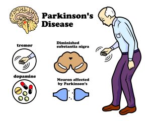
A new study describes how a spinal cord implant provides help to walk for Parkinson’s disease patients. The original study was published in Nature: . CNN published a simplified article that describes how a Parkinson’s patient with gait problems could walk again, when a spinal cord implant was inserted. This patient’s name is Marc Gauthier from a town near Bordeaux, France. He was 36 years old when he was diagnosed with Parkinson’s disease. For many years he could control his Parkinson’s disease symptoms with Dopamine replacement therapy.

Insertion of deep brain stimulator

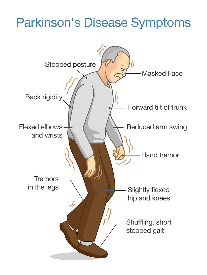
But by 2004 the medication did not control his gait anymore. His doctors inserted a deep brain stimulator, which helped with tremors and muscle stiffness. But as the disease progressed, he developed a severe walking disorder. The drugs and the deep brain stimulator no longer helped his walking problem. His muscles stiffened up, and often he fell 4-times per day. He had to give up his work as an architect. Finally, in 2021 his doctors inserted a nerve stimulator with electrodes connecting to the lower spinal cord.

Spinal cord stimulation

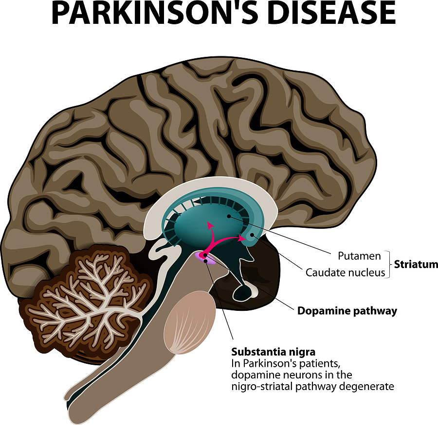
Researchers from France, Switzerland and other parts of the world determined which part of the spinal cord were in need of electrical stimulation to improve his gait and balance problems. Dr. Eduardo Moraud is an author of the study and researcher at Lausanne University Hospital. He said: “The stimulation here is focused on the spinal cord. We target the region of the spinal cord that will control all the leg movements.” The team managed to develop an electric stimulator for implantation under the skin over the abdomen with electrodes going to the lower spinal cord. This way his physicians stimulated muscle groups and also relaxed them in sequence so that his gait improved and steadied. His balance stabilized as well and he could walk stairs again.

Future routine surgery to implant spinal stimulator

Marc Gauthier’s case is a first in approaching Parkinson’s disease with a spinal stimulator. The researchers stressed that they will improve the technology and learn from other patients how to individualize this technique. But eventually the spinal cord stimulator could become a routine approach for end stage Parkinson’s disease. Dr. Moraud said: “Addressing deficits of gait and balance in Parkinson’s disease is extremely challenging. These deficits can be very heterogenous. They can be variable across patients. They can affect walking but also symmetry, balance, posture. The neuroprosthetic approach that we have developed here allows for the first time to target and address these problems individually in a highly specific manner for each patient. It operates in real time, and importantly, it is complementary to other existing therapies.”

Marc Gauthier’s progress since his spinal cord stimulator

The researchers identified hotspots in the lower spinal cord with connection to the gait and balance problem of the patient. The surgeon connected the neuroprosthesis to these hotspots to stabilize gait, balance and muscle strength. Following the surgical procedure Mr. Gauthier had to undergo a few months of rehabilitation with the neurostimulator to practice walking, develop strength and coordination between the right and left leg. His body was no longer stiffening or freezing in place. He could now take a 3-mile lakeside stroll without stopping. He can now manage stairs going up or coming down smoothly. In the past his gait would suddenly freeze or he lost hist balance. None of this

Is happening now. He simply is walking normally.

The final word of Dr. Moraud

“The stimulation here is focused on the spinal cord. We target the region of the spinal cord that will control all the leg movements.” Inserting a neurostimulator is not a cure, simply a procedure to help the patient lead a normal life.

Help to Walk for Parkinson’s Disease Patients

Help to Walk for Parkinson’s Disease Patients

Conclusion

A new surgical procedure, namely inserting a neurostimulator under the abdominal wall, is the latest approach to treat Parkinson’s disease. This is help to walk for Parkinson’s disease patients. The case, which I described here is the first case where doctors performed this new procedure. They identified hot spots in the lower spinal cord and connected them to the neurostimulator with electrodes. After a lengthy rehabilitation period the patient learned how to use the stimulator for optimal walking, balancing, and going stairs up and down. There are no more falls or gait problems and no freezing. At the end the patient can walk normally and even manage a walk of 3 miles. Researchers anticipate that this method will become an accepted treatment modality for gait problems of Parkinson’s disease patients.

Sep
09
2023

How the Immune System affects Parkinson’s Disease

This article explains how the immune system affects Parkinson’s disease (PD). Notably, in the past physicians thought that Parkinson’s disease was due to a degenerative change of the substantia nigra. This explained why balancing was a problem, why shaking of the hands occurred and why falls happened often. It it important to realize that nobody thought about the immune system.  And no-one knew that an autoimmune process could be behind Parkinson’s disease.

T cells that react to a damaged protein called alpha-synuclein

There are specific changes in the immune system approximately 10 years before Parkinson’s disease symptoms occur in patients who come down with the disease. Researchers from the La Jolla Institute for Immunology showed that T cells play a key role in causing PD. They react to a damaged protein called alpha-synuclein build up in the dopamine-producing brain cells. Laboratory physicians can assay this through a simple blood test, which becomes a screening tool for early Parkinson’s disease. The reactive T cells stay around for about 10 years, then fade away. There seem to be other immune factors that weaken the initial aggressive phase of the T cells.

The role of inflammation in Parkinson’s

When the immune system malfunctions chronic inflammation can develop. In farmers exposed to pesticides the later development of Parkinson’s disease was observed. The researchers thought that the pesticides caused an irritation of the immune system leading to chronic inflammation. There is evidence that the gut bacteria are different in Parkinson’s disease patients when compared to normal controls. The gut absorbs the metabolites of the abnormal gut bacteria and causes chronic inflammation. In an attempt to stop the inflammatory process, the immune system can develop autoimmune antibodies, which can cross react with cells of the substantia nigra. This in turn can cause Parkinson’s disease.

Lifestyle factors that people can change to prevent PD

Dr. Rebecca Gilbert, vice-president and chief scientific officer for the American Parkinson Disease Association (APDA) commented on the importance of lifestyle changes. She said: “It makes intuitive sense that instituting lifestyle modifications that potentially decrease inflammation may decrease the risk of Parkinson’s disease. Exercise, for example, has been shown to reduce inflammation and is probably one of the many reasons that exercise reduces the risk of Parkinson’s disease and also improves established Parkinson’s disease.” She commented further: “Also, we should avoid things like excessive alcohol and nicotine that we know have negative effects on the immune system,” she added. “And managing our stress as best as possible can slow and help maximize outcomes of many diseases.”

Changing diet can help postpone Parkinson’s disease

With regard to the best diet that will help Parkinson’s disease patients she said: “The MIND diet emphasizes whole grains, vegetables, nuts, legumes, and berries. Fish is the preferred protein and olive oil is the preferred fat. Recently a study showed that adherence to the MIND diet and the Mediterranean diet had an association with later onset of Parkinson’s disease.”

The gut connection to Parkinson’s disease

According to the WHO the global prevalence of Parkinson’s disease has doubled in the last 25 years. At this point we do not know why this is so. But many investigations have shown that there is a significant difference in the gut bacteria composition of healthy controls and Parkinson’s disease patients. There is a 30% difference between the bacterial composition of healthy controls and patients with Parkinson’s disease. This has led to Braak’s Hypothesis of Parkinson’s Disease. This hypothesis says that an unknown pathogen enters through the nose, the person swallows it and it ends up in the gut. Absorption gets it into the gut wall and it migrates through the vagus nerve into the central nervous system where it leads to accumulation off alpha-synuclein in the substantia nigra. This destroys the dopamine producing cells in that region causing the symptoms of PD.

Can any diet fight gut dysbiosis?

  • In 2022 study they found that flavonoids, the pigments of fruit were associated with a lower mortality of patients with Parkinson’s disease.
  • In an earlier study of 2018 researchers determined that a protein from fish with the name parvalbumin helped Parkinson’s patients to stop producing alpha-synuclein. PD patients suffer from clumping of alpha-synuclein, which causes their symptoms.
  • Restriction of refined carbohydrates “especially diets with a low glycemic index, rich of vitamins and polyphenols, a Mediterranean diet for example, can be recommended”.

Regular exercise to prevent Parkinson’s disease

Regular physical exercise maintains body function and muscle strength. Dr. Emer MacSweeney said: “Being physically active is one of the best things you can do for your body. Exercise helps protect against many diseases and keeps the heart, muscles, bones, and brain in optimum condition. Exercise promotes the oxygenation of the brain and stimulation of multiple neurochemicals.”

Several studies showed that patients with PD deteriorate slower, if they exercise regularly. Part of that response is due to the release of endorphins and serotonin, but we do not know all of the positive mechanisms of exercise at this time.

How the Immune System affects Parkinson’s Disease

How the Immune System affects Parkinson’s Disease

Conclusion

Recent research changed what we know about Parkinson’s disease (PD). Braak’s Hypothesis of Parkinson’s Disease states that an unknown pathogen enters through the nose, gets swallowed and ends up in the gut. From there it gets taken up into the gut wall and migrates through the vagus nerve into the central nervous system. There it leads to accumulation of alpha-synuclein in the substantia nigra. This destroys the dopamine producing cells in that region causing the symptoms of PD. But we also know that chronic inflammation can aggravate the symptoms of PD patients. When the composition of the gut bacteria deteriorates, this too will make PD patients worse.

Lifestyle changes help to postpone Parkinson’s disease

A healthy diet, like the MIND diet, DASH diet or the Mediterranean diet have beneficial effects on PD patients. Many studies also found that regular physical exercise is a stabilizing factor in PD patients. There are still many gaps in what we know about the causation of PD. But the above summarized factors are a good start.

Aug
24
2019

Connection Between Parkinson’s Disease And The Removal Of The Appendix

In the first place, a large study from Cleveland Medical Center found a connection between Parkinson’s disease and the removal of the appendix. It is important to realize that Dr. Mohammed Z. Sheriff who was the main investigator assembled 62.2 million patients’ records. Of these 488,190 (0.78%) patients had an appendectomy done. 4,470 (0.92%) among these patients went on with a diagnosis of Parkinson’s disease. Of the remaining 61.7 million patients physicians eventually diagnosed 177,230 individuals (0.29%) with Parkinson’s disease. This means that the patients with a prior appendectomy had a 3.17-fold higher risk to develop Parkinson’s disease. Whoever did not have their appendix removed had a lower risk to develop Parkinson’s disease.

Older, smaller studies in the past were ambiguous

A 2016 Movements Disorders study with 1.5 million people in Denmark found that patients who had an appendectomy were a slightly higher risk of developing Parkinson’s disease later in life.

In contrast, a 2018 Science Translational Medicine study found the opposite among 1.6 million Swedes. Patients who had an appendectomy had a lower risk of developing Parkinson’s disease and there was a delay in the onset of it.

It was this discrepancy what motivated Dr. Mohammed Z. Sheriff to design the much larger study that he carried out. He presented the findings at the 2019 Digestive Disease Week meeting that took place May 18–21 in San Diego, CA.

Alpha-synuclein associated with Parkinson’s disease

At the same time other investigators found that alpha-synuclein, an abnormal protein in nerve cells, is important for the development of Parkinson’s disease. It plays a role in patients with appendicitis who need surgery. But it also is present in the substantia nigra in the brain of patients with Parkinson’s disease. Notably, patients with Parkinson’s disease often have problems with their balance, muscle rigidly, tremor and slowness of movements.

Indeed, alpha-synuclein has become an important marker for the development of Parkinson’s disease. This publication indicates that monocytes that are activated by the presence of alpha-synuclein can cause significantly more inflammation in the nerve cells of the substantia nigra in the brain. Other authors postulate that oxidative stress in vagal neurons may lead to alpha-synuclein, which can be transported from foci such as in the appendix to the substantia nigra in the brain. More recent studies have shown alpha-synuclein in the gut of patients with Parkinson’ disease according to Dr. Sheriff. In particular, it clearly shows that our gut biome plays a role in the development of disease.

Older people affected by Parkinson’s disease

Certainly, it is mostly people above the age of 60 that come down with Parkinson’s disease. Another key point is that there is no known effective treatment for Parkinson’s disease. However, the new information that alpha-synuclein is a factor in the early stages of Parkinson’s disease is most compelling evidence. By and large, this process leads to toxic clumps of Lewy bodies in nerve cells of the brain and in the gut wall including the appendix. Nobody knows exactly the meaning of Lewy bodies and alpha-synuclein. But if they are playing a part in the initiation of Parkinson’s disease, medications that fight oxidative stress in the nervous system may be a new approach to treating Parkinson’s disease. There is a need for more research.

New approaches to treat Parkinson’s disease

Here is a publication that points out that existing treatment for Parkinson’s disease does not include targeting alpha-synuclein. In the future, these authors state, this will change, as immunotherapy directed at alpha-synuclein will interrupt the development of Parkinson’s disease. Here is another publication that stresses the importance of alpha-synuclein in the development of Parkinson’s disease. It points to testing new therapies in animal experiments and in clinical trials.

Connection Between Parkinson’s Disease And The Removal Of The Appendix

Connection Between Parkinson’s Disease And The Removal Of The Appendix

Conclusion

The observation that people who had an appendectomy have a 3.17-fold higher risk to develop Parkinson’s disease led to more investigations. It became obvious that an alpha-synuclein accumulation in the nerve cells of the gut wall was playing a role in the disease and formed Lewy bodies. It confirms the concept that disease processes can start with our gut biome. These cells can travel via the vagal nerve into the part of the central nervous system, called substantia nigra. It is there that the dopamine producing nerve cells reside. Destruction of these dopaminergic nerve cells leads to the symptoms of Parkinson’s disease. One of the new approaches to treat Parkinson’s disease is by immunotherapy where antibodies destroy the alpha-synuclein protein. However, clinical trials will have to follow that are testing this new treatment possibility.

Mar
10
2018

Dementia From Excessive Alcohol Abuse

A new study from France documented dementia from excessive alcohol abuse. One million patients who needed admission to hospitals in France for early dementia took part in that study. Dementia is a clinical syndrome, where the cognitive ability deteriorates progressively.

The study from France regarding dementia from excessive alcohol abuse

According to the French National Hospital Discharge database it was especially relevant that alcohol use disorders were present in 16.5% of the men and 4% of the women with dementia. Those who did not have the diagnosis of dementia had only half the amount of alcohol abuse disorders in both sexes.

There are other diseases that can lead to dementia like Parkinson’s disease and Huntington’s disease. These cases were not part of this study.

The association with alcohol abuse was particularly evident in early dementia cases at an age below 65.

Furthermore, alcoholics also have a shorter life expectancy.

Several mechanisms responsible for dementia development

Alcohol can cause dementia in several ways.

  • First of all, alcohol and the metabolic by-product acetaldehyde have toxic effects on the brain. They cause long-term toxic effects and functional brain damage is the result.
  • Furthermore, heavy alcohol abuse leads to liver damage with resulting changes in metabolism. Ammonia production from the cirrhotic liver causes brain damage in a condition called hepatic encephalopathy.
  • In addition, heavy drinking is a strong vascular risk factor. This leads to diabetes, high blood pressure, heart attacks and strokes.
  • Often it is the population group that is uneducated, people who smoke and people with depression who suffer from the effects of alcohol abuse.
  • Finally, heavy drinking is more frequent in men than in women. In men the dementia risk was 4.7-fold, in women 4.3-fold.
  • Obesity, smoking and high blood pressure are also risk factors for dementia. If these cases were not part of the study, heavy alcohol abuse caused an increase in dementia in both sexes by a factor of 3-fold.

Possible biases of the study

Dr. Kostas Lyketsos, a neuropsychiatry professor and director of the John Hopkins Memory and Alzheimer’s Treatment Center did not take part in the study and its investigation. A non- biased observer, Dr. Kostas Lyketsos stated that there is a problem with precision of the data the larger the study population is. In this case there were more than 1 million participants, so a number of biases can influence the outcome of the study. For instance, one problem is that these patients with mild cognitive impairment were inpatients in a hospital. Normally such patients would not be in a hospital. Another fact was that none of the participants had received questionnaires of the amount of alcoholic drinks they consumed, and as a result it is difficult to knows exactly how high the amount of alcohol was that caused the damage to the brain.

Country bias

The large sample size was from only one country, France. This means that we do not know whether there would be ethnic differences between countries. Nevertheless the findings of this study are important. In a 2014 review by the WHO the average person in France consumed 12.2 liters of pure alcohol per year. In contrast the average person in the US consumed only 9.2 liters of pure alcohol in this year. Apart from these concerns it is important to realize that alcohol has toxic effects on the brain. This can also result in dementia.

Other studies regarding alcohol abuse and dementia

The media has praised alcohol for preventing heart attacks. On the other hand, there are other articles in which we hear about alcoholic hepatitis and liver cirrhosis, both of which can be killer diseases. To get some clarification, let us examine the various facts.

Dr. Finnel mentioned that 7.9% of all emergency room visits in the US are due conditions which have an association to alcohol(Ref.1). When the causes of deaths  that are a consequence to alcohol are listed, they are: cancer of the mouth and pharynx, alcohol abuse disorders, coronary heart disease causing heart attacks, cirrhosis of the liver, traffic accidents, poisonings, falls and intentional injuries. You don’t get that from the news. Instead you read about the one glass of red wine per day that is good for women and up to two glasses of red wine that is good for men to prevent heart attacks and strokes.

Bioflavonoids

It is the bioflavonoids and among those in particular resveratrol, that are the active ingredient responsible for heart health.

Resveratrol is a powerful antioxidant that protects against ischemia-reperfusion injuries.  It is responsible for the cardio protective properties of red wine known as the “French paradox” (Ref.2). According to this reference resveratrol contributes to at least 3 processes that stabilize the metabolism.

Toxicity of alcohol

According to the WHO 5.9% of all deaths worldwide is a consequence of alcohol overconsumption.  In 2012 the WHO recorded that 7.6% of deaths in males, but only 4% of female deaths were due to alcohol. Toxicity comes from the breakdown product acetaldehyde, which all cells can convert from alcohol. Liver cells are especially able to do that. According to Ref. 3 alcohol diffuses easily through all of the cell membranes and reaches every organ in the body. The toxicity of acetaldehyde is shutting down the mitochondria, which affects the energy metabolism and causes cell death. The immune system reacts with inflammation, when it attempts to repair the damage.

So, what are the major damages which alcohol can cause? First there is fat accumulation (steatosis), next chronic inflammation followed by necrosis (dying of cells) and finally fibrosis. An example of fibrosis is liver cirrhosis, where non-functioning connective tissue replaces liver cells.

Different tissue sensitivity to alcohol

Certain tissues are more susceptible to alcohol toxicity than others. As the concentration of alcohol is highest in tissues that are in direct contact with alcoholic drinks, cancers related to alcohol consumption develop in the oral cavity, pharynx, larynx, esophagus, and in the colon and rectum. The pancreas is particularly vulnerable to inflammation and fibrotic changes with degeneration into cancer of the pancreas. The heart tissue and the arteries are very sensitive to alcohol.  Hypertension, heart attacks, stroke, cardiomyopathy and myocarditis as well as irregular heart beats (arrhythmias) can develop. The brain is very sensitive to toxic effects of alcohol as well. This causes major depression, personality changes with violent behavior, car accidents and injuries.

Other toxic effects of alcohol on organs

Kidney disease (alcoholic nephropathy) is another illness due to too much alcohol. Five percent  of breast cancers in northern Europe and North America are a direct consequence to the toxic effects of alcohol (Ref.3). Finally, the liver being so active in detoxifying alcohol, will be not functioning and finally develops liver cirrhosis, as described before. This accounts for a lot of premature deaths at a relatively young age (typically in the mid to late 50’s).

Ref. 3 goes on to say that literature exists which claims that 1 to 2 drinks per day would be useful for prevention of heart disease. But the observation of the authors is that people will not discipline themselves to stick to these limits and very quickly enter into the zone of alcohol toxicity. The authors further noted that with regard to causing any kind of cancer there is no safe lower limit; the risk is directly proportional to the amount of alcohol consumed and the risk starts right above the zero point.

The pathologist has the last word

When I studied medicine at the University of Tübingen, Germany I attended lectures in the pathology department where Professor A. Bohle, M.D. demonstrated pathology findings of deceased patients. Dr. Bohle had a special interest in Mallory bodies. These are alcohol inclusion cysts within liver cells that can be stained with a bright red dye.

Histological documentation of toxic effects in livers of corpses

I will never forget when Prof. Bohle pointed out that the livers of this most diverse population, whose bodies we had the privilege as medical students to study, had a rate of 25% positive Mallory bodies. He wanted to impress on us as medical students to watch out for the alcoholics that are usually missed in general practice. Obviously 25% of the pathology population was affected by the consumption of alcohol. It was Prof. Bohle’s hope that we could perhaps interfere on the primary care level before things went out of control. Many of these corpses were the sad results of traffic accidents that could have been prevented. (In 2018 things have changed: seat belts and alcohol limits are standard, in 1968 in Germany they were not).

Alcohol as an aging substance

Consistent use of alcohol on a regular basis will slow down cell metabolism and hormone production significantly. The major effect of alcohol leads to poisoning of the mitochondria in multiple organs, which translates into faster aging and a shortened life expectancy. This in turn results in a change of appearance. An older person who has abused alcohol for a number of years may look 5 to 10 years older than their chronological age.

50% of people above the age of 65 drink daily (Ref.4). Some more statistics: alcohol abuse in elderly men is 4-times higher than in elderly women. 5% to 10% of all dementia cases are related to alcohol abuse. About 15% of older adults are experiencing health risks from abusing alcohol. And about 90% of older adults are using medications. Close to 100% of medications can adversely interact with alcohol (Ref.4).

Social pressure

These are the scientific facts, and then there is social pressure, when you are invited to a party.

When you are young and believe that you are invincible, do you care what the science says? You want to have a “good time” and not worry about consequences. The data about long-term exposure and a slowly increasing cancer risk is there. The wine industry will remind you that 1 drink for women and two drinks for men will protect you from heart attacks. They will withhold the cancer information from you, as they don’t really want to hear about that (yes, it’s bad for their business!).

Resisting social pressure and doing what is good for you

Can you have a good time at a party without drinking alcohol? Yes, you can. You can talk and you can listen; you are probably more with it than those who had too much to drink. I like mineral water and hold on to a glass of that.

I explained in a blog before how I was convinced by three speakers at an A4M conference to join those who abstain from alcohol.

Socializing without alcohol is doable. You may at times miss it, but you can warm up even to a crowd that had a few drinks too much. It is about choice: we can choose what we want out of life.


Dementia From Excessive Alcohol Abuse

Dementia From Excessive Alcohol Abuse

Conclusion

Alcohol is a cell and nerve poison. The medical need for “one glass of wine for women and two glasses of wine for men to prevent heart attacks and strokes” has been vastly exaggerated. Fact is that resveratrol and other antioxidants like vitamin C and vitamin E can also prevent cardiovascular disease. They are alcohol-free! The risk of dementia development as a long-term result of alcohol exposure is something that is only now getting attention by the medical profession. We live longer these days, and this makes alcohol exposure over the decades a real threat to our mental wellbeing. Consumption of alcohol needs to be re-evaluated by every one of us. What risks are we willing to take? Is the stress-relieving effect of alcohol worth the risk of losing our mind to Alzheimer’s disease? If we care about our future the answer should be clear!

References

Ref. 1: John T. Finnell: “: Alcohol-Related Disease“ Rosen’s Emergency Medicine, Chapter 185, 2378-2394. Saunders 2014.

Ref. 2: “Hurst’s The Heart”, 13th edition, The McGraw-Hill Companies, Inc., 2011. Chapter 54. Coronary Blood Flow and Myocardial Ischemia.

Ref. 3: Ivan Rusyn and Ramon Bataller: “Alcohol and toxicity”, 2013-08-01Z, Volume 59, Issue 2, Pages 387-388; copyright 2013 European Association for the Study of the Liver.

Ref. 4: Tom J. Wachtel and Marsha D. Fretwell: Practical Guide to the Care of the Geriatric Patient, Third Edition, Copyright 2007 by Mosby.

Jan
27
2018

Bacterial Toxins Threatening The Brain

Dr. Robert G. Silverman gave a talk about bacterial toxins threatening the brain. He spoke at the 25th Annual World Congress on Anti-Aging Medicine in Las Vegas on Dec. 15, 2017. First of all, he pointed out how changes in the gut flora can affect the integrity of the gut wall. In addition this can eventually this lead to a leaky gut syndrome. But it does not end here. As a result the toxins enter the blood stream and affect the blood/brain barrier. Consequently in the end various neurological diseases can develop from this.

Here I am giving a brief overview of the talk by Dr. Silverman. But he was not the only one speaking to this subject. Several other speakers also brought up this subject throughout the conference. They stressed the importance of rectifying any gut dysbiosis to stop leaky gut syndrome and a leaking blood/brain barrier.

Leaky gut syndrome

When the gut flora changes there are often enteropathogenic E. coli strains, Shigella and Salmonella that invade the lining of the gut causing leaky gut syndrome. When toxins enter the blood stream, the body is starting to form antibodies against various proteins. Antibodies are acting against various targets: bacterial cytotoxins, cytoskeletal proteins, tight junction proteins and food antigens. Lipopolysaccharides (LPS) from toxins of gram-negative gut bacteria can also leak into the blood. This affects key organs like the liver, the heart, lungs, the joints, the immune system and the thyroid. When this process has gone on for some time, the blood/brain barrier is breaking down next. The intestinal inflammation causes the release of inflammatory cytokines that circulate in the blood stream. The cytokines cross the blood/brain barrier and activate the support cells in the brain, called microglia. This in turn causes inflammatory degenerative changes in the brain.

Blood/brain barrier

LPS circulating in the blood from gut bacteria endotoxins increase the permeability of the blood/brain barrier. This is bad news for the brain as it becomes vulnerable to attacks from the antibodies mentioned and from food particles. Dr. Silverman cited papers showing that circulating antibodies that cause inflammation in the brain can be the starting point for early Parkinson’s disease. Autoimmune antibodies can cause even depression.

Intestinal permeability can be assessed by various antibody constellations. For instance IgA antibodies point to an ongoing issue/early leaky gut syndrome. IgM antibodies indicate early onset and IgG antibodies chronic issues of leaky gut syndrome. If you add various antigens like LPS, zonulin and actomyosin you can pinpoint which structure of the gut wall is affected by leaky gut syndrome, and the antibody type adds more information about the timing of the onset of leaky gut syndrome.

Bacterial toxins threatening the brain when BBB damaged

As I already mentioned the blood/brain barrier (BBB) is often simultaneously affected when there has been leaky gut syndrome. There may be a delay, but eventually the BBB breaks down also, and the brain will be in jeopardy. Dr. Silverman gave an example of how depression can develop as result of a breakdown of the BBB. Chronic intestinal inflammation can suppress the sensitive hippocampus cells from regenerating. Physicians call that impairment of hippocampal neurogenesis. Inflammatory cytokines damage the neuronal cell progenitors. As a result patients with inflammatory bowel disease can have mood disorders and cognitive impairment. Sophisticated BBB blood tests can pinpoint whether the BBB is intact or establish whether there is impairment. The important thing to remember: there is a gut brain connection.

Fixing the gut to stop bacterial toxins threatening the brain

In order to fix the BBB, you must first concentrate on fixing leaky gut syndrome.

  • Avoid gluten, as gluten is causing inflammation of the gut wall.
  • Start taking probiotics that contain more than 30 Billion lactobacillus plantarum, lactobacillus acidophilus and Bifidobacterium lactis per daily dose.
  • Do a heavy metal detox involving phytonutrients, hops, turmeric, Andrographis, zinc, polyphenols, omega-3 fatty acids, and watercress plant extract. Andrographis, also known as the “King of Bitters”, is an Ayurvedic medicine used to promote digestion and stimulate appetite.

Nutrients to fix the blood/brain barrier

Dr. Silverman uses the following nutrients to repair the blood brain barrier.

  • Acetyl L-Carnitine: this helps to protect the mitochondria from oxidative damage
  • Berberine: reduces inflammation in brain injuries
  • Alpha-lipoic acid: preserves the integrity of the BBB by controlling oxidative stress
  • Curcumin: decreases brain swelling, preserves the BBB and increases tight junction protein in brain cells
  • Vitamin D3 (5000 IU or more): protects the BBB by various mechanisms
  • Omega-3 fatty acids: they increase cell membrane fluidity and protect the BBB
  • Resveratrol: reduces inflammation and restores the BBB

Neuroplasticity

In order for the brain to adapt to changes, it must be flexible, which means on a cellular level that nerve cells form new synapses, neurological pathways etc. This is what neuroplasticity means. Here are the factors that Dr. Silverman listed as facilitating neuroplasticity.

  • Regular exercise
  • DHA from fish oil capsule supplements
  • Turmeric
  • Whole coffee extract
  • Alpha-lipoic acid
  • Lactobacillus brevis and Bifidobacterium longum
  • Bifidobacterium animalis Lactis 420 (B420)
  • Probiotics: they feed the healthy gut bacteria (e.g. apple cider vinegar)
  • Elevate magnesium in the brain through L-threonate
Bacterial Toxins Threatening The Brain

Bacterial Toxins Threatening The Brain

Conclusion

In the last few years it has become abundantly clear that leaky gut syndrome is not an isolated matter. It is invariably connected to a breakdown of the blood/brain barrier (BBB). Leaky gut syndrome alone is bad enough as it can lead to a number of autoimmune diseases, like Hashimoto thyroiditis and others. But when the BBB is affected, antibodies can now affect nerve cells, can cause Parkinson’s disease, depression, and even Alzheimer’s disease. There is no reliable database for what can happen to the brain when the BBB breaks down.

Because of these connections it is important to sanitize the gut, re-establish a healthy gut flora and overcome leaky gut syndrome. This will at the same time repair the broken down BBB. It will also prevent further possible damage to the brain in the future. Your gut health is your brain health. Take care of both your gut as well as your brain!

Sep
30
2017

Parkinson’s Disease May Be Stopped

Parkinson’s disease is common in the US; new research shows that the use of an old anti-depression medication can stopParkinson’s disease The use of nortriptyline, a 50-year old antidepressant has shown to normalize a nerve cell protein. In rats nortriptyline dissolved toxic alpha-synuclein clusters in brain cells. These toxic protein clusters seem to be happening in the brain of Parkinson’s disease patients also. It is the protein by the name of alpha-synuclein that research first found in rats to cause the toxic protein clusters in nerve cells of the substantia nigra, a part of the brain stem.

But nortriptyline was able to normalize the concentration of the protein. In preliminary studies in humans the investigators found that there was a significant improvement of Parkinson’s disease with the use of nortriptyline.

Placebo controlled trial with nortriptyline

Now a research team from Michigan State University in Grand Rapids conducted a larger clinical placebo-controlled trial. The lead researcher Collier of the study group found that Parkinson’s patients who received treatment for depression with the tricyclic agent nortriptyline needed less dopamine, the main drug used to treat Parkinson’s disease. This indicated to the researchers that nortriptyline was preserving brain cells that were still making their own dopamine. In rat experiments they could show that it was the dissolving of toxic alpha-synuclein proteins by nortriptyline that was the key to therapeutic success.

Lisa Lapidus, a co-worker on the Michigan State University research team summed up their research: “What we’ve essentially shown is that we are dealing with a drug that the FDA approved already 50 years ago. Patients tolerate the medication relatively well. This could be a much simpler approach to treating the disease itself, not just the symptoms.”

Parkinson’s disease may be stopped also by old diabetes drug

Thomas Foltynie found that the diabetes drug exenatide helps patients with Parkinson’s disease. Dr. Foltynie is a professor of neurology at the University College London and co-author of the study.

Exenatide is an injection drug. When preliminary studies showed that this drug was effective in helping Parkinson’s disease patients lose their problems with walking and balance, a formal study followed.

Professor Foltynie designed a study where 60 people with Parkinson’s disease either got injections of exenatide or placebo injections. Patient exams followed regarding their musculoskeletal system and balance at baseline and every 12 weeks. A score system of 132 points assessed their Parkinson’s disease. After 48 weeks those who had been taking exenatide had a gain of 1 point on that scale while the placebo group dropped 3 points. After 48 weeks the drug administration (exenatide) finished. But after another 12 weeks another scoring and assessment of the Parkinson’s disease symptoms took place. The experimental group on exenatide scored 3.5 points higher than the placebo group. This suggests that exenatide is helping to treat the cause of Parkinson’s disease, not just the symptoms.

Parkinson’s disease may also stop through the use of caffeine

Parkinson’s disease was in the news again because of another study that involved breaking up misfolded alpha-synuclein through caffeine.

Misfolded alpha synuclein forms clumps inside dopamine producing cells in the substantia nigra of the brain stem. Misfolded alpha synuclein acts like a toxin to the dopamine producing cells and eventually these cells die off. This is the brain region that is responsible for making muscle movements smooth and stabilizes balance. The cells that have misfolded alpha synuclein clumps in them also go under the name of “Lewy bodies”.

Dr. Jeremy Lee from the University of Saskatchewan (Saskatoon, Saskatchewan, Canada) has isolated two compounds from coffee. They are called C8-6-I and C8-6-N. They can bind to alpha-synuclein and prevent clumping, which stops the toxic effects on dopamine producing nerve cells. Like with nortriptyline the caffeine effect is a curative approach to Parkinson’s disease.


Parkinson’s Disease May Be Stopped

Parkinson’s Disease May Be Stopped

Conclusion

There is a new therapeutic approach to Parkinson’s disease. Researchers have detected a protein called alpha-synuclein to cause toxic protein clusters in nerve cells of the substantia nigra, a part of the brain stem. When these cells die from the accumulation of these misfolded proteins, patients come down with Parkinson’s disease. But three different methods of treatment can improve Parkinson’s disease by dissolving the protein alpha-synuclein.

  1. Nortriptyline was able to normalize the concentration of the protein. In preliminary studies in humans the investigators found that there was a significant improvement of Parkinson’s disease with the use of nortriptyline.
  2. Exenatide, an injection drug for diabetes, has been described to help Parkinson’s patients get better.
  3. Caffeine can also dissolve misfolded alpha synuclein (two compounds from coffee called C8-6-I and C8-6-N). This helps patients with Parkinson’s disease to stabilize.

This is only the beginning of a new approach to Parkinson’s disease and an attempt to cure the disease by dissolving the underlying mechanism. So far the drugs that are in use for Parkinson’s disease are only attempting to stimulate dopamine producing nerve cells to produce more dopamine. But the underlying pathology of accumulating misfolded alpha-synuclein clumps is not yet in the treatment protocol. The new research is different, as it takes this into account in an attempt to prevent the condition.

Incoming search terms:

Oct
08
2016

Vitamin D3 Protects Your Brain

More and more studies are showing that vitamin D3 protects your brain. It protects against MS, but also against Parkinson’s disease and Alzheimer’s disease. In the following I will review what evidence there is to support each of these topics.

Vitamin D3 protects your brain from multiple sclerosis (MS)

It has been known for some time that in the northern hemisphere MS is more common because of the lack of sunshine, which in turn produces less vitamin D3 in the skin.

MS is an autoimmune disease where immune cells attack the lining of nerves. Both nerve cells and immune cells have vitamin D receptors. It appears that immune cells are calmed down by vitamin D3 and remission of an MS relapse is more likely.

There are two forms of MS, the relapsing-remitting MS and the progressive MS. The first one (relapsing-remitting) is more common. After a bout of active MS, the illness calms down and the condition of the patient is stable for some time until the next relapse occurs.

With progressive MS there are two forms, primary progressive MS and secondary progressive MS. The primary form is a case of MS where symptoms steadily worsen, without any remission. The secondary form of progressive MS occurs at the end of fairly stable relapsing-remitting MS. Symptoms become more pronounced and the condition deteriorates steadily from there.

Progression and disability in MS patients with various vitamin D3 levels

Dr. Fitzgerald and colleagues published a study in JAMA Neurology in 2015.

They took 1482 men and women who were on interferon beta-1b treatment. This treatment utilizes the immunomodulator interferon beta-1b and reduces the number of relapses in patients with MS. The study took place between November 2003 and June 2005. Results were analyzed between June 2013 and December 2014. The researchers measured vitamin D levels (as 25-hydroxy vitamin D). The vitamin D levels were obtained at baseline, at 6 months and 12 months.

The number of brain lesions were measured by MRI scans. All of the patients also underwent a functional test, called expanded disability status scale. This measured impairment of ambulation, ability to communicate and activity levels.

Results of this study showed marked differences between patients with high and low vitamin D levels. Those patients who had the highest vitamin D blood levels (more than 40 ng/mL) had the lowest rates of new MS lesions. Previous studies had found that a low blood level of vitamin D (less than 25 ng/mL) in patients was associated with a much higher risk of developing MS. Dr. Fitzgerald’s study showed that a 50.0-nmol/L increase in serum vitamin D levels associated with a 31% lower rate of new MS lesions. Patients with the highest vitamin D level of more than 100 nmol/L had the lowest amount of new MRI lesions (47% less than the patients with the lowest vitamin D levels).

Another study showed that a low-dose vitamin D level accelerated MS. There was a 5.9-fold risk converting the initial relapsing-remitting form of MS into the secondary progressive form of MS.

All these studies show that vitamin D3 can decrease the risk of getting MS. In addition vitamin D3 also delays progression in those who have MS.

Vitamin D3 protects your brain from Parkinson’s disease

Vitamin D3 plays a role in preventing Parkinson’s disease.

Parkinson’s disease is a neurodegenerative disease that causes tremor in muscles, causes balancing problems and eventually can lead to dementia. A metaanalysis was done in 2014 and 7 studies where identified to be relevant. The authors were looking for correlation of vitamin D levels with Parkinson’s disease. The study included 1008 patients in the metaanalysis with 4,536 controls.

  • Patients with a vitamin D level of less than 75 nmol/L had a 1.5-fold higher risk of developing Parkinson’s disease than the controls.
  • Patients with a vitamin D level of less than 50 nmol/L were at a 2.2-fold higher risk of developing Parkinson’s disease.

Another metaanalysis utilized 5,690 Parkinson’s disease patients and 21251 matched controls.

It found that vitamin D levels of less than 20 ng/ml were associated with a risk of 2.08-fold to develop Parkinson’s disease. Interestingly, vitamin D3 supplementation reduced the risk of Parkinson’s disease by 38%. Outdoor work reduced the risk of developing Parkinson’s disease by 28%.

Vitamin D3 protects your brain from Alzheimer’s disease

Alzheimer’s disease is a neurodegenerative disease of old age. We know that it is much more common in patients with type 2 diabetes where insulin levels are high. Studies have shown that Alzheimer’s disease can be termed type 3 diabetes.

The resulting neurofibrillary tangles and amyloid-beta deposits damage nerve cells, which are responsible for the memory loss and the profound personality changes in these patients.

What does vitamin D3 have to do with this?

A 2014 study showed that a low vitamin D level was associated with a high risk of dementia and Alzheimer’s disease.

Specifically, the researchers found the following observations.

  • Vitamin D level of less than 10 ng/ml: 122% increased risk of Alzheimer’s
  • Vitamin D level 10 to 20 ng/ml: 51% increased risk of Alzheimer’s

The same research group found in two trials that vitamin D deficiency leads to visual memory decline, but not to verbal memory decline.

Vitamin D3 combined with metformin suppresses cancer

The newest development with respect to vitamin D3 is the finding that it also has anti-cancer effects. Dr. Li demonstrated that vitamin D reduced prostate cancer cell line growth by 45% while metformin alone reduced it by 28%.

But when both vitamin D and metformin were present in the cell cultures there was growth inhibition of 86%. Dr. Li explained that vitamin D potentiated the growth inhibitory effect of metformin.

Vitamin D3 protects your brain: guidelines to proper vitamin D3 dosing

For years the medical profession stated that 400 IU of vitamin D3 would be enough supplementation. It may be enough to prevent rickets in children. But these low doses will be insufficient in many patients who are deficient for vitamin D to prevent MS, Parkinson’s disease, Alzheimer’s disease or cancer.

A study on medical staff in Northern India showed that 85% of the staff had very low vitamin D levels of less than 10 ng/ml.

It took high doses of vitamin D3 to increase the vitamin D level in the blood.

Generally supplements of vitamin D3 of 5000 IU to 8000 IU are the norm now. But some patients are poor absorbers and they may require 15,000 IU per day. The doctor can determine the patient’s requirement for vitamin D by doing repeat vitamin D blood levels (as 25-hydroxy vitamin D). The goal is to reach a level of 50-80 ng/ml. The optimal level with regard to nmol/L is 80 to 200 (according to Rocky Mountain Analytical, Calgary, AB, Canada).

Vitamin D3 Protects Your Brain

Vitamin D3 Protects Your Brain

Conclusion

Many people are deficient with regard to vitamin D, and they do not know it. The most important thing is to do a vitamin D blood test to assess your vitamin D status.

We know for a long time that vitamin D plays a role in bone metabolism and this is why women approaching menopause often need vitamin D3 supplementation. But it may come to you as news that vitamin D3 also protects from MS, Parkinson’s disease and Alzheimer’s disease. In addition, as indicated above, we know that vitamin D3 when taken regularly suppresses many cancers.

When you realize that all body cells have vitamin D receptors on their surface, it is no surprise that vitamin D3 is so important to take. The vitamin D3 receptors must be there for a reason. When you deprive your body of this valuable vitamin, the high risk of degenerative diseases will be the consequence.

Apr
25
2015

Rejuvenate With Stem Cells

We all age; but can we rejuvenate with stem cells? There is a limit to detoxification, to eating organic food, to exercising, to the effects of vitamins and supplements and even to the effect of bioidentical hormone replacements. The limit comes from our telomeres and from stem cells that get depleted in our body as we age. Some researchers report that in regions where we suffer from a disease stem cells are even more depleted than in the rest of the body.

We do not have all the answers yet. We would like to know why our stem cells in the fatty tissue or in the bone marrow do not migrate on their own into an aching back or a sore shoulder. There are all the aches and pains associated with old age. So, why do our own stem cells not help us? They seem to be locked away in fatty tissue and in bone marrow.

At the 22nd Annual World Congress on Anti-Aging Medicine in Las Vegas (Dec. 10-14, 2014) I learnt that there is a group of stem cell experts in California with affiliates all over the US. They simply take stem cells from the fatty tissue and sometimes also from the bone marrow, isolate the stem cells through a stem cell separator and infuse the stem cell rich fraction (minus fatty and connective tissue) in a bit of saline solution back into the vein of the patient. When the stem cells are in the blood stream, they get activated by the growth factors that are present in blood and can now find where they are needed and start the healing process.

Studies have shown that when stem cells are in circulation in the blood, they are very sensitive to signals from tissues that indicate that there is an inflammatory process. This is why stem cells will repair arthritic changes. The can repair a torn meniscus, a rotator cuff tear in the shoulder or repair a weak immune system. The interesting observation is that stem cells from fatty tissue, also termed mesenchymal stem cells, are pluripotent. This means they can develop into cartilage building cells (chondrocytes) and build up cartilage; this is badly needed in a person with severe osteoarthritis. But stem cells are flexible: they can turn into meniscus cells in a knee with a torn meniscus. They also can repair the damage and relief the patient of the chronic pain. In a shoulder with a rotator cuff tear they can turn into a tough ligamentous material mending the tear.

Some data even indicates that circulating stem cells can repair vital organs like the brain, heart, liver, kidneys and bone marrow; these latter observations were mostly done in animal experiments, but human data is starting to be published in the medical literature.

So, let’s examine what has been found useful with regard to stem cells that are taken from your fatty tissue or your bone marrow and injected into one of your veins.

Here is a website from Arizona that I am only showing as a typical example (I have no conflict of interest and no commercial connections to this group) of what I described above.

With websites like this it is also important to read the disclaimer: “Even though our treatments are done using autologous cells, our Stem Cell Therapies are not approved by the FDA. Stem Cell Treatments are not a cure for any condition, disease or injury, nor a substitute for proper medical diagnosis and care…” Another website from La Quinta, CA describes the use of mesenchymal stem cells for regenerative therapies.

Stem cell treatments are in flux. There is a large body of knowledge that has accumulated showing that with proper technique and aseptic conditions it is a safe procedure. The FBA has been watching this. There are publications regarding the safety of procedures with adipose mesenchymal stem cells; here is one example.

The next step is to show in clinical trials that a certain procedure with stem cells is effective in treating a certain condition.

Below I did a literature review, which are only a few examples, but does not claim to be complete; it highlights some of the problems with stem cell treatments.

Stroke treatment with intravenous administration of bone marrow mononuclear stem cells

This study from India showed no statistical difference of stroke patients treated intravenously with bone marrow derived mononuclear stem cells (the experimental group) and the control group that did not receive such treatment. The investigators examined both groups with functional brain tests and performed PET scans to look at the healing of the brain lesions. Unfortunately the tests showed no statistical difference, but did show that the stem cell procedures were safe. It may be that the wrong stem cells were used (mononuclear bone marrow stem cells) when adipose derived mesenchymal stem cells may have done better. In stark contrast to the study from India is the stem cell treatment for a severe stroke in the former hockey player, Gordie Howe that has gone through the media recently. His procedure was done in Mexico. The stem cells were administered via a lumbar puncture approach as well as intravenously. As you can see from this case, stem cell treatment is even possible in patients who are in their mid 80’s with impressive results.

Parkinson’s disease

Here is a feasibility study from March 2014. A 71-year-old Asian man with progressive supranuclear palsy, an aggressive form of Parkinson’s disease was treated with adipose tissue-derived mesenchymal stem cells that were administered intravenously and intrathecally (to get stem cells into the cerebrospinal fluid that bathes the brain). A remarkable functional recovery took place.

Possible side-effects

This is a report of pulmonary embolism after administering intravenous adipose tissue-derived stem cell therapy. The blood clots in the lungs were treated with anticoagulant therapy. Repeat CT scans of his lungs showed later that the emboli were dissolved spontaneously. It is not clear whether this was a case where familial clotting problems pre-existed as a relative of this patient experienced a similar occurrence after stem cell therapy as well.

A case of chronic autoimmune thrombocytopenic purpura

A rare form of autoimmune disease exists where the body forms antibodies against platelets that help your blood to clot. Here is a paper from June 2009 that describes how a man with this disease was cured using adipose tissue-derived mesenchymal stem cells that were injected intravenously.

Renal transplant survival in type 1 diabetes patient

This case report from India shows that adipose tissue derived mesenchymal stem cells that were given at the time of a kidney transplant to treat end stage kidney disease. The treatment stabilized the condition of this patient after a kidney transplant. At the same time some of the mesenchymal stem cells differentiated into insulin producing cells, which made it much easier to control this patient’s diabetes. In this case stem cells were providing stability following an organ transplant (kidney) and some stem cells turned into insulin producing pancreatic cells.

Osteonecrosis of hip treated with adipose tissue derived MSC

In this study from South Korea dated January 2012 two cases of osteonecrosis of the hip, where the hipbone died (osteonecrosis) are described. The following stem cell protocol helped: The fraction that contained the stem cells (called stromal vascular fraction) was mixed with platelet rich plasma and hyaluronic acid. Using a long needle this mixture was injected into the affected hip joint. Conventional medicine has nothing to offer except a total hip replacement. But here are two cases that showed complete resolution of their pain, regained hip function completely, and healing could be documented with the help of MRI scans.

Treating heart attack patients with stem cells

Here is a paper from The Netherlands, published in June 2014 that describes the problems with stem cell treatment in humans. It points out that much has been learnt from animal experiments. The problem following a heart attack is that there is a massive inflammatory response in the infarcted heart muscle, which makes it difficult for stem cells to establish themselves in the injured heart muscle. However, stem cells have been shown to prevent the development of cardiomyopathy that follows a massive heart attack and often is the cause of death. More refinements are needed for successful treatments, such as the ideal timing of stem cell injections in relationship to the time of the heart attack, the best treatment approach and what number of stem cells to inject are all questions that still need to be answered.

MS model in mice shows promise with adipose mesenchymal stem cells

Experimental encephalitis in mice is used as a model for MS in humans. It helps to preselect potentially effective treatments for MS in humans. In this 2013 paper from Australia researchers used mesenchymal stem cells from adipose tissue and injected them intravenously. To their surprise the mesenchymal stem cells were able to penetrate the blood/brain barrier and end up in the myelin lesions inside the brain. In contrast, bone marrow derived stem cells were unable to do that. The researchers stated that adipose mesenchymal stem cells should be considered “as a cell therapeutic that may be used to treat MS patients”.

A group from Iran published this paper in February 2015 further emphasizes that mesenchymal stem cells would be a logical way to treat MS in humans.

Immunosenescence

As we get older the immune systems weakens because of a process called immunosenescence.

A research group from Austria published a paper in December 2011 that is typical for the thinking that mesenchymal stem cells from fatty tissue have properties that help the immune system to get stimulated. Based on this human data it should be possible to stimulate the immune system by giving stem cells from the fatty tissue to the same person intravenously. This publication shows that this process, which would benefit people above the age of 50 or 60 when the immune system gets weaker, will indeed stimulate the immune system. However, at this point we do not have the data of large clinical trials where this would have been done with measurements of the immune function before and on several occasions after stem cell injection to get a feeling for how long the effect would last. We also do not know whether this procedure is associated with longevity.

Rejuvenate With Stem Cells

Rejuvenate With Stem Cells

Conclusion

Stem cell therapy is definitely coming and many applications are already established as I discussed in a prior blog. It is only recently that physicians are no longer worried about creating tumors with stem cell transfer. Now we are in a phase where various stem cell transfer methods (intravenous, intrathecal, interstitial) are being tested as a treatment for various illnesses. It looks like stem cells from fatty tissue may soon be used intravenously, but I have not seen any such trials when checked on PubMed. The activation of stem cells by laser light has only been mentioned sparingly in the literature. This combination (laser activated, intravenous mesenchymal injection) has the potential for being useful for a multitude of chronic illnesses like fibromyalgia, MS, generalized arthritis, just to mention a few. Mesenchymal stem cells are anti-inflammatory, and they can mend defects without leaving scars.

Mar
07
2015

Drink Your Coffee, But…

I have blogged about coffee drinking several times in the past. Coffee consumption and health benefits have become a news item again because of yet another study. The recent media reports are based on a South Korean study that involved 25,138 men and women with a mean age of 41.3 years.

Here I like to concentrate on aspects regarding coffee consumption that are often lost in the media when studies regarding coffee consumption are discussed. I will break it down into points and then conclude at the end with my recommendations.

1. Calcification of coronary arteries and osteoporosis

The South Korean study published online on March 2, 2015 showed that with up to 4 cups of coffee there was a direct linear relationship between consumption of coffee and prevention of heart attacks. Coronary artery calcium (CAC) deposits were measured by a CAT scan as they are known to be a good measure for a future risk of heart attacks. Less than 1 cup of coffee per day resulted in a 23% reduction of CAC in the coronary arteries compared to controls without coffee consumption. 1 to 2 cups of coffee reduced CAC’s (meaning the risk of heart attack rates) by 34%, while 3 to 4 cups prevented CAC’s and thus heart attacks by 41%. The fun stops at 5 cups of coffee per day as only 19% of CAC’s (heart attacks) were saved. Clearly there is something in coffee that shows detrimental effects, if the dosage is too high.

In the past there was a question as to whether coffee consumption would lead to osteoporosis in women. However, a study showed that there was no correlation between coffee consumption and osteoporosis.

Other studies have clarified this and found that vitamin D3 and K2 are important to remove calcium from the arterial wall and transport calcium into the bone and deposit it there. Vitamin D3 and vitamin K2 seem to override all the other nutrients when it comes to osteoporosis prevention. The other factor in older women is hormone deficiency as they age necessitating bioidentical hormone replacement in addition to vitamin K2 and vitamin D3 to prevent osteoporosis.

2. Whether or not you put sugar into your coffee

is an important question. This is routinely done in Germany where I grew up. The addition of sugar changes the entire game plan, as it is sugar that oxidizes LDL cholesterol, which is directly deposited under the arterial walls. This is the root cause of hardening of the arteries. Coffee alone is beneficial; coffee with sugar is not. I use a tiny amount of KAL Stevia (which does not have the bitter aftertaste) instead of sugar to sweeten my coffee. This sweetens it to the equivalent taste of sugar, but without the detrimental oxidizing effect of sugar. Somebody like me who was conditioned to eat sugar from childhood on in Germany has been left with a “sweet tooth”; so I need to have this tiny bit of stevia as a crutch. Purists may disagree with me. Keep in mind that the Korean study was done without sugar.

3. What’s the difference between real and decaffeinated coffee?

The recent study showed that you need to drink the real thing (caffeinated coffee), if you want to reduce your risk to get the dreaded pigmented skin cancer, melanoma. Decaffeinated coffee did not have this melanoma protective effect. This points to the fact that there are several substances in real coffee and decaffeinated coffee that have different effects. Ref. 2 shows that there was a clear reduction in the risk of developing type 2 diabetes in people who drank either coffee, decaffeinated coffee or tea. Unfortunately many studies do not distinguish clearly between caffeinated coffee and decaf coffee.

4. Micronutrient components of coffee

As this link shows there are many micronutrient components in coffee such as caffeine, diterpenes, chlorogenic acids, and melanoidins. There is about 100 mg of caffeine contained in a tall (240 ml) Starbucks cup of coffee. This will stimulate the nervous system and your adrenal glands getting that energy rush.

Diterpenes consisting mainly of cafestol and kahweol are substances that have been found to increase the LDL cholesterol. The fact that we are dealing with a concoction of mostly beneficial, but also some less beneficial micronutrients in coffee is responsible for the lower beneficial effect of 5 cups of coffee mentioned in the South Korean study. Filtered coffee seems to largely remove these undesirable substances.

This link explains more details about the micronutrients in coffee.

5. Clinical conditions that are partially prevented by coffee consumption

The last link mentioned a study where a large group of people were followed and monitored for Parkinson’s disease. Those who had consumed only 1 cup of coffee per day were compared to controls without coffee consumption. This one cup of coffee per day prevented Parkinson’s disease by 40 to 60%. Similarly, in a study that investigated prevention of type 2 diabetes 4 to 6 cups of coffee per day prevented 28% of type 2 diabetes. In postmenopausal women decaf coffee was also significantly effective in reducing the risk to develop diabetes.

The Linus Pauling Institute link summarized that there were several studies that showed that colorectal cancer could be partially prevented by consuming real coffee (4 or more cups), which lowered the risk by 24% compared to non-coffee drinkers. Another study noticed that 1 to 2 cups per day of decaf coffee reduced the risk for colorectal cancer by 48%.

Cirrhosis of the liver, often due to excessive alcohol use can be prevented by 40% when at least 2 cups of coffee were consumed. More astounding than that is that the risk of death from liver cancer can be reduced by 50% when at least 1 cup of coffee was consumed compared to those who never consumed coffee.

However, liver and colon cancer are not the only ones that can be prevented to a large extent by drinking coffee. Breast cancer, prostate cancer, endometrial cancer, uterine cancer, oral cancer, brain cancer and lung cancer can also be significantly prevented by a regular cup of coffee. As there is a risk of increasing miscarriages in pregnant women, it is best not to consume coffee during pregnancy or at the most limit it to one cup per day. Also, nursing mothers should avoid coffee (even decaffeinated coffee) as caffeine gets transmitted into mother’s milk.

People with high blood pressure may be better off to not drink coffee or to drink decaf coffee, because caffeine has been shown to elevate blood pressure substantially.

6. What are the risks of drinking coffee?

Seeing that coffee is an effective drug-like compound with many benefits, it is worthwhile asking the question: what are the side effects of coffee consumption? There are people who are very sensitive to caffeine. They get over stimulated and experience heart palpitations, a lack of sleep and anxiety. They should refrain from coffee. They may even be over sensitive to decaffeinated coffee that still contains about 3% of caffeine. People with rheumatoid arthritis have been shown to deteriorate with coffee consumption, making this another subgroup of people who should stay away from coffee.

7. What is the process of decaffeinating coffee?

Essentially there are 4 processes of decaffeination that have been developed over time. As this link shows, all of the decaffeination processes are done with the green coffee beans. There are two solvent-based processes and two non-solvent based processes. The latter two are the healthiest: the Swiss water process and the carbon dioxide process. The problems with the older solvent-based processes are the chemicals used to extract the caffeine. They can be harmful to the body.

Organic decaffeinated coffees are manufactured with the environment-friendly Swiss water process.

Drink Your Coffee, But…

Drink Your Coffee, But…

Conclusion

There are some people who simply are too sensitive to caffeine. They should refrain from drinking coffee. Pregnant women and nursing mothers should either severely reduce coffee consumption to one cup per day or refrain from coffee altogether. Those with high blood pressure and rheumatoid arthritis patients better refrain from drinking coffee as well. The majority of us will benefit from coffee consumption, if this is your taste. You may prefer green tea or Oolong tea instead. As I explained above there is compelling evidence in the literature that many cancers, heart attacks, strokes and diabetes can be partially prevented by regular coffee consumption. Decaffeinated coffee can prevent type 2 diabetes to some extent and colorectal cancer as well. The majority of evidence shows that coffee drinking is healthy. So, go ahead and enjoy!

References:

Ref. 1: Ding, Ming; Bhupathiraju, Shilpa N; Satija, Ambika; van Dam, Rob M; Hu, Frank B. “Long-term coffee consumption and risk of cardiovascular disease: a systematic review and a dose-response meta-analysis of prospective cohort studies.” Circulation – February 11, 2014; 129 (6); 643-59.

Ref. 2: Huxley R, Lee CM, Barzi F, Timmermeister L, Czernichow S, Perkovic V, Grobbee DE, Batty D, Woodward M. “Coffee, decaffeinated coffee, and tea consumption in relation to incident type 2 diabetes mellitus: a systematic review with meta-analysis.” Arch. Intern. Med. – December 14, 2009; 169 (22); 2053-63

Incoming search terms:

Feb
15
2014

Melatonin More Than A Sleeping Aid

Melatonin has been available to the public in the US since 1992. It is usually used as a sleeping aid or for jet lag related sleeping problems. However, in the last decade much more data about melatonin has come out that has proven that melatonin is a major hormone. The pineal gland contains another brain hormone, serotonin, which is converted into melatonin within that gland. Melatonin is a key hormone that regulates the sleep/wake cycle. It works in concert with cortisol, which has the highest level in the morning while melatonin has its highest level in the evening and during the night. Melatonin also regulates the menstrual cycle and determines when women get into menopause.

Lately new information has come to the forefront showing that there are connections to Alzheimer’s disease, Parkinson’s disease, stroke size and recovery from strokes. Even traumatic brain injury can be minimized when enough melatonin is present. In addition melatonin is an important anti-oxidant.

Finally, there is evidence that melatonin helps to determine how well we age.

In the following I like to review some of the evidence for all of these claims.

1. Melatonin as a hormone

Melatonin levels were found to be very low in breast cancer and prostate cancer patients. It has been determined that the immune cells have melatonin hormone receptors and need melatonin for stimulation. Because of the immune stimulatory effect of melatonin, it is often given as a cancer adjuvant treatment to other cancer treating modalities. Ref. 1 describes that melatonin regulates the female hormones (LH, FSH), which then determine when a woman has her menstrual period and also when she eventually enters menopause. The pineal gland is the master gland for the diurnal hormone rhythms.

Melatonin More Than A Sleeping Aid

Melatonin More Than A Sleeping Aid

2. Melatonin levels decline with age

Melatonin levels in both men and women decline as we age. This figure shows that the highest melatonin levels are reached by the age of 10; by the age of 40 only 15% of the youthful levels remain while by the age of 55 only 5% or less of the original youthful levels are left. This explains why older people are more prone to infections (missing immune stimulation) and why the sleep pattern in older people is changed (shorter periods of sleep, less restful sleep). Ref. 1 points out that with insulin resistance (from diabetes or due to excessive sugar and starch consumption) cortisol levels are chronically elevated, which in turn inhibits melatonin production.

3. Melatonin protects from neurodegenerative diseases

A newer application of melatonin is as a preventative in the neurological field, particularly in the area of Alzheimer’s disease, Parkinson’s disease and the prevention of strokes. With respect to Alzheimer’s disease studies have shown that patients with Alzheimer’s have much lower melatonin blood levels when compared to age matched normal controls. In ischemic stroke patients it was found that stroke patients had much lower melatonin levels when compared to normal age-matched controls. Other studies have shown that pineal gland calcification was associated with low melatonin levels and a high risk for ischemic stroke. This risk was even higher when the patients had high blood pressure, diabetes and high cholesterol/triglycerides. When a stroke has occurred, it is important that the free radicals are removed as quickly as possible, which is where the antioxidant properties of melatonin fit into a rehabilitative program. The presence of melatonin enhances brain plasticity. However instead of using melatonin after a stroke, it is much better to use melatonin regularly before a possible stroke, as this gives a better chance reducing the size of the stroke. This in turn will lead to a faster and more complete recovery after a stroke.

Another important disease of the elderly is Parkinson’s disease. Melatonin helps to prevent oxidative damage to the dopamine producing cells in the basal ganglia thus preventing Parkinson’s disease. As with Alzheimer’s disease, there is a correlation of low melatonin levels and this neurodegenerative disease, which goes beyond the age-related reduction of melatonin levels. In experimental Parkinson’s disease models in mice melatonin was highly effective in preventing deterioration of Parkinson’s disease.

4. Melatonin may extend life

The combination of being a free radical scavenger, an immunostimulant and an integral key hormone allow melatonin to have beneficial effects in the aging process. When melatonin supplements are given, the stimulation of the immune system can cut down infection rates in the elderly, prevent and mitigate degenerative diseases of the brain (Alzheimer’s, Parkinson’s), re-establish sleep/waking rhythms and help reduce arthritis.

Conclusion

Melatonin is a widely used sleep aid. As it is practically absent in people beyond the age of 55, it makes sense to supplement with melatonin in that patient group. However, there are side effects particularly in people on blood thinners as coumadin competes with melatonin in getting eliminated through the cytochrome P450 liver enzyme system. This will result in longer bleeding times in patients on blood thinners who also take melatonin supplements. It is important that patients discuss this with their doctors. However, given all of the benefits described above, for the vast majority of the baby boomers melatonin supplementation would be very beneficial. Doses as a sleep aid vary between 1mg and 5mg at bedtime for most people. Cancer patients require higher doses (10 to 20 mg per day).

More information on melatonin, which is at the center of the circadian hormone rhythm as the key hormone switching from day to night and welcoming the day by switching its secretion from the pineal gland off in the morning: https://www.askdrray.com/how-to-cope-with-time-switches/

Reference

1. Datis Kharrazian: “Why isn’t my brain working?” Copyright 2013, Elephant Press, Carlsbad, CA, USA (pages 306-310).

Last edited Nov. 7, 2014