Nov
28
2021

Marijuana and Uncontrolled Vomiting

Marijuana was considered safe in the past, but now marijuana and uncontrolled vomiting suddenly make the news. Recently there also were reports of marijuana causing heart attacks and schizophrenia.

Abdominal pain with cannabis hyperemesis syndrome

Cannabis hyperemesis syndrome or cannabinoid hyperemesis syndrome (CHS) is a relatively new disease entity. It occurs in people who use marijuana daily for several years. But people affected by this condition often do not realize that it is an overdose of marijuana that brings on the nausea and vomiting of CHS. When they started marijuana use, they may have used intermittent doses of marijuana to treat nausea and vomiting. In intermittent doses marijuana may have been helpful, however constant use is a different story! During several years of use of marijuana, patients never had abdominal pains or vomiting, until one day CHS started.

Possible mechanisms regarding marijuana and uncontrolled vomiting

The body has its own endocannabinoid system with cannabinoid receptors that are distributed throughout the body. There are two cannabinoid receptors, CB1 and CB2. In the central nervous system, there are mainly CB1 receptors, in the rest of the body CB2. It appears that stimulation with intermittent small doses of marijuana suppresses nausea and vomiting in the hypothalamus area. However, constant stimulation of CB2 receptors in the gut with higher doses of marijuana are the cause of CHS. When a person develops cannabis hyperemesis syndrome, the only permanent cure is to stop marijuana use completely. This eliminates the CB2 receptor stimulation and allows the body to heal the gut. Researcher believe that Tetrahydrocannabinol, or THC is more powerful than the endocannabinoids. THC overwhelms the CB1 and CB2 receptors. When people who were cured of CHS restarted marijuana, their symptoms of nausea and abdominal pain returned.

In some people hot bath and hot showers help uncontrolled vomiting

Researchers noted that people reported how sometimes having a hot shower or a hot bath stopped the vomiting. The hypothalamus controls both body temperature and vomiting. A hot bath may send a signal to the hypothalamus, which interrupts the vomiting for a period of time. But with continued use of marijuana the vomiting reoccurs.

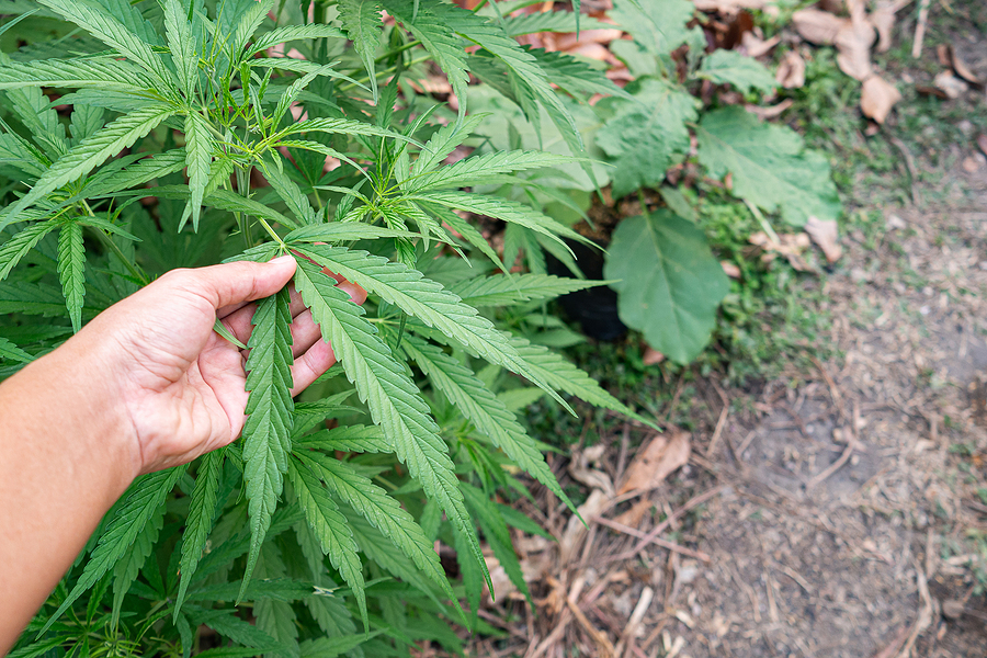
Increased strength of marihuana preparations

Dr. Wang, an associate professor of pediatrics at the University of Colorado Anschutz Medical Campus in Aurora, Colorado noted: “It’s been well documented that the amount of THC that now comes in cannabis is increasing substantially. In the ’90s the average was like 4% or 5%. Now in Colorado, it’s anywhere from 15% to 20%.” This means that THC causes more and more toxicity in patients.

In Colorado medical marijuana was legal since 2009 and recreational marijuana was legal since 2014. Dr. Wang researched the cannabis hyperemesis syndrome in Colorado. He found over 800,000 cases of vomiting in Colorado between 2013 and 2018. This was an increase of 29% from the time before marijuana became legal.

Symptoms of cannabis hyperemesis syndrome

The 5 most common symptoms of cannabis hyperemesis syndrome are continuous nausea, repeated vomiting, abdominal pain, weight loss because of decreased food intake and dehydration from fluid loss. Many people have several showers a day because it diminishes their nausea.

Three phases of cannabis hyperemesis syndrome

The cannabis hyperemesis syndrome often presents in 3 stages: the prodromal phase, the hyper emetic phase, and the recovery phase.

During the prodromal phase symptoms consist of nausea and abdominal pain early in the morning. The eating pattern is still normal in this phase. Some people increase their marijuana consumption as they hope to treat the nausea this way. This phase can last for months or years.

During the hyper emetic phase all of the 5 symptoms mentioned above can occur. This phase often continues until the patient gives up all marijuana consumption. This is when the recovery phase starts.

In the recovery phase the patient returns to a normal eating pattern. All of the symptoms gradually disappear. This phase lasts between days to months. But if the patient starts marijuana again, the symptoms return very quickly.

Treatment of cannabis hyperemesis syndrome

Patients who have severe vomiting require treatment in a hospital. The doctor starts intravenous fluids to treat dehydration. Some medicine to stop vomiting helps in the beginning. The physician must convince the patient to completely stop marijuana use, which helps to treat nausea and abdominal pain. Antacid medication like proton-pump inhibitors is useful to treat stomach lining inflammation. Frequent hot showers help to tone down nausea and vomiting. Hot showers seem to work via the hypothalamic brain center, which is responsible both for nausea/vomiting and heat perception. The physician may prescribe small amounts of benzodiazepams to treat anxiety.

By avoiding marijuana in any form THC, which is a powerful stimulus for CB1 and CB2 receptors gets washed out of the system. This allows the endocannabinoid system to rebalance itself. As long as the patient stays away from marijuana there is usually a complete recovery.

Marijuana and Uncontrolled Vomiting

Marijuana and Uncontrolled Vomiting

Conclusion

Since marijuana is legal in many jurisdictions the cannabis hyperemesis syndrome (CHS) occurs more and more often. Emergency teams at hospitals are kept busy treating these types of patients. It appears that CHS develops in patients who use marijuana regularly and use it in higher concentrations. Nausea and vomiting are the most common symptoms. Some patients can cope for a period of time by taking frequent hot showers. But eventually this home remedy does no longer work. A brief hospital stay may help the patient to recover from this syndrome. The patient needs to stay away from marijuana products to recover from CHS completely, otherwise there will be a relapse.

Oct
16
2021

Marijuana Causes Schizophrenia and Heart Attacks

Two new studies showed that marijuana causes schizophrenia and heart stacks. Marijuana use has been increasing significantly in the general population since the 1990’s. Initially many believed that marijuana would be harmless. But increasingly there are medical publications showing the opposite.

In the following I present data how the use of marijuana causes these two documented side effects, heart attacks and schizophrenia.

Schizophrenia increased since the 1990’s

A July 21, 2021 study from Denmark included all people of Denmark who were older than age 16 from January 1, 1972 to Dec. 31, 2016. The number of participants were 3 ,595 ,910 women (50.0%) and 3 ,590 ,924 men (50.0%). The total number of individuals in the study were 7,186 ,834.

Here are the figures of the percentages of cannabis related schizophrenia cases in Denmark according to the study:

  • 1995: 2% of all schizophrenia cases related to cannabis use in Denmark
  • 2000: 4% of schizophrenia cases due to cannabis use
  • 2010: 8% of all schizophrenia cases from cannabis use

The researchers found that the risk of coming down with schizophrenia heightens with increased cannabis use. Heavy users are more likely to develop schizophrenia than light users.

Cannabis use disorder

One subgroup of cannabis users are people with a cannabis use disorder. They use cannabis, but they become tolerant to it. This requires a higher dose of cannabis to achieve satisfaction. But they are unable to reduce cannabis. They spend more and more time to obtain cannabis, use it and recover from the effect. They give up other activities in favor or cannabis and they continue the use despite negative consequences. Researchers found that this group of individuals had the highest risk to develop schizophrenia. The authors of the Danish study found that patients with cannabis use disorder over the past 2 decades have increased their risk for developing schizophrenia by 3- to 4-fold. They feel it is because of the increase in use and the increase in potency of cannabis.

Heart attacks increased with cannabis use

A Canadian study assessed a cross-sectional study of 2017 and 2018.

The study in question was the American Behavioral Risk Factor Surveillance System survey of US adults. The authors zeroed into young adults (aged 18–44 yr.) with recent cannabis use and a history of a heart attack. Among 33,173 young adults there were 4,610 respondents with recent cannabis use (17.5%). 61 respondents among the recent cannabis users reported that they had a heart attack, which is 1.2%. In comparison non-cannabis users had a heart attack rate of only 0.8%. The adjusted odds ratio for cannabis users compared to non-users was 2.07. This means that the probability of getting a heart attack when using cannabis was 2.07-fold higher when compared to non-users. Also, the investigators found that a history of a heart attack had a probability of being due to cannabis use with a probability of 2.31-fold.

Cannabinoid receptors

Researchers found endogenous cannabinoids in the brain that function as messenger molecules. They need to activate their targets, the cannabinoid receptors, called CB1 and CB2. CB1 receptors are found mainly in the central nervous system. CB2 receptors are located mainly in the immune system. Reproductive organs have their own cannabinoid receptors. The lining of the uterus contains only CB1 receptors. Ovaries and testicles both use CB1 and CB2 receptors. Tetrahydrocannabinol (THC) from smoking or ingesting marijuana is the main psychoactive compound in cannabis. It stimulates these cannabinoid receptors also. But compared to the body’s own cannabinoids THC is much stronger. This leads to more pronounced effects that concern many physicians.

The overwhelming response of the cannabinoid receptors to THC leads to a blunting of the signals of the body’s own cannabinoids. This causes a breakdown in communication between neurons and body cells.

Marijuana Causes Schizophrenia and Heart Attacks

Marijuana Causes Schizophrenia and Heart Attacks

Conclusion re. marijuana causes schizophrenia and heart attacks

Legislators in Canada and many of the states in the US legalized the use of marijuana. As a result, the cannabis use in the younger age group of adults (between 18 to 44 years) has increased significantly. This means that more and more people are exposed to THC from cannabis, which overstimulates the body’s own endocannabinoid system. As explained this leads to a breakdown of communication between neurons and body cells. In time diseases like heart attacks and schizophrenia can develop, a fact overlooked by the media and the public. The overuse of marijuana leads to more than a 2-fold risk to get a heart attack and a 3- to 4-fold risk of coming down with schizophrenia. These are the facts right now. But with further increased use of cannabis researchers will likely find many other diseases that THC can trigger.

Nov
28
2020

Mental Illness and Covid-19

A topic less discussed is mental illness and Covid-19. Covid-19 has been noticeably around us since March of 2020. With the various social distancing and quarantining methods people become more or less isolated emotionally. Social gatherings are outlawed depending on where you live. This helps to interrupt the spread of the virus, but it makes people feel more stressed as their relationships get interrupted.

Symptoms of stress

The CDC sums up that stress during a pandemic can cause the following:

  • Fear of your own health and the health of your loved ones
  • Loss of your job or financial support
  • Changes in eating habits
  • Changes in sleep pattern with lack in concentration
  • Increase in use of tobacco, alcohol or other substance consumption
  • Chronic health problems may deteriorate
  • Worsening of Mental health conditions

The more isolated we are, the more stress we feel. This undermines our stress coping mechanisms, makes us more anxious and more depressed.

Develop coping skills

Taking care of your family and friends may relieve your stress, but it needs to be balanced by taking care of yourself. Although you are physically isolated from others when you are in quarantine, you can stay in contact with others by phone or video chats. This makes you less lonely and isolated.

Take care of yourself. This includes eating well balanced meals, exercising regularly and getting enough sleep. Avoid the consumption of alcohol, tobacco and drugs. Share with a family member or friend how you feel. Maintain friendships through the phone, emails, social media and computer chats. This builds a strong support system for you.

Avoid too much exposure to news stories. Seeing negative stories about Covid-19 over and over again can be undermining, so take a break from the news media. Go for a walk. Take a nap.

Suicide

Sometimes depression can get out of control to the point of driving a person to the brink of suicide. In the US major depression is the leading cause of disability for ages 15-44.  At any given year 16.1 million American adults (about 6.7% of the U.S. population age 18 and older) suffer from major depression. 10.3% of Americans have thought about suicide. If you feel that way, do not act on this impulse. Call 1-800-799-4889. That is the National Suicide Prevention Lifeline & Chat.

Medical literature about the psychological impact of Covid-19

In a July 2020 publication the authors pointed out that Covid-19 infection is a significant psychological stressor. There is the fear of getting the virus and uncertainty about the future. Pre-existing anxiety and depression disorders get accelerated from the stress. In addition, mental disorders like schizophrenia with psychosis may worsen. This may require more antipsychotic medication to control it.

A Kaiser Family Foundation poll found in March 2020 that the Covid-19 pandemic has caused a major impact on US citizens. 45% of respondents said that the pandemic has affected their mental wellbeing significantly. If you are scared, depressed or anxious, you are tense and you have problems falling asleep, you are not alone. You can seek and receive help!

Prevention of mental disease from Covid-19

Here is some practical advice from the World Health Organization.

  • Stay informed.
  • Have a daily routine. Get up and go to sleep at regular times. Eat healthy balanced meals. Exercise regularly. Have your work routine and also find time to rest. Do things that you enjoy.
  • Watch the news only to be informed, but limit news watching to a minimum. This will keep anxiety and depression in check.
  • Stay in contact with close friends by phone and computer technology.
  • Keep your alcohol consumption low. If you did not drink before at all, don’t start drinking in an attempt to deal with boredom, fear, anxiety or social isolation.
  • Take breaks from screen time. This reduces tension.
  • Video games: doing this may relax you for a short time. But longer video game activities can make you tense; you should balance this with the rest of your daily activities.
  • Social media. Use this for honest communication. Keep it brief. If you see misinformation, point out the truth.
  • Helping others: if you can, help others in the community with shopping or other chores.

Treatment for Mental disease from Covid-19

When you notice anxiety, depression or a flare-up of psychotic symptoms from schizophrenia, it is important to see your health professional right away. You may need some counselling. Others may need an antidepressant (for depression) or an adjustment to your antipsychotic medication. Some patients with milder depression or anxiety may benefit from cognitive/behavioral therapy. Most importantly, know that help is available! More info about cognitive therapy for suicide prevention.

Mental Illness and Covid-19

Mental Illness and Covid-19

Conclusion

Mental illness and Covid-19 is a huge topic. People definitely have experienced more anxiety and depression since the Covid-19 epidemic. This is because our lifestyle is suddenly restricted. We may have to go through a 14-day quarantine. But we also cannot participate in large gatherings with more than 50 people. Often, we have to wear masks in stores. In areas with high cases of Covid-19 there is also a severe restriction regarding with whom you can visit, leading to feelings of isolation. All of this can lead to mental illness. Essentially there are two ways of coping with this. First, we need to get a routine that makes us more resistant to mental illness. Secondly, if this fails, seek the advice of a health professional right away.

Seek mental illness treatment right away

When you catch mental illness early, it is much easier to treat. Milder cases of depression or anxiety may benefit from cognitive/behavioral therapy. More severe cases often require medication. In this outline I have discussed mental illness during the Covid-19 epidemic. I have not discussed the cases where people came down with Covid-19 coronavirus and got affected in their brain function from the virus. These cases need treatment by a neurologist and psychiatrist, and they are much more complicated.

Apr
18
2020

Changes of Metabolism by Inflammation

Dr. James LaValle gave a presentation about changes of metabolism by inflammation in Las Vegas. I listened to this lecture on Dec. 15, 2020. The 27th Annual World Congress on Anti-Aging Medicine in Las Vegas took place from Dec. 13 to 15th, 2019. His original title was: “Innovations in Metabolism and Metaflammation”. This talk was complex and as a result it may not be easy reading. But it shows how various factors can affect our metabolism and our life expectancy.

In the first place he understands “metabolism” as all of the chemical reactions together that make you feel the way you feel today. In the same way metabolism is the chemistry that drives you toward future health. It is equally important to note that disregulation of your metabolism occurs from global metabolic inflammatory signalling. As has been noted he called this “metaflammation” (inflammation affecting your metabolism).

Dr. LaValle said that understanding disruptors of your metabolism can lead to renew your health on a cellular level. The key to achieve this is to remove inflammatory signals.

Factors that accelerate aging and damage your metabolism

It is important to realize that several factors interfere with the normal aging process. Oxidative stress and inflammation are major factors. But hormone disbalance and increased blood sugar values and insulin resistance can also contribute to accelerated aging and damage your metabolism. Certainly, with a disturbance of the immune balance, autoimmune reactions can take place, which also does not help. In addition, pollutants from the environment derange the metabolism due to heavy metals that block important enzymatic reactions. In the minority there are also genetic factors that can interfere with a normal metabolism.

Many of the metabolic changes can lead to chronic inflammation. One source of inflammation can be lipopolysaccharides that stimulate the immune system to start an inflammatory process.

Many conditions are associated with inflammation such as diabetes, obesity, stress, the SAD diet (standard American diet), and liver or kidney damage.

How Metaflammation is developing

Metaflammation can start in the gut with microbiota alterations. The wrong types of bacteria can release lipopolysaccharides, and low grade endotoxemia develops. With obesity inflammatory kinins start circulating in the body. Stress can activate inflammatory substances in the brain and the rest of the body. Major contributors to inflammation in the body come from faulty diets. The Western diet contains too much sugar and refined carbs; it is too high in trans fats and saturated fats. It contains too many artificial additives, preservatives, salt, sweeteners and dyes. And it is too low in nutrients, complex carbs and fiber.

More problems with metaflammation

Kidney and liver illness can contribute to metaflammation. Several diseases come from chronic inflammation, like cardiovascular disease, type 2 diabetes, chronic kidney disease, depression, cancer, dementia, osteoporosis and anemia. Metaflammation alters the methylation patterns, which can slow down your metabolism. Increased blood lipids and chronic inflammation of the blood vessels lead to cardiovascular problems. The liver and kidneys are the major detoxification organs, and their disease leads to more metaflammation. Metaflammation also leads to hormone disbalances, sleep disorders and dysfunction of the immune system. The brain reacts to metaflammation with cognitive dysfunction and mood disorders. Muscle loss (sarcopenia) is another issue, so is osteoporosis. Finally, chronic metaflammation can cause cancer.

Major causes of metaflammation

The three major causes of metaflammation are changes of the gut microbiome, obesity and chronic stress. When the gut bacteria change because of a Western diet, the wrong bacteria release lipopolysaccharides that are absorbed into the blood. The gut barrier is breaking down and a low grade endotoxemia develops. With obesity adipokines, which are inflammatory substances secreted by the fatty tissue, circulate in the blood. Chronic stress activates inflammation in the brain and in the body.

Two major conditions are common with metaflammation: hyperlipidemia (high fat levels in the blood) and hyperglycemia. Both of these conditions change the metabolism and lead to cardiovascular disease (hyperlipidemia) or to type 2 diabetes (hyperglycemia). Both of these metabolic changes lead to one or more of the conditions mentioned above, accelerate the aging process and lead to premature deaths.

Interaction of various organ systems can cause metaflammation

Dr. LaValle stated that it is vital that your hormones stay balanced. With chronic stress cortisol production is high. This causes increased insulin production, reduced thyroid hormone and lowered serotonin and melatonin production in the brain. It also leads to autoimmune antibodies from the immune system and decreased DHEA production in the adrenal glands. In addition, growth hormone production and gonadotropin hormones are slowing down. We already heard that cortisol levels are up. The end result of these hormone changes is that the blood pressure is up and abdominal visceral obesity develops. The brain shows cognitive decline, with memory loss as a result. The bones show osteopenia, osteoporosis and fractures. The muscles shrink due to sarcopenia, frailty is very common. Heart attacks and strokes will develop after many years. The immune system is weak and infections may flare up rapidly. There are also higher death rates with flus.

Other mechanism for pathological changes with hormone disbalances

When Insulin is elevated, inflammatory markers are found in the bloodstream. This elevates the C-reactive protein and leads to damage of the lining of the blood vessels in the body. A combination of insulin resistance and enhanced atherosclerosis increases the danger for heart attacks or strokes significantly.

There is a triangle interaction between the thyroid, the pancreas and the adrenals. Normally the following occurs with normal function. The thyroid increases the metabolism, protein synthesis and the activity of the central nervous system. The pancreas through insulin converts glucose to glycogen in the liver. It also facilitates glucose uptake by body cells. The adrenal hormones are anti-inflammatory, regulate protein, carbohydrate and lipid metabolism and contribute to energy production.

Change of thyroid/pancreas/adrenals triangle when cortisol is elevated

When cortisol is elevated the balance of the thyroid/pancreas/adrenals’ triangle is severely disturbed. Cortisol is high, the T4 to T3 conversion is limited and, in the brain, there is hippocampus atrophy with memory loss and brain fog. The immune system will change with production of inflammatory kinins (IL-6 and TNF alpha). Insulin sensitivity is down, sugar craving up and weight gain develops (central obesity).

Change of thyroid/pancreas/adrenals triangle when the thyroid is depressed

The thyroid activity can be lower because of autoimmune antibodies (Hashimoto’s disease) or because of hypothyroidism developing in older age. This leads to decreased pregnenolone synthesis from cholesterol. As pregnenolone is the precursor for all the steroid hormones, the metabolism slows down profoundly. Mentally there is depressed cognition, memory and mood. The cardiovascular system shows reduced function. In the gut there is reduced gastric motility. The mitochondria, which are tiny energy packages in each cell, are reduced in number, which causes a loss of energy. There is increased oxidative stress, increased lactic acid production and decreased insulin sensitivity.

Cardiovascular disease not just a matter of high cholesterol

Dr. LaValle stressed that a heart attack or stroke is not just a matter of elevated cholesterol. Instead we are looking at a complicated interaction between hypothyroidism, diabetic constellation and inflammatory gut condition. The inflammatory leaky gut syndrome causes autoimmune macrophages and Hashimoto’s disease. The end result is hypothyroidism. The inflammatory kinins (TNF-alpha, IL-6) affect the lining of the blood vessels, which facilitates the development of strokes and heart attacks. You see from this that cardiovascular disease development is a multifactorial process.

Microbiome disruption from drugs

Drugs affecting the intestinal flora are antibiotics, corticosteroids, opioids, antipsychotics, statins, acid suppressing drugs like protein pump inhibitors (PPI’s) and H2-blockers. Other factors are: high sugar intake, pesticides in food, bactericidal chemicals in drinking water, metformin, heavy metals and alcohol overconsumption. Chronic stomach infection with H. pylori, stress and allergies can also interfere with the gut microbiome.

The microbiome disruption affects all facets of metabolism. This means that there can be inhibition of nutrient absorption and this may affect the gut/immune/brain axis. There are negative effects on blood glucose levels and insulin resistance. A disturbance of the sleep pattern may be present. A significant effect on the hormonal balance can occur (thyroid hormones, sex hormones and appetite related hormones). When liver and kidney functions slow down, there is interference of body detoxification.

Dr. LaValle talked more about details regarding the gut-brain-immune pathology. I will not comment on this any further.

Changes of Metabolism by Inflammation

Changes of Metabolism by Inflammation

Conclusion

Dr. LaValle gave an overview in a lecture regarding changes of metabolism by inflammation. This took place at the 27th Annual World Congress on Anti-Aging Medicine in Las Vegas from Dec. 13 to 15th, 2019.

This article is complex and contains a lot of detail, but there is one simple truth: oxidative stress and inflammation are major factors that influence our health on many parameters and lead to a list of illnesses. They lead to hormone disbalance and increased blood sugars and insulin resistance, which can also contribute to accelerated aging and damage of your metabolism. Dr. LaValle explained how high cortisol from chronic stress can lead to low thyroid hormones and in the brain, there is hippocampus atrophy with memory loss and brain fog. With alterations of the immune system there is production of inflammatory kinins (IL-6 and TNF alpha). Insulin sensitivity is down, sugar craving up and weight gain develops (central obesity). It does not stop there! We put our hope in medications, but the sad truth is that there are

Drugs that change the gut biome

Many drugs that are common also change the gut biome with resulting increased permeability of the gut wall (leaky gut syndrome). This overstimulates the immune system and leads to autoimmune diseases like Crohn’s disease and rheumatoid arthritis. Whenever there is an injury to the gut barrier, the blood brain barrier is following suit. This is how brain disease can develop as a result of a change in the gut biome. Impaired cognition, memory and mood can result from this. Alzheimer’s disease is one of the worst conditions that may be related to a combination of gut inflammation, chronic stress and inflammatory kinins.

Feb
29
2020

Celiac Disease in Various Disguises

Dr. Tom O’Bryan gave a lecture in Las Vegas on Dec. 13, 2019 about celiac disease in various disguises. This was at the 27th Annual World Congress on Anti-Aging Medicine. The title of his talk: “An Ounce of Prevention Is Worth a Pound of Protocols: Halting Our Brains Slow Deterioration”.

Case # 1: 44-year old male with an assumed diagnosis of ALS

In the first place a 44-year old male had a history of a right leg weakness that developed over the last 6 months. He had intermittent spasms in his right quadriceps muscle. In addition, over the last few months he noticed a weakness develop in his right arm with difficulties writing. Significantly, his family history revealed that a maternal aunt had celiac disease. Moreover, a sister had Crohn’s disease and his maternal grandmother had multiple sclerosis. Electromyographic studies showed widespread acute denervation. An MRI scan of the spine showed hyperintensity in the corticospinal tracts. A diagnosis of amyotrophic lateral sclerosis (ALS) followed as a result based on the MRI scan findings.

Further tests

At the same time blood tests revealed that his anti-endomysial antibodies were elevated.  Duodenal biopsy demonstrated villous atrophy, crypt-hyperplasia and increased intraepithelial lymphocytes consistent with gluten-sensitive enteropathy (celiac disease). An MRI scan of the brain also showed some hyperintense lesion in the left-brain hemisphere.

Gluten free diet instituted

It was clear with these test results that the initial diagnosis was a misdiagnosis. The real diagnosis was celiac disease. 7 months after the onset of his symptoms he started on a gluten free diet. He received no medications. Notably, his right arm function returned to normal after 9 month of the gluten free diet. Although there was some improvement in his right leg function, he still had some muscle wasting and spasticity in his right leg. However, he could now walk without any aid. His hand-writing was back to normal, and he could button his shirts again. Repeat MRI scans followed 2 months and 9 months after the start of the gluten free diet. At two months after initiation of the gluten free diet the brain lesion in the left brain was somewhat larger than before, but at 9 months it was half the original size.

Case #2: Autism in children, youth depression and Alzheimer’s patients

The autism spectrum disorder (ASD) has significantly increased from 1 in 166 in 2004 to 1 in 40 in 2018. In addition, Dr. O’Bryan also mentioned that in 2017 statistics showed that 13.3% among youth aged 12 to 17 in the US suffered major depressive episodes. 1 in every 12 youth suffer from severe behavioral and emotional problems. According to the CDC since 1994 the number of children on psycho-stimulants increased 5-fold. In the same time children under 18 with bipolar disorder have increased 40-fold. There has been a 6-fold increase of prescriptions for antipsychotic medications for children in the same time frame.

Other effects on adolescents

However, I like to point out that there are other powerful factors that can explain increased depression and emotional problems in adolescence. The Canadian Medical Association published an article about social media and smart phones and the effects they have on adolescence.

On the other end of the life cycle 1 in 3 seniors die with Alzheimer’s disease or dementia. Between 2000 and 2015 death from Alzheimer’s disease has increased by 213%.

Breakdown of the blood brain barrier

According to Dr. O’Bryan autism in children, behavioral and emotional problems in teenagers and dementia from Alzheimer’s disease are all related to the same process, namely a breakdown of the gut barrier, often called leaky gut syndrome. It is important to realize that this leads to a secondary breakdown of the blood brain barrier. The end result is a compromise of the brain, where antibodies attack the brain protein. In young children this causes lower adaptive and cognitive function and behaviors typical for autism. Teenagers are more likely to present with depression or schizophrenia. In older people the breakdown of the blood brain barrier can result in Alzheimer’s disease and other forms of dementia.

25 to 30% of protein in wheat are non-gluten. Antibodies can be directed against gluten, but also against non-gluten protein.

IgG antibodies against gluten cross placenta

In the later stages of pregnancy IgG antibodies cross the placenta easily. They provide passive immunity from various viral infections. Unfortunately, antibodies against gluten also cross through the placenta, which can lead to a breakdown of the fetal gut lining, in the sense of leaky gut syndrome. In this study 211 children were found to have a risk of 1.7-fold to develop psychosis later in life. The mothers were positive for anti-gliadin IgG antibodies in the last 4 weeks of pregnancy. Anti-casein antibodies did not have this psychosis effect (risk only 0.8-fold). The investigators felt that an allergy to wheat in the mother set up general inflammation. Psychosis in the offspring only develops when inflammatory mediators reached the brain of the fetus. It was the brain inflammation, which caused the subsequent psychosis later in the child.

Blood brain barrier and healthy gut barrier

Another key point is that the barrier both in the gut and in the blood brain barrier consists of a single epithelial layer. The cells are held together by zonulin and occludin proteins. Autistic children were exposed already in the uterus to mother’s wheat induced anti-gliadin antibodies. This led to a break-down of the children’s blood brain barrier and the symptoms of the autism spectrum disorder. These children have a lot of brain inflammation and in addition often have impaired gut barrier integrity. It must be remembered that they require a comprehensive program to improve the gut flora, build up the gut barrier integrity and re-establish the blood brain barrier.

Effects of phthalates on young children

A 2014 study measured urinary metabolites of phthalates and related this to the children when they were 7 years old.

The investigators did several cognitive tests and measured the IQ (Wechsler Intelligence Scale). Children of mothers with the highest quartile of phthalates had an IQ, which was on average 7.0 points lower than the control group of the lowest quartile of phthalates. Dr. O’Bryan showed a slide taken from this study.

With this in mind, it points out that a pregnant woman has an intact blood brain barrier, which prevents antibodies from entering. However, the immature brain of the fetus has not developed an efficient blood brain barrier yet. This allows maternal gliadin antibodies from wheat intolerance to enter the fetal brain and cause autism spectrum disorder (ASD).

PCB’s disrupt the blood brain barrier

In mouse experiments the effects of PCB’s were investigated. By the same token, researchers found that the blood brain barrier was broken down by PCB’s that are known to have carcinogenic and neurotoxic properties on the brain. The researchers injected melanoma cells into the animals and found that the PCB pretreated mice sustained brain metastases. However, the control animals that did not have PCB pre-treatment did not develop brain metastases. They concluded from this that PCB’s are breaking down the blood brain barrier.

Maternal brain antibodies causing autism in children

This publication examined antibodies to two different brain proteins. The researchers found that 86% of the children from mothers with two different fetal brain antibodies were diagnosed with autistic regression. According to this publication there are at least 50 different epitopes of gluten peptides that exert cytotoxic, gut permeating and immunomodulatory activities.

DNA microarray technology can now detect many subtypes of food disorders and gluten sensitivities. The tests for celiac disease have a sensitivity of 97% for IgG and 99% for IgA. With regard to specificity the test is now 98% accurate for IgG and 100% for IgA.

Case #3: 34-year old female vegan patient with depression and mild cognitive decline

A 34-year old woman has followed a Vegan lifestyle for 10 years. She has been working long hours and had a lot of stress. In addition, her thyroid was borderline low with high TPO antibodies. A blood test for vitamin D showed vitamin D deficiency. For the past year her energy level was low and she had developed chronic depression. Her physician did a genetic test that found she carried the gene that converts GABA into glutamate. She thinks that she has small intestinal bacterial overgrowth (SIBO). A review of her dietary habits revealed that she ate more cooked foods and less raw food. Her memory is slightly off, her speech not as fluid and she has some cognitive decline.

Her blood tests showed anti-immunity to RAGE peptides. To put it another way, RAGE stands for “receptor for advanced glycation end products”. When you eat too much overcooked foods you ingest advanced glycation end products. This can have adverse effects on your body, particularly the brain.

More tests regarding this woman

Another specific test revealed a blood brain barrier disruption with the presence of anti-brain antibodies. A stool sample was obtained. It showed low Akkermansia, low Faecali bacterium, low Bifido longum and low Bifido adolescentis bacteria. A chemical analysis revealed low butyrate, propionate and acetate. The treating physician concluded that she had a gut dysbiosis and a dysfunctional gut barrier. This has also affected her blood brain barrier. The constellation of symptoms and blood tests explain her clinical condition. She has developed autoantibodies that affect her thyroid gland and her brain because of the antibodies against her RAGE peptides. People can develop Alzheimer’s disease given enough time with exposure to these antibodies. The leaky gut has led to a break-down of her blood brain barrier and exposed her brain to autoimmune antibodies directed against brain cells.

Treatment of gut dysbiosis

This patient started a gluten free diet (GFD). But one of the problems of the GFD is that wheat is removed that normally provides 69% inulin and 71% oligofructose, both important prebiotics that are necessary for probiotics to work with. Inulin is contained in beets, leeks, asparagus, onions, garlic and bananas. Oligofructose is contained in chicory root, bananas, onion, and garlic.

When people consume a typical Western diet, they get between 1 and 4 grams of inulin daily. But others who eat balanced diets get up to 25 to 100 grams of inulin per day. Dr. O’Bryan explained that going on a GFD leads to an altered microbiome.

Experiment with volunteers to measure the effects of a gluten free diet

He discussed an experiment on 10 healthy volunteers who were fed a GFD for 1 month.

The researchers ordere stool samples in the beginning and at the end of the experiment. There was less of the good bacteria and more of the the bad bacteria. This led to a less protective and more inflammatory environment. The remedy for that is to eat 1 root vegetable and 2 other prebiotics per day. The patient on a GFD must supplement with prebiotic-rich foods to prevent this from happening.

Non-digestible oligosaccharide supplement

Inulin and oligosaccharides support the intestinal microbiota.  Dr. O’Brien suggested to add a supplement, called Precision Prebiotic™, non-digestible oligosaccharides that can increase microbial diversity. This supplement supports the growth of the healthy bacteria. These are keystone bacteria like Akkermansia muciniphila, Faecal bacterium prausnitzii, and Bifidobacteria.

Other supportive measures for the gut

  • 1 tablespoon of fermented vegetables like sauerkraut once per day
  • The ingestion of fermented foods increases the beneficial gut bacteria by a factor of 10,000-fold!
  • A 100% spore-based probiotic supplement increases diversity of the gut flora and helps to maintain the gut barrier
  • Sodium butyrate, which comes from fermented food is an important modulator of the central nervous system
  • In addition, sodium butyrate also inhibits pathological gut bacteria and maintains the gastrointestinal balance
  • In neurodegenerative disorders sodium butyrate provides anti-inflammatory and neuroprotective effects
  • Sodium butyrate restores the blood brain barrier
  • Following heart attacks or strokes sodium butyrate promotes tissue repair and recovery through cell survival
Celiac Disease in Various Disguises

Celiac Disease in Various Disguises

Conclusion

Dr. Tom O’Bryan delivered a lecture at the 27th Annual World Congress on Anti-Aging Medicine in Las Vegas on Dec.13, 2019. Wheat allergies have increased in the last decades. Researchers have found that in many people there is a deterioration of the gut flora and a breakdown of the gut barrier. This leads to antibody formation against gluten or gliadin (wheat proteins). This exposes the body to many proteins from the gut. The body reacts by producing antibodies to them. These are also affecting cells in the body as they cross react with body proteins. The inflammation from the autoantibodies cause the blood brain barrier to break down. Now the immune system can produce antibodies against brain tissue. In the past  with an intact blood brain barrier this was not possible.

Autoantibodies in various life epochs

At a young age autism can develop because of antibodies against gliadin from wheat. In our youth schizophrenia and depression can occur from gut dysbiosis and a subsequent break down of our blood brain barrier. In old age Alzheimer’s disease develops in 1 out of 3 people due to gut dysbiosis and a breakdown of the blood brain barrier with anti-brain antibodies. Dr. O’Bryan explained how a person can turn this negative spiral around and start a new life without these problems. You can avoid a lot of these diseases by eliminating wheat and processed food from your diet.

Oct
28
2017

Take Enough Vitamin D3

Many people supplement with 300 to 400 IU of vitamin D3, but do they take enough vitamin D3? There is a simple way of finding out: ask your doctor to order a 25-hydroxyvitamin D blood test.   This will show whether the gut absorbed enough of the essential vitamin. It will also show whether or not your vitamin D3 capsules or tablets were strong enough. It is now generally accepted that a good range of the vitamin D blood level is between 50 and 80 ng/ml. Unfortunately many Americans who come down with various diseases have blood levels of less than 30 ng/ml. Here are some facts about what a lack of vitamin D3 can cause.

Increased risk of mortality with lower vitamin D levels in ICU patients

  1. A New England Journal study from 2009 reported about 1100 patients in Intensive Care Units (ICU). Their average vitamin D blood level was only 16 ng/ml. They tracked the mortality rates depending on the vitamin D blood level. Insufficient vitamin D levels showed an association with a mortality rate of 45%. An intermediate level had a mortality rate of 35%. And a satisfactory level of vitamin D had a mortality of only16%. Between the low level of vitamin D and the normal level there was a 3-fold difference in mortality!
  2. Another study from 2015 repeated the mortality study with 135 ICU patients. Researchers correlated Vitamin D blood levels with mortality rates of patients. When vitamin D levels were below 12 ng/ml, there was a mortality rate of 32.2%. Patients with higher levels of vitamin D had a mortality rate of 13.2%. The authors concluded that vitamin D blood levels were an independent risk factor for mortality. Patients less than 12 ng/ml had a 2.4-fold higher risk of dying than patients with normal vitamin D levels.

Do patients with multiple sclerosis take enough vitamin D3?

Perhaps one of the earliest results of vitamin D3 research was the following observation. More than 90% of patients with multiple sclerosis were deficient in vitamin D blood levels. Their levels were below 20 ng/ml. Other researchers showed that vitamin D could directly tone down the aggressiveness of the immune cells of MS patients. These were the ones that attacked the myelin sheath. As a result of this knowledge it is important for MS patients to take high enough vitamin D3 supplements. When they reach good vitamin D blood levels their MS is better controlled.

Canada as a northern country has 291 MS patients per 100,000 people. Contrast this to 110-140 MS patients per 100,000 people in the northern US (between the 37th parallel and the US/Canadian border). In addition south of the 37th parallel there are only 57-78 cases of MS per 100,000 people. Researchers have concluded that the less sun light people get, the higher the rate of MS in the population will be. However, instead of sun exposure you can supplement with vitamin D3 capsules to get the blood vitamin D levels up to the range of between 50 and 80 ng/ml.

Do stroke patients take enough vitamin D3?

Strokes are very common. About 6.8 million Americans survive a stroke and live with various disabilities. 15% die shortly after their stroke. 40% are left with moderate to severe disabilities. Many require special care.

  1. Studies have shown that patients with the lowest level of vitamin D have the poorest functional outcomes. Moreover, for every 10 ng/ml decrease in vitamin D levels the odds of a healthy recovery 3 months after the stroke fell by about half. This was independent of age and the initial stroke severity.
  2. In another 2015 study from South Korea 818 stroke patients took tests to evaluate whether they had adequate vitamin D blood levels. There was a clear division between those whose levels were higher than 10 ng/ml or lower. When the vitamin D level was higher, there was a 90% better recovery from their stroke after 3 months. In comparison those whose vitamin D levels were below 10 ng/ml had poor recovery rates. Experts say that vitamin D levels should stay in the range between 50 and 80 ng/ml. This will prevent numerous diseases.

Do diabetics take enough vitamin D3?

  1. Vitamin D3 can silence diabetes genes in connection with the right diet and cofactors of zinc and magnesium. A Mediterranean diet can stabilize the metabolism and fight inflammation. Zinc and magnesium are important cofactors in enzymes necessary to prevent diabetes. Vitamin D3 and omega-3intake are helping to control inflammation and preserve beta cells in the pancreas in diabetes patients. This is important for continued production of insulin.
  2. A Chinese research team found that vitamin D3 protects beta cells in the pancreas from dying off. The finding was that vitamin D3 receptors in the insulin producing cells prevented the dying off of these cells, as long as there was enough vitamin D available. Insulin production by the pancreas remained effective. And insulin is vital for long-term survival of diabetes patients. The key for diabetes patients is to take adequate doses of vitamin D3 to protect their insulin producing beta cells.
  3. A 2015 Italian study showed that micro vascular complications in diabetes patients were high, if the vitamin D3 blood levels were low. If patients had high levels of vitamin D3, there were no complications such as retinopathy or nephropathy. But if levels were below 20 ng/ml, damages were significant in the capillaries of the eyes and kidneys.

Do patients with inflammatory conditions take enough vitamin D3?

What do the lining of the arteries, the inflamed joints, a degenerative meniscus and heart attacks and strokes have in common? It is the inflammation that changes the body chemistry. It gets even more complicated, because the extra calories that we consume get stored as visceral fat. This is done automatically when you eat too much sugar and starchy foods. When the glycogen stores are full, any surplus sugar gets metabolized by the liver into triglycerides, fatty acids and LDL cholesterol and gets stored as body fat. The most active fat is the visceral fat between our guts and around our body organs. This produces interleukins and other inflammatory cytokines that circulate in the blood causing inflammation in all our arteries. Interleukin-6 is an inflammatory cytokine. High interleukin-6 levels contribute to causation of various cancers.

This 2015 study from Seattle University followed 218 obese postmenopausal women with a body mass index of larger than 25.0 for 12 months. Both received weight loss intervention and either 2000 IU of vitamin D3 daily or a placebo pill. Both groups lost about 5 to 10% of weight in 12 months. However, the interleukin-6 level of the vitamin D3 group had a reduction of 37.3%. This was in stark contrast to the placebo group where the interleukin-6 level reduction was only 17.2%. This type of research shows the incredible power of vitamin D3. This likely is the reason why several cancer frequencies can show a reduction with regular vitamin D3 supplementation.

Attention deficit disorder and vitamin D3

  1. Other research compared a group of 37 children with attention deficit hyperactivity disorder (ADHD to 37 normal children. Blood levels of vitamin D were 19.11±10.10 ng/ml in the ADHD group and 28.67±13.76 ng/ml in the normal group. Other researchers have found similar findings, establishing that very low vitamin D levels have a connection with ADHD.
  2. A prospective study from Spain involving 1,650 mother-child pairs investigated the effect of mother’s vitamin D level during her pregnancy with the risk for ADHD by the time the child was 4 to 5 years old. Schoolteachers followed the standard test procedures to establish the ADHD diagnosis. The study showed that for every 10-ng/ml increment of the mother’s blood vitamin D level during her pregnancy the children had 11% less ADHD-like symptoms. The authors cautioned that it takes mega doses of vitamin D3 to reach these kinds of results. The usual 400 IU of vitamin D3 per day will not achieve the desired increase of vitamin D3 levels, but amounts of 5,000 IU to 8,000 IU are necessary to achieve this.

Schizophrenia and vitamin D3

A 2014 Meta analysis found that low vitamin D levels have an association with a 2.16-times higher probability of having schizophrenia than controls with normal vitamin D levels. Another study examined whether those patients who had an acute psychosis would have lower vitamin D blood levels than schizophrenia patients in remission or control patients without schizophrenia. Studies compared 40 patients with an acute psychosis to 41 patients in remission and 40 healthy controls. Patients with an acute psychosis had extremely low vitamin D blood levels, while patients in remission had much better vitamin D levels. Healthy controls had the best vitamin D levels.

Absorption and metabolism of vitamin D3

Magnesium plays a central role in activating vitamin D3. This publication points out that magnesium is also necessary for absorption of vitamin D3 in the gut. The activation of vitamin D3 is also partially responsible for vitamin D absorption. Both vitamin D3 and magnesium play an important role in bone and calcium metabolism. The fact that every body cell has vitamin D3 receptors shows how important it is for the maintenance of the body. Many researchers say that vitamin D3 qualifies as a hormone because of the specific effects on cells via vitamin D3 receptors.

Take Enough Vitamin D3

Take Enough Vitamin D3

Conclusion

Vitamin D3 is an important signaling hormone and vitamin that regulates the body’s calcium absorption and is responsible for bone metabolism. Research has shown that the lack of vitamin D3 causes several unrelated diseases, like rickets, multiple sclerosis, and schizophrenia. But other diseases, where a lack of vitamin D3 was present, were diabetes, attention deficit disorder and strokes. When patients with elevated inflammatory markers take vitamin D3 their interleukin-6 levels dropped by 37.3%. To achieve this, patients needed to consume at least 2000 IU. We all should have our vitamin D blood level measured from time to time. It should be between 50 and 80 ng/ml. Too many Americans are deficient in vitamin D3 and come down with the diseases mentioned! Prevention and supplementation go hand in hand. You can prevent a lot of diseases this way.


Incoming search terms: